Combined heart defects are also called combined. Damage to one heart valve with the development of stenosis of the orifice and valve insufficiency leads to an increase in the volume of blood entering the chamber and to the creation of increased pressure in the chamber, overcoming the narrowed opening. With each defect, different changes occur in the heart.
- Instrumental diagnostics
- Treatment of valvular heart defects Treatment of aortic stenosis
- Treatment of aortic insufficiency
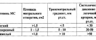
- Treatment of mitral stenosis
- Treatment of mitral regurgitation
- Treatment of tricuspid valve disease
When mitral stenosis and mitral valve insufficiency are combined, dilatation and hypertrophy of the left atrium and dilatation of the LV of the heart are recorded. Therefore, the symptoms will be the same as with both of these defects. Stenosis of the left atrioventricular orifice and mitral insufficiency can be expressed to a greater or lesser extent, therefore remodeling of the heart chambers can occur in several ways. Therefore, when diagnosing, doctors indicate the predominance of one of the defects, for example, mitral heart disease with a predominance of valve insufficiency.
With mitral-aortic disease, the combination of mitral stenosis with aortic insufficiency or mitral stenosis with aortic stenosis is important. In the first two cases, mitral stenosis impedes the flow of blood into the left ventricle.
What is a combined heart defect and how does it differ from other types?
In cardiological practice, it is customary to distinguish between the concepts of acquired (ACH) and congenital heart defects (CHD):
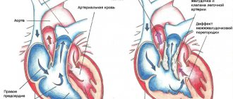
- A congenital defect develops during fetal development or childbirth, and is characterized by defects in the septa between the chambers of the heart, as well as the origin of the great vessels and additional shunts (for example, the ductus ductus);
- PPS is exclusively damage to the valve apparatus, which occurs against the background of concomitant systemic or cardiovascular diseases.
There are two types of acquired valve defects, depending on the anatomical basis of impaired hemodynamics: stenosis (narrowing of the lumen due to the deposition of calcium salts or replacement with dense connective tissue) and insufficiency (incomplete closure of the valves).
Diagnosis of isolated damage does not cause difficulties when combining clinical examination and echocardiography (Echo-CG) data. However, a burdened somatic history contributes to the development of combined variants.
Combined heart disease is a combination of two forms of disorder in one valve structure (combined mitral disease, in which there is evidence of stenosis and insufficiency), the treatment of which involves surgery to install an artificial valve.
It is necessary to distinguish this concept from the combined variant of the defect. The latter represents simultaneous or gradual damage to several structures of the valve apparatus (for example, mitral-aortic stenosis).
In clinical practice, there is no consensus on the use of these terms, so it is permissible to use both options with the diagnosis detailed in parentheses.
Classification of heart defects
Today there are quite a few classifications of heart defects. According to the etiology of occurrence, they distinguish: - Congenital defects that develop as a result of pathology of intrauterine development as a result of external or internal causes. External causes include environmental, medicinal, chemical, viral, and so on. Internal causes include various abnormalities in the parents’ bodies. For example, hereditary background, hormonal imbalance in a woman during pregnancy, and others. — Acquired defects occur at any age, that is, after birth, as a result of heart injuries or some diseases. The most common cause of acquired defects is rheumatism. Also, heart defects can develop as a result of atherosclerosis, myocardial injury, coronary artery disease, syphilis and other diseases. According to localization they distinguish:
- Mitral valve disease is a change in the mitral valve (also called the bicuspid valve), which is located between the left ventricle and the left atrium.
- Aortic valve disease is a pathology in the aortic valve, which is located in the vestibule of the aorta.
- Tricuspid valve disease is a disorder of the tricuspid (three-leaf) valve. Sometimes this is called “tricuspid valve disease,” but this is not a completely correct name, since there is another tricuspid valve in the heart - the aortic valve. The tricuspid valve regulates the flow of blood from the right atrium to the right ventricle.
- Pulmonary valve disease is a pathology of the pulmonary valve, which is located in the vestibule of the pulmonary trunk (artery).
- Unclosed ductus arteriosus (foramen ovale) is a congenital pathology when the ductus arteriosus does not fuse in newborns. Quite often, in the first year of a girl’s life (this type of defect is usually found in girls), the duct itself grows together and, accordingly, the defect goes away.
Atrial septal defect is a pathological abnormality of the septum between the left and right atria. Atrial septal defects can occur in three places:
- upper - sinus venosus,
- middle - ostium secundum
- and the lower part - ostium primum.
Ventricular septal defect is a disruption of the septum between the left and right ventricles. It can also appear in three sections of the septum:
- upper - membranous,
- medium - muscular
- and the lower part - a trabecular defect.
Transposition of the great arteries (vessels) is a critical congenital heart defect, which is characterized by ventricular-arterial discordant and atrioventricular concordant connections. Dextrocardia is an abnormal location of the heart, that is, most of it is located on the right side of the chest, completely symmetrical to normal. In this case, a “reverse” arrangement of outgoing and incoming vessels occurs. Combined defects also occur. The most common of them are:
- Tetralogy of Fallot is a combination of pulmonary stenosis, ventricular septal defect and aortic dextraposition. With this defect, hypertrophy of the right ventricle develops over time.
- Fallot's triad is a combination of pulmonary stenosis, right ventricular hypertrophy and ventricular septal defect.
- Pentade of Fallot is a combination of atrial septal defect and tetralogy of Fallot.
- Ebstein's anomaly is a very rare congenital defect characterized by tricuspid valve insufficiency as a result of the atrioventricular valve, instead of originating from the atrioventricular ring, originating directly from the walls of the right ventricle, which leads to a decrease in the internal volume of the right ventricle and patent foramen ovale.
Based on anatomical changes, they are distinguished:
- Stenosis is a narrowing of the opening of blood vessels or valves and, as a result, an obstruction to normal blood flow.
- Atresia is the absence or narrowing of a vessel, cavity or opening, leading to obstruction or disruption of normal blood flow.
- Coarctation is the same as stenosis.
- Insufficiency or defects are insufficiently developed valves, walls or blood vessels of the heart.
There are two main types: 1. defects of obstruction - these are when valves, veins or arteries are atretic or stenotic 2. and defects of the septum separating the right part of the myocardium from the left. Hypoplasia is underdevelopment, usually of one part of the myocardium, for example the left or right. As a result, the second part (side) of the myocardium performs double the load. A combination of pathologies when two or more of the above anatomical changes are combined. For example, stenosis defect. A combination is when several defects with different localizations are combined, for example, mitral-aortic and so on. In accordance with hemodynamics, they are distinguished: I degree - minor changes. II degree - moderate changes. III degree - sudden changes. IV degree - terminal changes. Also, according to hemodynamics, they distinguish: White defects, when there is no mixing of venous and arterial blood, that is, with a left-right direction of discharge. Among white defects there are four types:
- When enriching the pulmonary (lesser) circulation. For example, with an open foramen ovale, ventricular septal defect, and so on.
- When pulmonary circulation is depleted. For example, isolated pulmonary stenosis.
- With the enrichment of the bodily (systemic) circulation. For example, isolated aortic stenosis.
- Without significant hemodynamic impairment. For example, the disposition of the heart.
Blue heart defects are when venous blood is refluxed into arterial blood when the right-left flow is disrupted. Blue defects are divided into two types: With enrichment of the pulmonary circle. For example, with the Eisenmenger complex or absolute transposition of the great vessels. With depletion of the pulmonary circulation. For example, Ebstein's anomaly or tetralogy of Fallot. Diagnostics For diagnosis, use according to indications:
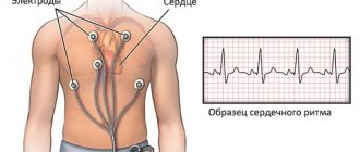
- ECG,
- Heart ultrasound and Doppler echo-CG,
- radiography of the heart and contrast radiography of the heart.
Symptoms and treatment The symptoms of each individual defect are different, as is the treatment, which can be either conservative or surgical (emergency surgery or planned surgery). Moreover, therapeutic treatment is auxiliary-symptomatic and only surgical therapy is considered more radical.
Prognosis The prognosis of treatment depends on many factors and is considered for each defect separately. But in any case, the main factor for a positive prognosis is early treatment and the appointment of radical adequate treatment.
Consultation with a cardiologist
Combined defect and its variant with predominant stenosis
The presence of a complex lesion implies heterogeneity of the clinical picture and the absence of specific complaints. The diagnosis is established based on the signs of an isolated disorder, therefore the following variants of a combined defect are distinguished:
- With predominance of stenosis. When the mitral valve is damaged, patients complain of shortness of breath and palpitations, which progressively increase. The borders of the heart predominantly shift upward and to the right (due to the left atrium).
- With a predominance of insufficiency. Aortic disease is characterized by visible pulsation of blood vessels (both carotid arteries and jugular veins), frequent fast and high heartbeat. The boundaries of the relative dullness of the heart are shifted down and to the left, the expansion of the vascular bundle occurs mainly due to the aorta.
- No obvious dominance. Symptoms and results of additional research methods are expressed to the same extent.
Combined aortic heart disease with predominant stenosis is characterized by signs of both pathologies, but a specific feature is a weak, slow and rare pulse, as well as a decrease in blood pressure, mainly systolic (upper).
The appearance of auscultatory features and displacement of the borders of the heart can also be a sign of other pathologies:
- Bacterial endocarditis, which is accompanied by severe symptoms of intoxication, high temperature, and when analyzing ECHO data, purulent lesions and growths on the valves are determined.
- Acute rheumatic fever is a disease caused by the work of beta-hemolytic streptococcus toxins in the blood. It is marked by high fever, ring-shaped erythema (redness), chorea (involuntary muscle contractions), and signs of damage to the heart muscle.
- Traumatic injury (chordal rupture), which causes mitral valve insufficiency. To diagnose this condition, a patient interview and echocardiography data play a role.
- Hypertrophic and dilated cardiomyopathies are diseases characterized by an increase in the size of the heart cavities and the formation of valvular insufficiency. The causes of the pathologies are not fully understood, the symptoms of heart failure increase at a high rate, and most often treatment is not effective.
Treatment methodology
After the appearance of an acquired heart defect, the body compensates for the impaired circulatory function. That is, a sick child does not feel discomfort and does not feel the need for treatment.
Drug therapy is prescribed to stabilize the heart rhythm, reducing the risk of heart failure, relapses and complications of the disease that caused the defect.
The main method of treatment is surgery. During it, the heart valve can undergo changes, that is, the defect is corrected and proper function is restored. Or the damaged valve is replaced with an artificial one (prosthetics). Source: E.I. Kinoshenko Emergency conditions for acquired heart defects // Emergency Medicine, 2013, No. 4(51), pp. 9-24
Diagnostics
Emerging hemodynamic disturbances have an unfavorable prognosis for the life and health of the patient. The danger of the pathology lies in the fact that part of the blood that should pass through the narrowed space into the systemic circulation is sent to the upper chambers. This phenomenon causes chronic hypoxia of all organs and tissues, as well as excessive physical stress on the heart muscle.
Diagnosis of combined heart defects is carried out using the following studies:
- Auscultation using a stethophonendoscope or recording a phonocardiogram reveals additional heart sounds characteristic of each pathology. For example, if the aortic valve is damaged: systolic murmur at the second point of auscultation and diastolic at the apex.
- X-ray of the chest organs. The processes of changes in the myocardium caused by defects and increased pressure in the great vessels lead to distortion of the cardiac shadow. The severity of individual arches on the right (right atrium and ventricle) or on the left (aorta, pulmonary trunk, left atrium, left ventricle) is noted.
- Electrocardiogram (ECG): registration of the occurrence and conduction of electrical impulses through the myocardium using sensors located on the chest and limbs. The development of hypertrophy and pronounced dilatation (expansion) of the heart chambers contributes to the appearance of arrhythmias that require special treatment.
- Echocardiography (echo-CG) is an ultrasound method for examining internal structures and blood movement in the chambers of the heart in different phases. Using the study, the diameter of the hole, the presence of valve damage, the degree of regurgitation (backflow of blood) and the speed of blood flow are determined.
To verify the diagnosis, cardiac catheterization and ventriculography (x-ray examination with intravenous contrast agent) are used. These methods allow you to measure the pressure in the chambers of the heart and great vessels, the size of the cavities and the severity of regurgitation.
Tricuspid valve defects.
The indication for surgery for tricuspid valve stenosis is an effective orifice area < 1.5 cm2, and in case of insufficiency, regurgitation of blood into the right atrium of II-III degree. When choosing a method for correcting a tricuspid defect, the presence of predictors of residual pulmonary hypertension in the patient is taken into account: PAP > 50 mm Hg, RV wall thickness > 7 mm, LA diameter > 55 mm, RV EF < 30%.
The main method for correcting relative tricuspid valve insufficiency is annuloplasty. Methods for reducing the diameter of the tricuspid valve ring include purse-string plasty and the use of rigid or flexible correction rings. In some cases, if it is impossible to perform corrective surgery, bioprosthetic valve replacement is used.
conclusions
Combined and associated defects are complex cardiac pathologies that require certain skills for timely diagnosis and initiation of treatment. Clinical signs of damage to a valve (or several) with two mechanisms of circulatory disorders occur latently, so most often doctors diagnose some kind of isolated defect. The use of an integrated approach and modern methods allows us to establish a reliable diagnosis and send the patient for surgery before irreversible changes occur in other organs.
Disease prevention
The most important measure to prevent acquired heart disease is careful prevention of the diseases that lead to it. It is necessary to diagnose and treat sore throat in time, to prevent rheumatism and its relapses.
A correct regimen of physical activity and nutrition is necessary. At the same time, the sport should not be difficult or involve competition. You should lead an active lifestyle and do not overload your child with mental work. Morning exercises should be included in the daily “rituals,” which parents can do together with their child, setting an example of the correct lifestyle. The most favorable sports are swimming, cycling and walking.
Advantages of SM-Clinic
- Our pediatricians and cardiologists are among the best in St. Petersburg.
- Diagnostics are carried out using modern equipment.
- There are no queues.
- The diagnosis is made in a short time, and the most effective treatment in a particular case is prescribed.
Make an appointment at the first sign of acquired heart disease in your child. SM-Clinic will do everything to ensure that he quickly and permanently returns to a healthy life.
Sources:
- A.N. Kalyagin. Features of the management of patients with rheumatic heart disease and chronic heart failure // Modern Rheumatology, 2009, No. 3, pp. 24-29.
- R.K. Georgikia, G.I. Kharitonov. Modern aspects of diagnosis and treatment of acquired heart defects // Kazan branch of Apteka-Holding CJSC, 2003, No. 2, pp. 25-26.
- E.I. Kinoshenko. Emergency conditions for acquired heart defects // Emergency Medicine, 2013, No. 4(51), pp. 9-24.
The information in this article is provided for reference purposes and does not replace advice from a qualified professional. Don't self-medicate! At the first signs of illness, you should consult a doctor.
Mitral-aortic pathologies
Mitral-aortic pathology is characterized by a combination of aortic and mitral defects.
Vice manifests itself in different ways. Most often diagnosed:
- double stenosis;
- insufficiency of one valve and stenosis of the second;
- two combined defects.
This type of disease refers to acquired pathologies and includes a combination of two defects:
- Reduction of the aortic duct at the exit from the heart. This is called aortic stenosis.
- Valvular damage occurs when the leaflets are unable to close completely during diastole. A reverse flow of blood towards the heart is formed.
Stenosis can cause increased blood pressure. With this deficiency, the patient's heart rhythm is disturbed and a systolic murmur appears in the heart.