General information
Mitral stenosis is an acquired monovalvular defect that impairs the activity of the heart muscle as a result of morphological and/or functional changes in the structure of the mitral valve.
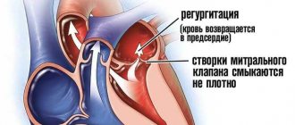
Normally, it has a bicuspid (bicuspid) structure and is located on the border of the left atrium and the left ventricle, preventing the reverse flow (regurgitation) of blood into the chamber of the left atrium in systole, that is, during ventricular contraction. However, infectious lesions, inflammation, autoimmune reactions, overload, and enlargement of the heart chambers can cause pathological changes in the valve system in the form of narrowing of the atrioventricular orifice, which leads to disruption of intracardiac hemodynamics and systemic blood flow. The disease is characterized by a slow course - the first symptoms may appear after 40-50 years, although the formation of pathological narrowing usually begins at a young age. A downward change in the diameter of the atrioventricular orifice occurs in approximately 0.05-0.08% of the population, more often in females.
Causes
Most often, mitral stenosis develops as a result of previously suffered rheumatism , in other cases the cause becomes:
- infective endocarditis ;
- chronic valvulitis ;
- carcinoid syndrome;
- mitral valve calcification
- thrombus;
- obstruction during the tumor process and the development of myxoma ;
- heart injuries.
Congenital anomaly of the mitral valve in the direction of narrowing of the atrioventricular orifice is extremely rare, for example, with Lutembasch syndrome .
Tests and diagnostics
In addition to echocardioscopy, Dopplerography, phonocardiography, X-ray, ECG (including daily monitoring to determine heart rhythm), 4 standard diagnostic methods are used:
- Upon examination, against the background of pallor of the skin, a purple, sharply defined so-called mitral flush of the cheeks and cyanosis of the lips, as well as the tip of the nose are visible, while it is possible to detect increased epigastric pulsation of the right ventricle (“heartbeat”) against the background of the absence or weakening of the apical beat, because the walls of the left ventricle are not enlarged and are displaced as a result of the process of hypertrophy of the right ventricle.
- Upon palpation, a cardiac hump is revealed, and in the area of the apex of the heart, especially after performing physical exercises in patients in a position on the left side, when exhaling, a diastolic tremor is “audible” in the form of the so-called “cat purr” caused by fluctuations in the blood during passage through the constricted mitral passage, to identify the symptom of Nesterov’s 2 hammers, it is enough to place the palm of your hands on the top, placing your fingers on the second intercostal space on the left of the sternum, you can detect the flapping initial first sound and feel it as the first beat of the “hammer”, while the second tone causes a recoil in the fingers in in the form of a second “hammer”.
- During percussion, a displacement of the border of absolute relative dullness of the heart muscle is revealed upward and to the right (caused by hypertrophy of the left atrium and expansion of the right ventricle, respectively), to a greater extent than the relative one, due to the expansion of the right ventricle, the heart and the expanded edges of the lungs - it is pressed against the chest wall with its right enlarged half.
- Auscultation makes it possible to detect a fairly strong flapping first sound in places above the apex of the heart, since during diastole the filling of the left ventricle with blood does not occur fully and rapid contraction begins, and auscultation also makes it possible to listen to an additional third sound at the apex, which is associated with the opening of the mitral valve and its sharp movements of the valves, this valve defect is characterized by diastolic murmurs and a three-part sound of two tones with a click of the opening of the bicupidal valve at the apex.
Classification
Taking into account the state of general hemodynamics, mitral stenosis is divided into compensated, subcompensated and decompensated. In addition, valve defects can differ depending on the etiology and be rheumatic (the most common type - in 50-80% of cases), atherosclerotic, syphilitic, as well as the outcome of bacterial endocarditis .
The most important factor in dividing mitral stenosis into types is the severity of the defect and its degree of influence on intracardiac hemodynamics. Violations can be moderate and moderately severe with a transmitral gradient of 1 – 25 mm Hg, and also not exceed 5 mm Hg. st and does not have any effect on the state of intracardiac hemodynamics.
Classification of mitral stenosis by severity
Mitral stenosis is usually isolated, but can also be combined with disorders of other valves or against the background of mitral valve insufficiency.
Pathogenesis
Stenosis is a persistent narrowing of any hollow structure in the body of humans and other living beings. In the case of a mitral valve consisting of two connective tissue leaflets, their thickening, fibrosis and subsequent calcification , fusion of commissures or shortening of the chords occur. This leads to a decrease in the size of the atrioventricular junction from normal - 4-6 cm square to 2.5 cm square or more, which causes pathological manifestations of functional class heart failure. As a result, blood does not have time to be completely pumped out of the left atrium during diastole and therefore systole is prolonged.
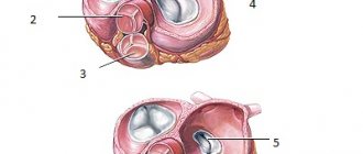
Mitral valve of a healthy person and in case of stenosis
Narrowing of the left atrioventricular orifice impairs the ability of the mitral valve to regulate blood flow from the ventricle to the atrium, and the diastolic flow of arterial blood from the left atrium into the left ventricular chamber is disrupted, which leads to circulatory disorders and causes blood stagnation.
The compensatory mechanism to ensure normal blood filling of the left ventricle is to increase the pressure in the atrium to 25 mmHg, creating a greater pressure gradient to facilitate blood flow through the mitral valve.
An increase in pressure in the cavity of the left atrium also has negative aspects, because it causes an increase in pressure not only in the right ventricle, but also in the pulmonary arteries and throughout the pulmonary circulation. In addition, high pressure in the left atrium leads to hypertrophy of its myocardium. Under conditions of increased work of the atria and the progression of hypertrophy, the volume and mass of the wall of the right ventricle increase, which creates a risk of increased pressure in the pulmonary arteries and in the lungs themselves. Pathogenesis is usually divided into several stages:
- compensation – observed with a slight decrease in the size of the atrioventricular orifice and is asymptomatic;
- subcompensation - emerging circulatory disorders caused by valve dysfunction lead to increased pressure in the pulmonary artery and the first symptoms appear in the form of shortness of breath , increased fatigue, heart pain , etc.;
- depletion of the body's compensatory forces is a condition in which pronounced signs of blood stagnation in the pulmonary circulation, heart failure and atrial fibrillation ;
- sclerotic or dystrophic changes - accompanied by a persistent decrease in myocardial function, severe pulmonary hypertension and arrhythmia .
Pathological specimen of a stenotic mitral valve