Every modern person must know the minimum practical medical actions that can be applied in an emergency situation. It is especially important to understand the location of the main organs, bones, and arteries.
In medical practice, cases are often described when people of any age lose consciousness or simply lie on the ground, without any signs of a normal condition. In this case, the person needs to feel for a pulse in the carotid artery. If it is absent, it is necessary to perform artificial respiration.
What is the carotid artery? It is important to understand the process that it carries out. The carotid arteries direct blood to the brain, and its amount accounts for 70% of the total volume. The name of these vessels is directly related to their ability to put the human body into a state of sleep, when compressed with a certain force for a period of 10 seconds.
What happens if you press on an artery: instructions
When you squeeze it lightly, you feel discomfort and sometimes mild pain. Strong compression easily puts a person to sleep. Receptors perceive pressure as a stimulus to increase pressure, so they do the opposite - they sharply lower it. A person’s heartbeat calms down on the neck, and due to the minimal supply of oxygen, the body experiences drowsiness. Long-term and severe compression of the carotid artery can be fatal.
Typically, a person's pulse is checked on their arm. But in complex traumatic cases this is not enough. Therefore, the pulse is felt in the neck. It should be noted that the pressing force must be controlled. On a thin body, the pulse will be clearly felt even with weak pressure, but if you have developed muscles, you will have to try.
Clinical significance[edit]
The common carotid artery is often used in impulse measurement, [3] especially in patients who are in shock and who lack detectable impulse in the more peripheral arteries of the body. The pulse is measured by palpating the artery deep to the anterior border of the sternocleidomastoid muscle at the level of the superior border of the thyroid cartilage.
The presence of a carotid pulse is estimated to indicate a systolic blood pressure greater than 40 mm Hg, defined by the 50% percentile. [6]
Carotidynia is a syndrome characterized by tenderness of the carotid artery near the bifurcation.
Carotid artery stenosis can occur in patients with atherosclerosis.
Intima-media thickness of the carotid artery wall is a marker of subclinical atherosclerosis and increases with age and with prolonged exposure to air pollution particles.[7]
Structure of the carotid artery
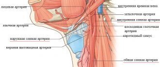
The common carotid artery and its branches are divided into two halves: on the right side of the body it passes in the neck from the brachiocephalic trunk, and on the left, it comes from the aortic arch in the thoracic region. To understand where the carotid artery is located in a person, you need to feel the pulse in the neck, you need to press the vertical depression in the neck on the right or left side. Everything is clearly shown in the photo.
Medical students often wonder where the carotid artery is located in the neck? To feel the pulse of the carotid artery in the neck, you need to lay the person down or sit on a chair. Place several fingers, usually the middle and index fingers, in the cavity between the Adam's apple and the lateral neck muscle.
Normal heart rate values are about 60-80 beats per minute. Each of the arteries runs diagonally upward; in the lower part they are connected by the trachea.
Atherosclerosis with thrombosis of the anterior cerebral artery
Atherosclerotic deposits in the lower segment of the anterior cerebral artery rarely give pronounced clinical symptoms of neurological deficit, since blockage of the lumen (occlusion) is compensated by collateral blood flow through the anterior communicating artery. The likelihood of a transient ischemic attack (TIA, microstroke) and stroke increases if the path of collateral blood flow is characterized by a congenital fusion (atresia) or if there are atherosclerotic changes in a higher section of the anterior cerebral artery.
External carotid artery
The paired organ provides brain tissue with 55 ml of blood per 100 g. tissue, and also saturates with oxygen molecules 3.7 ml per minute. Such provision is considered normal. The external artery in humans is located slightly higher from the larynx, closer to the face, and is part of the front of the head.
In the area of the Adam's apple, the artery diverges to the front and back of the head. The first directs blood flow to the eyeballs, the second to the brain cells. The external artery consists of:
- front end;
- medial part;
- rear end;
- terminal branches - a system of capillaries directed to the eyes and into the oral cavity.
Proof of the presence of a capillary network is the redness of the face. In stressful situations, excessive physical strain, and hot weather, blood rushes to the skin. For some, this effect is practically unnoticeable, for others it is clearly pronounced. This is influenced by genetic predisposition, skin properties, etc.
The external artery directs blood to the following organs:
- thyroid;
- auricles, tympanic cavity;
- eyes;
- facial muscular system, ligaments;
- skin of the face and head;
- some parts of the meninges, the occipital vessel;
- roots of teeth, soft palate, lingual vessels.
The branches of the external artery do not send blood directly to the brain. However, pathologies in the functioning of blood vessels can lead to negative consequences. All that can be done if the external artery is malfunctioning is to contact a neurosurgeon.
In some cases, plastic surgery is required. What diseases can appear if the artery malfunctions:
- Benign tumors in the facial and cervical regions.
- Arteriovenous pathology is the connection between an artery and a vein (blood does not flow into many capillaries).
- Congenital malformation of blood vessels. Signs are external facial defects, migraines and regular pulsation in the head at night, bleeding from large arteries that cannot be stopped.
As a rule, such diseases arise due to dangerous facial injuries, plastic surgery on the nose, ears, throat, etc. Also, there is a high probability of pathologies occurring due to improper implementation of basic procedures.
For example, aesthetic punctures, rinsing the sinuses, tooth extraction, cosmetic injections in the eye area. Rare cases of the disease can cause hypertensive complications.
Internal carotid artery
The branch of the internal artery is directed through the temporal bone. The strength of blood flow increases due to external and internal factors. The number of oxygen molecules entering the brain with the blood exceeds the norm and a person feels a surge of strength, energy for work and other vigorous activities.
But a long process can lead to the opposite - a person feels a loss of strength, drowsiness and fatigue. Oxygen must be supplied normally - excess or deficiency harms the body. Components of the internal artery:
- cavernous - in the dura mater;
- cervical - located deep under the muscle layer;
- part in a torn hole;
- brain;
- stony - laid in the bone canal.
The artery runs laterally, along the edge of the pharynx, parallel to the ear gland. It is divided into small vessels. The complex system supplies brain cells well with oxygen. In the cranium, the branch of the clivus and the pituitary vessel depart from the internal artery. Small arteries are directed to:
- gray matter;
- nuclei of the medulla oblongata;
- white matter;
- cortical parts.
Pathological deformations of the carotid artery Lack of treatment for diseases such as atherosclerosis, syphilis, tuberculosis can lead to deformation of the artery. Against the background of inflammatory processes, internal tissues begin to grow, and the lining of the artery ruptures, letting blood into the area between the walls.
As a result, medical intervention can lead to narrowing of the artery, as a result, a lack of oxygen molecules provokes an ischemic stroke. The narrowing can also cause other health problems - an internal artery that twists incorrectly, or splits into three, a blood clot, or an aneurysm.
Symptoms
Since atherosclerotic lesions of the carotid arteries develop slowly and are often asymptomatic, the first clinical manifestations of this disease may be a stroke (acute cerebrovascular accident - stroke) or transient ischemic attack (TIA), sometimes called a microstroke.
Treatment of atherosclerotic lesions of the carotid arteries usually includes a set of measures, such as lifestyle changes, drug therapy and, in some cases, surgical treatment (open surgery or stenting).
In the early stages, atherosclerotic lesions of the carotid arteries are usually asymptomatic. You and your doctor may not be aware that you have a narrowing of the carotid artery until an acute disruption of cerebral blood supply develops as the first and very serious manifestation of the disease.
Clinical manifestations of stroke or transient ischemic attack may include:
- a sudden feeling of numbness or weakness in the face or limbs, usually on one side.
- Impaired speech or understanding
- sudden loss of vision in one or both eyes
- dizziness or loss of balance
- sudden, causeless severe headache
If you have risk factors for developing atherosclerotic lesions of the carotid arteries, you should consult your doctor. Your doctor may order an examination to determine the condition of these vessels. Even if you do not have clinical manifestations of the disease, your doctor may recommend a number of measures aimed at reducing the severity of risk factors and reducing the likelihood of developing a stroke.
Seek immediate medical attention when you experience symptoms of a transient ischemic attack or stroke.
Even if the duration of symptoms is short, usually less than an hour, but possibly longer, tell your doctor right away. The appearance of these symptoms indicates that you have suffered a transient ischemic attack - a short-term decrease in blood flow to the brain. The presence of transient ischemic attacks is the most important sign that you are at high risk of developing a stroke if preventive measures are not taken in time. A timely visit to the doctor increases your chances that atherosclerotic lesions of the carotid arteries will be identified and eliminated before an acute cerebrovascular accident develops.
A transient ischemic attack may also indicate a decrease in blood flow through other blood vessels in the brain. To clarify the diagnosis, your attending physician will prescribe the necessary examination.
Make sure your family and close friends are aware of the clinical manifestations of a stroke and that it is important to act quickly if they occur.
Concept of carotid artery aneurysm
An aneurysm is an increase in the size of a small area of an artery with an accompanying thinning of its lining. This is a very dangerous disease that can be fatal if the walls of the vessel rupture. Aneurysm can be congenital or acquired, due to muscle atrophy, prolonged inflammatory processes, etc.
What can cause a wall rupture:
- stress, prolonged nervous and physical tension;
- sudden surges in pressure;
- injury to the cervical spine or head.
Blood gradually builds up after the rupture, putting pressure on the surrounding brain tissue. Very often death occurs, but in the case of a small hematoma, the situation can be corrected with surgery.
Links[edit]
- DOO
2nd edition, 1989. - Entry for "carotid artery" in the Merriam-Webster online dictionary
. - ^ ab Ashrafian H (March 2007). "Anatomically specific clinical study of the carotid arterial tree." Anatomical Science International
.
82
(1): 16–23. DOI: 10.1111/j.1447-073X.2006.00152.x. PMID 17370446. - ^ ab Manbachi A, Hoi Y, Wasserman BA, Lakatta EG, Steinman DA (December 2011). "On the shape of the common carotid artery with implications for blood flow velocity profiles". Physiological measurements
.
32
(12): 1885–97. DOI: 10.1088/0967-3334/32/12/001. PMC 3494738. PMID 22031538. - J. Kreiza; M. Arkushevsky; S. Kasner; J. Weigele; A. Ustimovich; R. Hurst; B. Cucchiara; S. Messe (April 2006). "Carotid artery diameter in men and women and relationship with body and neck size". Stroke
.
37
(4):1103–1105. DOI: 10.1161/01.STR.0000206440.48756.f7. PMID 16497983. - Jump up
↑ Deakin CD, Low JL (September 2000).
"Accuracy of advanced trauma life support guidelines for predicting systolic blood pressure using carotid, femoral, and radial pulses: an observational study". BMJ
.
321
(7262):673–4. DOI: 10.1136/bmj.321.7262.673. PMC 27481. PMID 10987771. - Provost, E; Madhloum, N; Int Panis, L; De Boever, P; Nawrot, T. (2015). "Carotid intima-media thickness, a marker of subclinical atherosclerosis and exposure to particulate air pollution: meta-analytic evidence". PLOS ONE
.
10
(5):e0127014. DOI: 10.1371/journal.pone.0127014. PMC 4430520. PMID 25970426.
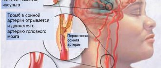
Artery disease - thrombosis: concept and characteristics
Thrombosis is a disruption of normal blood circulation in the brain. As a rule, a thrombus forms at the site where the artery branches into external and internal. In this area, blood does not move as intensely, which allows platelets to form deposits on the walls of blood vessels. What can affect platelet formation:
- congenital or acquired heart disease;
- brain damage;
- increased blood clotting;
- an autoimmune hypercoagulable state associated with the production of antiphospholipid antibodies;
- a disease associated with heart rhythm disturbances - sudden excitation and slowdown.
How does thrombosis manifest itself, symptoms:
- without any symptoms;
- sharp pain;
- subacute;
- chronic.
The patient experiences tinnitus, migraines in the head, constant pain in the cervical spine, blurred vision, the inability to chew normally due to muscle problems, and possible loss of consciousness.
Thrombosis of the internal carotid artery is characterized by a lack of sensation in the limbs, nervousness, hallucinations, inability to speak normally, and skin pain on the head. If a section of the intracranial artery is damaged, vomiting, lack of sensation in the limbs, and sleep disturbances are possible.
How to identify problems with the carotid artery When visiting a clinic, an accurate diagnosis can only be made based on research:
- Magnetic resonance angiography.
- Rheoencephalography.
- Electroencephalography.
- Computer tomography.
- Ultrasound examination of blood vessels.
Examination before carotid endarterectomy
Before hospitalization, the patient must have the following results of laboratory and instrumental examinations with him:
- Clinical blood test
- General urine analysis
- Blood biochemistry (AST, ALT, total bilirubin, direct bilirubin, urea, creatinine, total protein)
- Blood test for hepatitis B, C, HIV, syphilis.
- Chest X-ray or fluorography
- ECG
- Already in the hospital, before the operation, the patient undergoes:
- Duplex ultrasound angioscanning
- Computed tomography of the brain
- Angiography
These examinations help to find out how narrowed the lumen of the vessel is, the size and location of atherosclerotic plaques, the speed characteristics of blood flow through the carotid arteries, and the characteristics of cerebral blood flow.