Definition
Diagnostics and examination of blood vessels is a set of diagnostic methods that allow us to study the condition of blood vessels and determine the pathologies present in them. Vascular examination is carried out to determine the cause of vascular diseases and effective treatment. There are a number of diagnostic procedures that make it possible to identify any deviations from the normal functioning of blood vessels even in the early stages. The examination of blood vessels includes a doctor’s examination, laboratory tests and instrumental techniques for examining blood vessels.
Which is better: MRI or ultrasound?
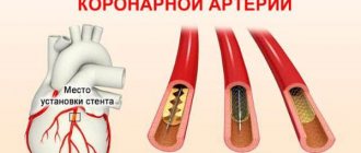
The doctor will tell you which is better: vascular ultrasound or MRI. It all depends on the indications, on which vessels need to be examined. When it comes to the neck, ultrasound diagnostics becomes the preferred method. If you need to diagnose atherosclerosis, venous stenosis, and evaluate the speed of blood movement, it is recommended to perform an ultrasound.
Magnetic resonance imaging is more informative when it comes to neoplasms or vascular malformations. Doctors often prescribe an MRI to clarify the diagnostic results if the results of ultrasound examination are suspicious or there are abnormalities.
In any case, the examination technique is chosen by the treating specialist. At the same time, he is guided by his own knowledge and experience, the patient’s complaints, and the anamnesis collected during the survey.
When to undergo a vascular examination?
Patients who observe symptoms of vascular diseases in their condition need vascular diagnostics. Here are some of them:
- Vascular diagnosis is required when pain syndrome occurs - angina pectoris. Angina pectoris is one of the symptoms of coronary heart disease. Caused by oxygen starvation of the heart, angina pectoris is fraught with myocardial infarction. How is angina diagnosed? The cause and extent of the disease is determined by hardware tests; risk factors for the development of the disease and laboratory tests are also taken into account. Read more about the symptoms and treatment methods of angina by clicking on the link.
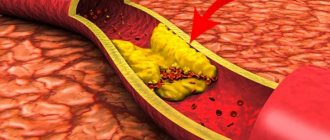
- An examination is prescribed if a person develops symptoms of vascular disease in the legs. Signs of the pathology include excessive sensitivity to cold, pale and dry skin, intermittent claudication or pain when walking. Unpleasant symptoms and pain occur with severe damage to the arteries of the legs, for example, with obliterating atherosclerosis of the vessels of the lower extremities. It is advisable to avoid complications and be attentive to the slightest changes in well-being. We will talk about what methods are best to examine the vessels of the legs in one of the following publications.
- Vascular diagnostics are mandatory if cerebral vascular diseases are suspected. Alarming symptoms - hypertension, headaches, cerebral ischemic attacks. The root cause of poor health is atherosclerosis of the BCA. This is a dangerous condition that leads to a stroke. In this case, the chosen research method will help to correctly diagnose and determine further treatment.
- Regular preventive examination of blood vessels is recommended for people who are at cardiovascular risk. These are patients with high blood cholesterol levels, elderly people, smokers, and patients with diabetes. People with a hereditary predisposition to heart and vascular diseases should also be more attentive to their health.
Prognosis and prevention
With early treatment and prevention of complications, the prognosis is favorable. Vascular pathologies today are successfully treated with conservative and surgical methods, while maintaining the patient’s high quality of life.
Vascular diseases are best prevented by using medications that lower cholesterol levels, as well as products that contain polyunsaturated fatty acids that can prevent the development of atherosclerosis. It is recommended to adhere to the principles of a healthy diet, do not consume fatty and fried foods, and alcohol. It is advisable to quit smoking and start exercising to strengthen the heart muscle and prevent the development of many diseases, including obesity and hypertension. Moderate physical activity has a positive effect on the entire cardiovascular system.
After 30-35 years, check your cholesterol levels and monitor your blood pressure numbers. If symptoms of vascular disorders appear, begin treatment promptly before complete damage to the arteries and veins occurs.
Consultations with a doctor online Taking care of your health is a life priority for everyone.
Communicate with doctors online and receive qualified assistance without leaving your home. Try it Please note! The information on this page is provided for informational purposes only. To prescribe treatment, you must consult a doctor.
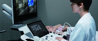
Doppler ultrasound
The Doppler effect, better known to physicists, suggests that ultrasound is reflected from moving objects. When special devices based on this effect were invented, determining the specificity of blood flow in the lower extremities became possible in any segment of interest.
Ultrasound ultrasound devices, as a rule, are portable, so it is possible to carry out diagnostics directly near the patient without transporting him.
This method covers vessels from the iliofemoral region to the foot, detecting anomalies in the early stages of their occurrence.
How is vascular MRI performed in the POMC?
The procedure is carried out without special preparation.
Bring with you to the study an extract from your outpatient card, the results of past diagnostic examinations and other certificates related to the disease. If your doctor ordered an MRI, be sure to bring a referral.
Before starting the procedure, you must remove any metal objects: watch, phone, clothes with metal buttons.
Vascular MRI is performed in the supine position. The head and arms will be gently secured using special straps.
The operation of the device is associated with the rotation of a massive scanning ring around the body, which makes a lot of noise. To prevent it from disturbing you, you will be offered earplugs.
To ensure that the resulting images are clearly visible, you will need to lie still during the entire vascular MRI scan.
The MRI examination is painless, but if you feel more comfortable, a relative or loved one may be with you during the procedure.
Based on the results of the procedure, a medical opinion is issued. You receive photographs and a copy of the report. They should be taken to the doctor who referred you for a vascular MRI. If desired, the results of the study can be transmitted to you electronically.
Contraindications for MRI with contrast agent
The drugs used in our center are low-toxic and almost do not provoke allergies. The contrast agent is administered intravenously. Then it moves through the vascular system and is retained in the tissues. This method is used to increase the accuracy of research. Often, vascular MRI can be performed without the use of a contrast agent. There are 3 contraindications to this procedure:
- Contrast material may harm the fetus or pass into breast milk. Therefore, this procedure cannot be carried out during pregnancy or breastfeeding.
- It takes at least 24 hours to properly remove the contrast from the body. Therefore, chronic renal failure is also a contraindication to MRI with contrast agent.
- And the last contraindication: individual intolerance to the drug. It is possible to develop an allergic reaction to the components of the contrast agent.
Diseases of the lower extremities
The legs perform the function of support and movement. The lower limbs have a complex structure. The main anatomical zones are the thigh, lower leg and foot. Birth defects and serious injuries to any area of the leg are fraught with irreversible consequences, including loss of the ability to move and disability.
The lower limbs consist of the following areas:
- gluteal;
- front and back of the thigh;
- knee;
- ankle joint;
- inner and outer areas of the ankle;
- dorsum of the foot and sole.
The leg skeleton consists of bones connected by joints. A complex system of muscles includes muscles, fascia, ligaments, tendons, etc. The trophism of soft tissues is provided by veins and arteries, the functions of the legs are provided by nerve fibers.
Liposarcoma (indicated by an arrow) of the lower thigh
The characteristics of the area under consideration predispose to:
- injuries of a traumatic nature (fractures, sprains and ruptures of ligaments, penetrating wounds, etc.);
- degenerative-dystrophic lesions of joints (arthrosis);
- bone diseases (osteoporosis, etc.);
- vascular pathologies (atherosclerosis, venous insufficiency, varicose veins, thrombosis);
- muscle diseases (myopathies, myositis, dystrophies);
- damage to neuromuscular junctions;
- infectious and inflammatory processes of any localization (tuberculosis, phlegmon, abscesses, etc.);
- benign and malignant tumors.
Pathological changes can affect any structural element of the limbs. Causes: genetic disorders, developmental abnormalities, infectious, autoimmune diseases, excessive stress, injuries, etc.
MRI image of the foot
CT images of neck vessels
CT angiography of neck vessels with contrast is the gold standard for diagnosis, but to get the maximum benefit from the study, coordinated work of professionals is necessary. The radiologist interprets the results. At the Magnit center in St. Petersburg, CTA is one of the most popular procedures, and our doctors will answer any questions. In difficult cases, tomograms are assessed collectively. We present to your attention several tomograms where pathological changes are clearly visible:
CT angiogram of blood vessels in the neck. The vertebral artery is located between the vertical lines. Arrow points to the spine
3D images of computed multislice tomography of the circle of Willis. Its contours can be seen on the tomogram in the center, the neck is below, the main arteries are white.
Coronal MSCT image demonstrates saccular contrast leakage (thick arrow) from the right internal carotid artery (pseudoaneurysm). The left carotid artery (thin arrow) is intact.
Interpretation of MRI photographs of the lower extremities
During the scanning process, monochrome images of the anatomical area are obtained in 3 mutually perpendicular planes. In conclusion, they note what the MRI of the leg showed:
- Injuries - fractures, dislocations, muscle and tendon sprains, ligament ruptures, intra-articular changes. When tracking the dynamics of tissue healing, the nature of regeneration and the presence/absence of complications (false joint, pinched nerve endings, etc.) are noted.
- Tumors - benign (lipomas, hemangiomas, etc.) and malignant (sarcoma, etc.).
- Vascular pathologies - atherosclerotic lesions of the arteries, disorders in the microvasculature, inflammatory changes in the walls, including autoimmune ones, congenital and acquired anomalies (aneurysms, malformations, etc.).
- Inflammatory changes in acute and chronic joint diseases (rheumatism, rheumatoid arthritis, gout, etc.), myositis, osteomyelitis.
- Nerve damage - compression, rupture, pinched fibers, tumor and degenerative changes are detected.
The radiologist indicates in the report the nature of the deviations, but does not make a diagnosis. The results are provided in written form along with the images on disk. The doctor who performed the decoding briefly explains the situation and can recommend where to go for treatment. The final diagnosis is made by a specialized specialist or doctor who referred for examination.
Scar-atrophic changes in the thigh muscles on an MRI image (indicated by arrows)
CT scan of neck vessels with contrast
Modern injection systems significantly improve the quality of diagnostics
The introduction of an iodine-based radiopharmaceutical improves the visibility of the smallest structures, which improves the quality of diagnosis. CT angiography of neck vessels with contrast in 95% of cases is not accompanied by extraordinary situations and proceeds without complications. As the drug enters the vein, the patient may experience slight nausea, an unpleasant taste in the mouth, dizziness, and a burning sensation at the injection site. These phenomena disappear on their own within a few seconds to a minute and are considered as normal. A generalized feeling of itching of the skin, difficulty breathing, swelling of the neck, cold sweat, and the appearance of a rash all over the body are signs of a generalized allergic reaction; these symptoms should be reported to the staff immediately via loudspeaker.
Contrast is performed using an injector through an intravenous catheter (the amplifier is injected automatically at certain phases of the study), or the drug is administered simultaneously.
CT scan of head and neck vessels
The tomograms clearly show the architecture of the blood vessels in the area under study.
CT angiography of neck vessels with contrast is indispensable for injuries: the speed of obtaining results and excellent visualization of vascular accidents, damage to bone structures and nerve tissues allow you to quickly determine the tactics of patient management. Most pathological processes occur in a generalized manner (atherosclerosis, autoimmune diseases), so it is necessary to simultaneously examine the vessels of the head and neck for the nature of the lesion and the severity of the disease. On tomograms you can see:
- vascular development abnormalities;
- aneurysms before and after rupture;
- acute disorders of cerebral circulation in the carotid and vertebrobasilar beds (hemorrhagic or ischemic strokes) and their causes;
- possible arteriovenous malformations, etc.