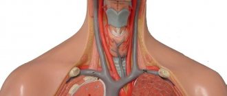
It is believed that catheterization of the internal jugular vein is accompanied by fewer complications (pneumothorax, thrombosis, bleeding) compared to catheterization of the subclavian vein. At the same time, in some cases it is more convenient to use the subclavian approach, for example: with hypovolemia, motor agitation, low blood pressure in the patient, etc.
Femoral vein catheterization is associated with an increased risk of infectious and thrombotic complications. And it is used as a backup option if it is impossible to perform central catheterization from another access. To facilitate the search for a vein and reduce the risk of complications, ultrasound examination allows us to clarify the individual characteristics of the location of the patient’s venous trunks.
Attention! If an attempt to catheterize a vein ends in failure, do not persist and immediately call a colleague for help - it often helps, if not to solve the problem, then at least to avoid troubles in the future.
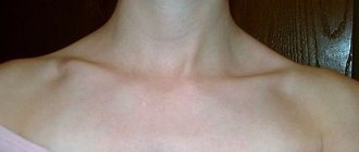
Puncture of the right internal jugular vein through central access
Place the patient on his back, arms along the body, turn his head to the left. To increase the filling of the central veins and reduce the risk of air embolism, place the Trendelenburg position (the head end of the table is lowered 15° downwards), if the bed design does not allow this - horizontal.
Determine the position of the right carotid artery. The internal jugular vein is located more superficially, lateral and parallel to the carotid artery. Treat the skin with an antiseptic and limit the puncture site with sterile wipes. Infiltrate the skin and subcutaneous tissue over the anterior edge of the sternocleidomastoid muscle at the level of the thyroid cartilage with 5 ml of 1% lidocaine solution. A search puncture is performed with an intramuscular needle in order to localize the location of the vein with minimal risk of significant bleeding due to unintentional puncture of the artery.
You should also use a “search needle” if there is coagulopathy, or the puncture needle from the set is inconvenient for you, or you need to insert a large-diameter catheter. If you have good manual skills, you can naturally refuse to use the “search puncture”. With your left hand, determine the course of the carotid artery. Insert the needle slightly lateral (approximately 1 cm) of the artery at a 45° angle to the skin towards the right nipple in men or the right superior anterior iliac spine in women. Advance the needle slowly, maintaining a vacuum in the syringe, until blood is drawn. The vein is located superficially, so you should not insert the needle deeper than 3-4 centimeters.
If you do not find a vein, slowly withdraw the needle under the skin, maintaining a vacuum in the syringe (since the needle could accidentally puncture both walls of the vein). If you are unable to obtain blood, try again, this time taking a slightly more medial direction. Once you are sure that you have found a vein, you can remove the search needle, remembering the direction of the puncture, or leave it in place, removing it after the needle from the set hits the vein. Venous puncture with a needle from the set is performed in the direction determined during the search puncture.
Puncture of the right subclavian vein
Place the patient on his back, arms along the body, turn his head to the left. To move your shoulders back and down, place a bolster between your shoulder blades. To increase the filling of the central veins and reduce the risk of air embolism, place the Trendelenburg position (the head end of the table is lowered 15° downwards), if the bed design does not allow this - horizontal.
Feel the jugular notch of the sternum, sternoclavicular and acromioclavicular joints. Next, treat the skin with an antiseptic solution and limit the puncture site with sterile wipes. The puncture point is located 2-3 cm below the clavicle, on the border of the middle and medial thirds of it. Infiltrate the skin and subcutaneous tissue around the puncture site with 5-10 ml of 1% lidocaine solution.
Insert the needle through the indicated point until it touches the collarbone. Gradually push the end of the needle down until it is just below your collarbone. Then rotate and point the needle at the jugular notch. Slowly advance the needle forward, maintaining a vacuum in the syringe, until blood is drawn. The cut end of the needle should be turned towards the heart - this increases the likelihood of correct installation of the catheter. Try to keep the needle parallel to the plane of the bed (to avoid puncture of the subclavian artery or pleura);
If you miss a vein, slowly withdraw the needle under the skin while maintaining vacuum in the syringe. Rinse the needle and make sure it is clear. Try again, taking the injection direction a little more cranial.
Complications
Installation of a catheter in the central veins may be accompanied by some complications - atrial and ventricular arrhythmia; hematomas; pneumo- and hemothorax; perforation of a vein; damage to the trachea, nerve trunks, heart.
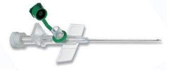
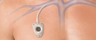
Catheter device for central venous access Certofix
Some complications can be managed with high-quality Certofix catheters. They have a soft tip (1) made of polyurethane, which prevents perforation of blood vessels and damage to the intima. Also scale (2) to determine the length of the intracorporeal section of the catheter. They are made of radiopaque material, which allows for X-ray control of its placement in the vessel. If there are multiple channels, they are color coded (3) to identify the distal, middle and proximal channels. In addition to the fixing wings, each channel has a movable clamp (4) - a clamp, which avoids turning or dislodging the catheter. There is also a self-closing system (5) which reduces the risk of air embolism or blood leakage.
Puncture of the right femoral vein
Position the patient on his back, with a cushion placed under the buttocks. The leg should be slightly abducted and turned outward. Determine the pulsation of the femoral artery below the inguinal ligament: the femoral vein is located more medially. Treat the skin with an antiseptic and limit the puncture site with sterile wipes. Next, infiltrate the skin and subcutaneous tissue with 5 ml of a 1% lidocaine solution. Incise the skin with a scalpel with a small blade.
2 cm below the inguinal ligament, determine the course of the femoral artery with two fingers of your left hand. The needle is inserted 1 cm medial to the femoral artery at an angle of 30° to the skin and directed along the vein, maintaining a vacuum in the syringe until blood is obtained. The vein is usually located at a depth of 2-4 cm from the surface of the skin. It is convenient to use a G14-16 peripheral venous catheter as a needle, after making sure that it passes the conductor.
If you do not find a vein, slowly remove the needle while maintaining a vacuum in the syringe. Rinse the needle and make sure it is clear. Try again, aiming the needle slightly to the right or left of the original puncture site.
Seldinger catheter insertion
Immediately after puncture of the vein, make sure that the blood flows easily into the syringe. Disconnect the syringe while holding the needle in place. Try to rest your hand on the patient’s body to minimize the risk of needle migration from the lumen of the vein. Close the needle pavilion with your finger to prevent air from entering;
Insert the flexible end of the guidewire into the needle. If there is any resistance to the conductor's advancement, carefully turn it and try to advance it. If this does not help, remove the metal conductor. Assess blood aspiration from the vein again. Change the angle of the needle or rotate it, check the flow of blood into the syringe. Try again. If it was not possible to insert a plastic conductor, in order to avoid cutting, it must be removed along with the needle.
After inserting the guidewire halfway into the vein, remove the needle. Before inserting the dilator, incise the skin with a scalpel with a small blade; Insert a dilator through the guidewire. Try to take the dilator with your fingers closer to the skin to avoid bending the conductor and additional trauma to the tissue, or even the vein. There is no need to insert the dilator over its entire length; it is enough to create a tunnel in the skin and subcutaneous tissue without penetrating the lumen of the vein. Remove the dilator and insert the catheter. Remove the conductor. Perform an aspiration test. Free blood flow indicates that the catheter is in the lumen of the vein.
Advantages and disadvantages of the technique
The undoubted advantage of catheterization is the provision of quick access to the patient’s bloodstream. In addition, when placing a catheter, the need for daily puncture of a vein for the purpose of conducting intravenous drips is eliminated. That is, the patient only needs to install a catheter once instead of having to “prick” the vein again every morning.
Also, the advantages include sufficient activity and mobility of the patient with the catheter, since the patient can move after the infusion, and there are no restrictions on hand movements with the catheter installed.
Disadvantages include the impossibility of long-term presence of the catheter in a peripheral vein (no more than three days), as well as the risk of complications (albeit extremely low).
- Catheterization of the subclavian vein
Installation of a catheter under ECG control
Installation of a catheter under ECG control allows you to optimize its position and reduce the likelihood of complications.
1. The catheter is washed with saline solution. A metal conductor is inserted into the catheter so that it does not extend beyond the catheter (some conductors have a special mark). Or a metal intramuscular needle is inserted through the catheter plug and the catheter is filled with a 7.5% sodium chloride solution. A plug is put on the needle;
2. Attach the chest lead “V” wire of the electrocardiograph or cardioscope to the needle or conductor using an alligator clip. And turn on the “thoracic abduction” mode on the recording device. Or connect the wire of the right hand to the distal electrode and turn on the second (II) lead on the cardioscope or cardiograph;
3. If the end of the catheter is in the right ventricle, we see a high-amplitude (5-10 times larger than usual) QRS complex on the monitor screen. Slowly tightening the catheter, we see a decrease in the amplitude of the QRS complex, but the P wave remains very high, which indicates that the catheter is in the atrium.
Catheterization kit
To insert catheters into venous tubes, the doctor uses standard kits for catheterization of central veins or peripheral vessels.
They differ in the diameter and configuration of the catheter tubes, as well as the presence of additional tools in the kit for inserting and fixing devices on the human body. The standard kit for catheterization of the subclavian and jugular veins (SJV) contains:
- a catheter tube made of polymer material, visible on x-rays, with a diameter of 1.2 to 2.3 mm and a length of 130 to 210 mm, with extensions;
- a round or triangular metal needle with a diameter of 1.1 to 1.6 mm and a length of 57 to 100 mm;
- conductors - straight made of polymer material or J-shaped made of metal;
- expanders;
- fixing elements;
- plug with membrane.
The standard set of catheters for the peripheral parts of the venous system differs from sets for catheterization of central veins in the absence of dilators and guides, as well as the size of the tubes: their thickness varies from 0.62 to 2.1 mm, and length from 19 to 45 mm.
The choice of catheter size depends on many factors, including the age and equipment of the patient, his anatomical and physiological characteristics. For example, the smallest sizes are used for catheterization of children, and larger ones are used for installation in large branches of the circulatory system.
Kinds
The official classification divides catheters into several types depending on the purpose of the devices, the materials from which they are made, sizes and design features. According to their purpose, they are divided into three types:
- CVCs, represented by sets for catheterization of central veins. Suitable for long-term installation in all large veins.
- PVK, represented by kits for catheterization of peripheral veins. Suitable for long-term installation in vessels of the upper and lower extremities.
- Butterfly catheters, which are a monolithic structure consisting of a tube and a needle, as well as a fixing element in the form of two round plates. In clinical practice, such a catheter is used for infusion into small veins with procedures lasting no more than an hour.
Butterfly catheter
By size, they are divided into 8 types and are marked with a two-digit number and the letter G (geich). The higher the number on the central venous catheterization kit, the thinner the tubing and needles.
According to their design features, catheters are divided into single-channel and multi-channel. Single-channel ones are used for administering drugs according to Seldinger during emergency care, for long-term administration of solutions and blood components. Multichannel designs are used for the simultaneous administration of drugs that are incompatible with each other.
Polyethylene and polyurethane tubes are considered the most common in kits for catheterization of subclavian veins. The industry also produces catheters made of polyethylene, PVC, silicone and Teflon.
Indications
Conditions requiring long-term administration of medicinal solutions, nutrients and blood components are considered absolute indications for central venous catheterization:
- Venous catheter - what is it? Vein catheterization
- the patient's inability to eat;
- oncological diseases (chemotherapy);
- renal failure requiring hemodialysis;
- administration of drugs that provoke irritation and spasm of peripheral vessels;
- the need to regularly monitor hemodynamics.
Catheterization of peripheral vessels is carried out if it is necessary to administer moderate amounts of medication over 3-5 days.