01 Aug 2021 at 10:37 MRI of the heart in Tushino 7911
“Aneurysm of the aorta of the heart - what is it?” Many patients in whom a specialist has just discovered this disease ask a similar question. An aortic aneurysm is a widespread or limited dilation of the largest vessel in the human body. With this disease, the diameter of the aortic lumen exceeds the norm by two or more times. Aortic aneurysm is one of the most dangerous heart diseases.
Definition:
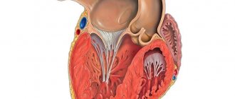
A cardiac aneurysm is a pathological condition that is accompanied by protrusion of the wall of the heart muscle. It is one of the complications after a myocardial infarction and can develop within several months after its onset. An aneurysm not only negatively affects the functioning of the heart and its contractile functions, but also poses a serious danger to life due to the possibility of sudden rupture.
Complications without surgery
Small LV aneurysms usually do not pose a threat to the patient’s life, although in rare cases they can provoke thromboembolic complications due to the formation of parietal thrombi in the heart cavity, which are carried by the blood flow to other arteries and can cause a heart attack, stroke, pulmonary embolism or mesenteric artery thromboembolism (PE). and mesenteric thrombosis).
Complications with aneurysms of medium and giant sizes are more common and are as follows:
- Thromboembolic complications,
- Progression of chronic heart failure, development of acute heart failure,
- Rupture of an aneurysm, leading to the rapid death of the patient.
Prevention of complications is the timely detection of aneurysm growth, regular examination by a doctor, as well as timely identification of indications for surgical treatment.
Cause
Usually the cause of the formation of a cardiac aneurysm is a heart attack, most often of the left ventricle of the heart. When they talk about the development of myocardial infarction, the damaged parts of the heart are weakened, and under the influence of blood pressure they can bulge. Blood trapped in them forms a blood clot. Aneurysms occur in patients with atherosclerotic cardiosclerosis and cardiosclerosis of other origins. Much less common are aneurysms that arise due to other reasons: infections, injuries, and congenital defects.
Diagnosis of aneurysm
In our department of vascular surgery, experienced phlebologists perform high-quality diagnostics of all forms of aneurysms using modern instrumental techniques:
· ventriculography and angiography are invasive research methods that involve the introduction of contrast into the blood vessels in the affected area. Such studies can detect vascular aneurysms, their dissection, signs of rupture or thrombosis;
· ECHO of the heart, ultrasound of the arteries – used to diagnose aneurysms located outside the brain;
· Ultrasound examination of blood vessels, Dopplerography;
· CT with contrast - allows you to identify signs of aneurysm dissection.
Signs of bleeding or arterial thrombosis can also be detected by laboratory blood tests.
Classification
Depending on the time period after myocardial infarction the aneurysm formed, the following forms are distinguished:
- Acute and subacute - develop in the early stages after a heart attack, often complicated by incomplete rupture.
- Chronic - formed after acute due to the replacement of a dead section of the heart muscle with connective tissue. They can be muscular, musculofibrous or fibrous.
1.General information
An aneurysm (the less correct term “aneurysm” is sometimes used) is a local bulge, a protrusion of any boundary surface in the form of a stretched sac, or a spindle-shaped expansion of the walls of a blood vessel (saccular aneurysms are also found on vessels, but much less frequently). Thus, an atrial septal aneurysm (ASA) is a curvature of the membrane separating the right atrium from the left. Normally, this septum is sealed, relatively straight and does not bend, at least significantly, in any direction.
Many authors qualify an MPP aneurysm as a pathology only if the protrusion in any direction (to the right atrium, to the left, or S-shaped) exceeds 10 mm and is combined with prolapse (sagging) of the mitral valve. In terms of the general population, such an anomaly is rare; more often it accompanies congenital connective tissue diseases. An MPP aneurysm is called a “minor cardiac anomaly” or “small heart defect”, since in most asymptomatic cases it does not pose a real danger and does not require treatment, and is usually discovered by chance. However, it does not at all follow from this that this anomaly can be forgotten without attaching any significance to it: the situation can sharply worsen with the onset of enlargement of the aneurysm, thinning of the aneurysm and its spontaneous breakthrough, after which the dynamics become unpredictable.
A must read! Help with treatment and hospitalization!
Symptoms
Symptoms of acute cardiac aneurysm are weakness, shortness of breath, fever, pulmonary edema, sweating, heart rhythm disturbances, and swelling of the veins in the neck. During the transition to the subacute form, symptoms of coronary insufficiency quickly develop.
With chronic cardiac aneurysm, signs of cardiovascular failure are observed: edema, angina attacks, rhythm disturbances. Complications of the disease can be thromboembolic syndrome with damage to peripheral vessels and arteries of the brain. If you experience such signs, immediately contact the cardiology center.
When an acute aneurysm ruptures, death occurs almost instantly. In this case, the patient experiences severe pallor, hoarse, difficult breathing, cold sticky sweat, congestion of the neck vessels, and cold extremities.
Post-infarction aneurysm: types of causes, diagnosis and treatment
Post-infarction aneurysm is a pathological deformation of a section of the myocardium with partial or complete loss of contractility, caused by disturbances in the functioning of the cardiovascular system and complications after an illness. The size of the damaged area depends on the extent of the infarction and can occupy most of the organ. A particular danger is that the scar tissue that replaces the dead tissue is almost always lined with blood clots on the inside. Modern diagnostic and treatment methods make it possible to detect the disease in a timely manner and take all measures to save a person’s life.
What happens
Heart attack refers to diseases, untimely detection of which in most cases leads to severe complications or death. According to medical statistics, an aneurysm is most often caused by a heart attack, but it can also be congenital or the result of a severe traumatic event.
The formation of a post-infarction aneurysm in 8/10 cases occurs in the upper region, and in the rest - on the anterior wall of the left ventricle. In this case, the deformed tissue does not contract and, as a result, the left ventricle is divided by the aneurysmal passage into 2 parts, one of them works, and the second is motionless.
Part of the blood in systole enters the sac of the non-contracting segment, resulting in circulatory failure. Until the moment when the dead area of the myocardium is completely replaced by connective tissue, there is a danger of rupture of the deformed wall and accumulation of blood in the pericardial cavity.
In addition to post-infarction cardiac aneurysm, there is a pseudoaneurysm. She appears after:
- infective endocarditis;
- rheumatism;
- syphilis;
- tuberculosis;
- as a result of a traumatic violation of the integrity of the vessel wall.
8
24/7
It is a blood-filled cavity between the connective tissue and the lumen of the artery. Leads to a decrease in the contractility of the heart due to loss of motor activity.
Aneurysmal disorders do not have a clear shape, a certain depth and extent of the lesion. There are:
- muscular;
- fibrous;
- fibromuscular.
An organ may have several deformed areas. Aneurysms are divided into:
- true, in which the violation covers 3 layers;
- false, limited to pericardial fusion;
- functional, formed by a piece of myocardium that has retained its activity, but has low contractility.
In an organ, formation takes several forms:
- flat or diffuse, which does not have a protrusion outside the heart, but inside there is a cup-shaped depression;
- sac-like, with a wide base and a convex rounded wall;
- mushroom-shaped: there is a narrow entrance followed by a large protrusion;
- An aneurysm within an aneurysm means that in the cavity of one deformed area there is another.
Despite the fact that the disease has been fairly well studied, it remains questionable why it develops only in 1/10 people who have had a myocardial infarction.
Causes
At the time of a massive myocardial infarction, the muscular structures of the heart wall die, and intracardiac pressure leads to stretching of the dead part of the organ. According to the moment of formation of the disease in the post-infarction period, there are several types
- Acute is diagnosed in the first 2 weeks. It is a necrotic, stretched area of the heart muscle protruding into or out of the organ.
- The subacute type appears between 3 and 8 weeks. In place of dead myocardial fibers, connective tissue begins to form. The inner layer of the organ shell is thickened, there are accumulations of immature reticular cells, fibroblast, collagen and elastic fibers.
- Chronic post-infarction aneurysm of the left ventricle develops after 2 months. It is a fibrous sac with a depleted wall. Loose parietal thrombi are often located inside the growth, the size of which varies and they can occupy the entire volume of the sac.
The main causes of any form of post-infarction aneurysm are considered to be:
- disorders of the cardiovascular system due to age-related changes;
- increased cholesterol levels in the blood due to poor nutrition;
- obesity, hypertension, diabetes mellitus and a number of other pathologies are a provoking factor;
- increased intraventricular pressure and disorders caused by scar compaction;
- motor or other form of physical activity in the acute and subacute stages of myocardial infarction.
The formation of an aneurysm can be motivated by uncontrolled consumption of alcoholic beverages, smoking and drug use.
Symptoms and diagnosis of the disease
The symptoms of the disease directly depend on the form and duration of its course.
| Type | Symptoms |
| Acute | Pulmonary edema, weakness with shortness of breath, increased sweating and prolonged fever, irregular heart rhythm. |
| Subacute | When moving, sweating, weakness, shortness of breath, rapid heartbeat with a feeling of interruptions in the functioning of the organ. Progression of symptoms characteristic of heart failure. |
| Chronic | Fainting and semi-fainting states, angina pectoris, swelling with swelling of the veins in the neck and shortness of breath, the occurrence of hydrothorax, pathological enlargement of the liver, possible pericarditis, adhesions. |
8
24/7
The chronic form of the disease is especially dangerous, as it is fraught with the development of gangrene of the extremities, strokes, renal infarction and a number of other pathologies that pose a threat to the patient’s life.
The main sign of the appearance and development of a post-infarction aneurysm is precordial pulsation that increases with each heartbeat, which is clearly visible on the anterior wall of the chest. Diagnosis is based on:
- based on the patient’s complaints of pain in the heart and behind the sternum, interruptions in the beat rhythm;
- when collecting anamnesis, information about a previous heart attack and coronary insufficiency indicates the need for additional research to detect a post-infarction aneurysm;
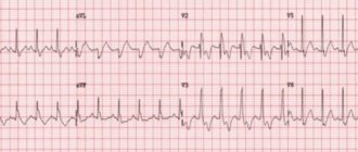
- An ECG with pathology shows a persistent rise in the ST interval, as well as a negative T wave and the presence of QS;
- The protrusion of the deformed part of the organ becomes clearly visible on the x-ray.
If there is a blood clot in the cavity, the pulsation will be noticeably reduced.
GSR and ventriculography help to make accurate conclusions necessary for choosing treatment methods:
- the localization of altered tissues is clarified;
- it is possible to assess the coronary bed;
- the exact size and shape of the aneurysm are determined;
- The contractility of the surviving left ventricular myocardium is assessed.
Collecting data is important because it is used to base a decision on the need for surgical intervention.
Treatment at different stages
Post-infarction cardiac aneurysm has indications for surgery in any form of development, since after its formation the life expectancy is no more than 2-3 years. The cause of heart failure is heart failure, embolism in the arterial system, or repeated myocardial infarction.
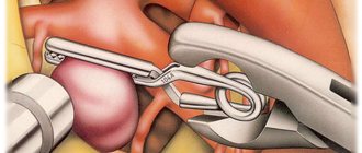
- The modified tissue is not completely removed during surgery. To preserve the cavity of the left ventricle, it is left 15 mm on each side.
- An aneurysm of the interventricular septum is corrected by shortening it or compensating it using plastic methods.
- If left atrioventricular valve insufficiency develops, it is removed and replaced with a prosthesis.
Indications for urgent intervention are:
- danger of rupture of the injured heart wall;
- numerous blood clots formed inside the aneurysm;
- partially hidden perforation of the aneurysmal sac;
- the appearance of ventricular tachycardia and other obvious signs of progression of cardiac disorders.
In post-traumatic and false forms of the disease, the heart muscle is sutured and the site of deformation is removed.
It must be taken into account that after excision and repair of the aneurysm, recurrent myocardial infarction may occur. But if the disease is left untreated, the chances of recovery are reduced to zero.
Prognosis, disease prevention
Without surgical treatment, predicting the further course of the disease and the degree of its impact on the patient’s health is completely pointless.
Flat, uncomplicated forms of pathology allow concomitant therapeutic treatment. So, using Warfarin for post-infarction aneurysm, you can:
- reduce the risk of recurrent heart attack;
- reduce the development of thrombosis;
- improve heart performance in a number of other factors.
However, the drug has a strict dosage and therefore can only be used as prescribed by the attending physician.
Saccular and mushroom-shaped post-infarction aneurysms are almost always complicated by the formation of blood clots and therefore cannot provide positive prognostic data.
The only acceptable form of preventing the development of the disease is timely diagnosis, prompt hospitalization and proper treatment of a patient with myocardial infarction. The rehabilitation period should include a gradual increase in physical activity, constant monitoring of thrombus formation in the post-infarction period, and control of heart rate.
8
24/7
General information about arterial aneurysms
An aneurysm is a local expansion of a vessel with gradual thinning of its wall. The process is irreversible; over time, under the influence of blood flow pressure, the protrusion only increases and, one day, may rupture. This leads to internal arterial bleeding. Without timely medical attention, a ruptured aneurysm leads to death.
Other possible complications include: thrombosis, thromboembolism (when a blood clot breaks off and clogs other vessels), infection of the aneurysm.
Risk factors for developing aneurysms
The development of aneurysms is predisposed by the loss of elasticity and strength of the vessel wall. Weakness of the vascular wall may be due to congenital characteristics or the influence of external factors. The main reasons for the development of the disease:
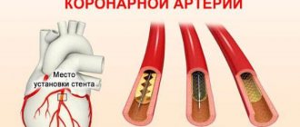
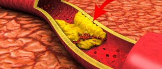
- atherosclerosis - formation of cholesterol plaques;
- inflammation of the aortic wall (occurs with traumatic injury, syphilis, fungal infection);
- autoimmune diseases;
- congenital pathologies of connective tissue (cystic fibrosis, Marfan syndrome);
- trauma during diagnostic and therapeutic procedures (coronary angiography, heart surgery).
Treatment methods
Surgery is considered the most effective treatment for aneurysms .
, drug treatment can be carried out . Also, medication may be indicated for inoperable processes, as well as for localization of the lesion in the abdominal aorta. During treatment, painkillers, antihypertensives, diuretics, and beta blockers are widely used.
Surgical intervention is necessary in cases where the aneurysm was discovered at the dissection stage. Our department successfully uses modern minimally invasive techniques, which allow us to perform a surgical operation with minimal consequences for the patient. Thus, with a dissecting aortic aneurysm, surgery to replace the vessel with a synthetic prosthesis or stenting with a graft may be indicated.
In case of a cardiac aneurysm, the patient is prescribed strict bed rest. If the disease progresses rapidly, signs of heart failure appear, and surgery is performed. Also, to avoid rupture, aneurysms of peripheral vessels are also treated surgically. During surgery for a cerebral aneurysm, the surgeon disconnects the aneurysm from communication with the vessel.