03 Jun 2021 at 09:50 MRI of the heart in Tushino 14791
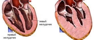
Left ventricular hypertrophy is a fairly common heart disorder. The disease in most cases begins to develop in patients suffering from hypertension. Hypertrophy provokes an increase in the size of the wall of the left ventricle. The disease can provoke a change in the size of the septum, which is located between the left and right ventricles. The development of hypertrophy in most cases occurs over several years.
Left ventricular hypertrophy: causes
The myocardium can increase in size under the influence of certain factors that force it to work harder. In this way, the heart cells try to compensate for the increased load. These factors include:
- High blood pressure. One of the most common causes of left ventricular hypertrophy.
- Aortic valve stenosis. It is located between the left muscular chamber and the aorta. If it narrows, the muscle has to work harder, causing it to increase in size.
- Intense physical exercise. During prolonged cardio and strength exercises, thickening of the myocardium is considered as an adaptation of the body to increased loads.
Sometimes left ventricular hypertrophy of the heart develops due to a genetic disease that causes structural changes in heart cells - hypertrophic cardiomyopathy. Another possible cause may be amyloidosis, which is accompanied by abnormal protein deposits in all organs, including the heart.
Prevention
Prevention is aimed at treating myocardial infarction, which causes the development of an aneurysm. It is important to take medications as prescribed by your doctor and not to violate the dosage regimen. Visit a cardiologist in a timely manner and do not ignore routine examinations.
Additionally, patients should:
- stick to a diet;
- control blood pressure;
- eliminate psycho-emotional stress;
- do exercises and physical warm-up;
- control weight;
- quit smoking.
Following simple rules of treatment and prevention will help improve your health and prevent relapse and progression of the disease. You can contact our specialist for advice and undergo examination in the cardiology department. Only comprehensive measures can help in the fight against the disease.
Diagnosis of the disease
At the first stage, the doctor will listen to the patient's complaints, take a medical history and conduct a thorough physical examination, including measuring blood pressure and a preliminary assessment of heart function (auscultation or auscultation). Further additional studies will be prescribed:
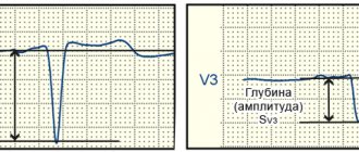
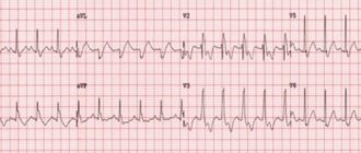
- Electrocardiogram (ECG). Thickening of the wall of the left heart chamber can lead to disruption of the electrical activity of the heart, which is reflected in the results of the ECG.
- Echocardiogram. Allows you to evaluate blood flow, detect myocardial hypertrophy and identify the causes that caused it, for example, aortic valve stenosis.
- MRI. It has a high discriminative ability when assessing soft tissues, including the heart muscle.
Laboratory tests and, in some cases, invasive tests such as coronary angiography and myocardial biopsy may also be performed.
Drugs for left ventricular hypertrophy
Medicines for high blood pressure help prevent further enlargement of the myocardium and, in some cases, reverse it. For this purpose, the following may be prescribed:
- ACE inhibitors (angiotensin-converting enzyme). They lower blood pressure and reduce the load on the heart by dilating blood vessels and improving blood flow. Examples: Captopril, Enalapril, Lisinopril, Ramipril.
- Angiotensin II receptor blockers (ARBs). Drugs such as Lozap, Closart, Sentor also relax blood vessels, reducing blood pressure.
- Calcium channel blockers. These drugs inhibit the penetration of calcium ions from the intercellular space into the muscle cells of the heart and blood vessels. The blood vessels relax, blood pressure decreases. Examples: Amlodipine, Diltiazem, Verapamil, Lercanidipine.
- Diuretics. Diuretics used for left ventricular hypertrophy are called thiazide diuretics. They work by decreasing the kidneys' ability to reabsorb salt and water from urine, thereby increasing urine production and output (diuresis). Also, diuretics of this type directly help relax the smooth muscles of blood vessels. Examples: Tonorma, Hypothiazide, Hydrochlorothiazide.
- Beta blockers. Drugs such as Metoprolol, Atenolol, Lokren, Betak help reduce heart rate and blood pressure, as well as prevent the negative effects of stress hormones. Beta blockers are generally not prescribed as the primary treatment for hypertension. Your doctor may recommend them if therapy with other drugs has not been successful.
In cases where the disease is caused by aortic valve stenosis, surgery may be required. The decision on the advisability of the operation is made by the doctor after a thorough diagnostic examination of the patient.
Treatment of acquired heart defects can be conservative or surgical
Conservative treatment is effective only in the early stages of the development of heart disease and requires mandatory follow-up by a cardiologist.
PPS should be treated surgically when:
- Heart failure progresses.
- Pathological changes in the valve significantly affect hemodynamics.
- The ongoing conservative therapy does not have the desired effect.
- And there are fears of serious complications.
Types of operations for heart defects
Open heart surgery is performed under artificial circulation through a median sternotomy. Median sternotomy creates optimal conditions for the cardiac surgeon to perform the necessary surgical interventions for various pathologies and to connect the heart-lung machine. The soft tissue incision is approximately equal in length to the length of the sternum (about 20 cm), and the sternum is dissected along its entire length.
The main two types of operations that are currently used for PPS are reconstruction of the affected valves (plastic) or their prosthetics.
Valve-sparing surgery
Performed to eliminate the cause of valve dysfunction.
If the valves do not close (valve insufficiency), then the cardiac surgeon during the operation achieves normalization of the closure of the valve leaflets, performing resection of the valve leaflets, annuloplasty, commissural plastic surgery, and chord replacement. If there is valve stenosis, then the sections of the valves that have fused due to a pathological process are separated and an open commissurotomy is performed.
Valve replacement surgery
If it is impossible to perform plastic surgery, when there are no conditions for this, valve replacement operations for prosthetic heart valves are performed. In case of intervention on the mitral valve, replacement is performed with full or partial preservation of the anterior or posterior valve leaflets, and if impossible, without their preservation.
In valve replacement surgeries, prostheses are used.
- Prosthetics can be made from animal or human tissue. Such prostheses are called biological. Its main advantage is that the patient does not need to take anticoagulant drugs during subsequent years of life, and their main disadvantage is their limited service life (10-15 years).
- Prostheses consisting entirely of mechanical elements (titanium and pyrolytic carbon) are called mechanical. They are very reliable and can serve without failure for many years, without replacement, but after such an operation the patient must always take anticoagulants for life, this is a negative aspect of using a mechanical prosthesis.
Minimally invasive surgeries
Modern surgery, thanks to the creation of new instruments, has the opportunity to modify surgical approaches to the heart, which leads to operations becoming minimally traumatic for the patient.
The point of such operations is that access to the heart is carried out through small incisions in the skin. During minimally invasive operations on the mitral valve, a right-sided lateral minithoracotomy is performed, with a skin incision of no more than 5 cm, this allows one to completely avoid dissecting the sternum and provides convenient access to the heart. To improve visualization, endoscopic video support with multiple magnification is used. With minimally invasive access to the aortic valve, the skin incision is approximately half as large (incision length 8 cm), and the sternum is incised lengthwise in its upper part. The advantage of this method is that the uncut portion of the sternum provides greater stability after surgery, as well as a better cosmetic effect by reducing the size of the suture.
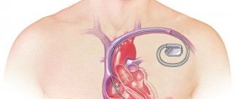
Endovascular operations - transcatheter aortic valve replacement (TAVI).
Methods of transcatheter aortic valve implantation:
- The entire operation is carried out through a blood vessel (femoral or subclavian artery). The meaning of the procedure is to puncture the femoral or subclavian artery with a guide catheter and deliver the stent valve against the blood flow to the site of its implantation (aortic root).
- Through the aorta. The essence of the method is to dissect the sternum over a short distance (ministernotomy) and puncture the aortic wall in the ascending section and implantation of a stent valve into the aortic root. The method is used when it is impossible to deliver the valve through the femoral and subclavian arteries, as well as when there is a pronounced bend in the arterial arch.
- Through the apex of the heart. The meaning of the procedure is to make a small incision in the fifth intercostal space on the left (minithoracotomy), puncture the apex of the heart with a guide catheter and install a stent valve. Once the new valve is implanted, the catheter is removed. The new valve starts working immediately.
There are two types of stent valves:
- Self-expanding stent-valve expands to the desired size after removing the restrictive sleeve sleeve from it.
- A balloon-dilatable stent valve that expands to the desired size when the balloon is inflated; After final installation of the stent valve, the balloon is deflated and removed.
To determine whether TAVI surgery is appropriate, the patient must undergo a series of tests, including an ECG, echocardiography, computed tomography (CT) scan, and angiography.
Currently, the TAVI procedure is increasingly used not only for aortic stenosis, but also for aortic insufficiency, as well as their combination. In addition, TAVI surgery is used for dysfunction of the biological prosthetic aortic valve.
TAVI surgery is performed under general anesthesia and requires a multidisciplinary approach. The procedure is performed by a specialized team, which includes an interventional cardiologist, cardiac surgeon, anesthesiologist, and radiologist.
The presence of a stent valve is not an indication for the patient to take the indirect anticoagulant Warfarin (in the absence of other indications).
Is left ventricular hypertrophy dangerous?
Without effective treatment, pathological thickening of the wall of the heart chamber can lead to serious complications such as:
- weakening of the blood supply to the heart;
- pumping dysfunction (heart failure);
- heart rhythm disturbance (arrhythmia);
- insufficient oxygen supply to the myocardium (ischemic disease);
- stroke and heart attack.
Timely consultation with a doctor and properly prescribed therapy can avoid serious consequences of the disease.
Differences between different types of LVH
Cardiomyopathy | Arterial hypertension | Sports heart | |
| Age | under 35 | over 35 | from 30 |
| Floor | both sexes | both sexes | more often men |
| Heredity | burdened with hypertension | burdened with cardiomyopathy | not burdened |
| Complaints | dizziness, shortness of breath, fainting, heart pain, rhythm disturbances | headaches, less often shortness of breath | stabbing pain in the heart, bradycardia |
| Type of LVH | asymmetrical | uniform | symmetrical |
| Myocardial thickness | more than 1.5 cm | less than 1.5 cm | decreases when the load stops |
| LV dilatation | rarely, more often a decrease | Maybe | more than 5.5 cm |
Popular questions about left ventricular hypertrophy
What medications should I take for left ventricular hypertrophy?
The choice of medications primarily depends on the causes of LVH. Therefore, drug therapy should be prescribed by a doctor. Most often, it includes taking ACE inhibitors, angiotensin II receptor blockers, calcium channel blockers, thiazide diuretics and beta blockers.
Why is left ventricular hypertrophy dangerous?
If the patient does not receive proper treatment for severe LVH, this can lead to the development of complications such as acute heart failure, arrhythmia, coronary artery disease, heart attack and stroke.
Is it possible to cure left ventricular hypertrophy of the heart?
You can achieve good results by eliminating the underlying cause of the disease, namely high blood pressure. Correctly selected antihypertensive therapy in many cases makes it possible to stop the progression of the pathology, and sometimes leads to a reduction in the hypertrophied heart wall.
What is concentric left ventricular hypertrophy?
It is a disease that occurs as a result of stressors on the heart, such as hypertension, congenital heart defects (such as tetralogy of Fallot), valve defects (narrowing of the aorta or stenosis), and primary myocardial defects that directly cause hypertrophy (hypertrophic cardiomyopathy). It is characterized by thickening of the myocardium without a corresponding increase in the size of the ventricles, and is often accompanied by symptoms such as chest pain and shortness of breath on exertion, general fatigue, syncope and palpitations.
Symptoms of the development of an abnormal condition
Dilatation of the left ventricle in most cases develops very slowly. The patient may not experience any unpleasant signs or symptoms, especially in the early stages of the disease. But as hypertrophy develops, the following may occur:
- shortness of breath;
- unexplained fatigue;
- chest pain, especially after exercise;
- sensation of fast, fluttering heartbeats;
- dizziness or fainting.
You should seek medical help if:
- there is a feeling of pain in the chest that lasts longer than a few minutes;
- there are serious breathing difficulties that interfere with daily activities;
- have severe, recurring memory problems;
- there are loss of consciousness;
- shortness of breath combined with rapid heartbeat is troubling.