A common cause of hemorrhagic stroke. Aneurysms are protrusions of the vascular wall and are usually located at the sites of vessel division. Under the influence of various factors (hypertension, smoking, hereditary predisposition), the aneurysm grows and its wall thins. These processes ultimately lead to rupture of the aneurysm and hemorrhage into the brain and surrounding spaces. More than half of the patients cannot tolerate hemorrhage and die despite intensive therapy even in the best clinics. That is why, in almost all cases, prophylactic surgery is recommended to exclude the aneurysm from the bloodstream.
General information. Diagnostics Choosing a method for switching off an aneurysm Treatment of patients in the acute period of the disease Treatment of large and giant aneurysms Microsurgical treatment of asymptomatic aneurysms Multiple aneurysms Treatment of aneurysms in pediatric patients
General information. Diagnostics
Surgical treatment of arterial aneurysms of cerebral vessels has been one of the main and most important areas of work of the institute’s vascular department for several decades. This is due to the fact that there are no conservative treatment methods for this pathology, and surgical interventions on aneurysms remain among the most complex neurosurgical operations. Despite numerous studies aimed at developing methods for preventing the rupture of aneurysms and treating complications of aneurysmal hemorrhages, arterial aneurysms of the cerebral vessels are still a disease with a high probability of death or permanent disability: in the absence of surgical care, up to 70% of patients die in the first year from onset illness, 25% of survivors remain disabled. The prevalence of arterial aneurysms in the population is quite high - at least 0.6% (about 600 people per 100,000 population). The incidence of SAH due to ruptured aneurysms is 10–15 cases per 100,000 population per year. Thus, in Russia, about 18,000 patients with aneurysmal SAH require hospitalization annually. The emergence of new diagnostic capabilities has led to an increase in the detection of asymptomatic aneurysms, which has further increased the number of patients seeking medical help.
| CT angiography mode |
Over a more than 40-year period, the institute has performed about 5,000 direct surgical interventions and more than 700 endovascular operations for arterial aneurysms. The number of patients admitted annually for surgical intervention has increased from 100 patients in the 70s–80s to 300 or more today. Constant analysis of this clinical material in dissertations, detailing the principles of treatment depending on the timing after hemorrhage, size, location, anatomical features of aneurysms, their number in one person, and the age of patients, led to the creation of separate areas in the surgery of cerebral aneurysms. The most important role in the surgical treatment of aneurysms is played by methods of diagnosing the disease. Currently, diagnosis of aneurysms can be performed using direct angiography, MRI and SCT angiography. The results of these three methods can be presented in a volumetric image (3D reconstruction). SCT-AG can currently be considered the method of choice in identifying the cause of SAH. A special role in examining a patient in the acute period of SAH is played by methods for diagnosing vasospasm. Transcranial Doppler ultrasound (TCUSDG), inferior to neuroimaging methods in sensitivity and specificity, allows one to assess the dynamics of vasospasm, since the non-invasiveness of the method makes it possible to conduct multiple studies. CT and MRI in various modes in patients with aneurysms are indispensable in the diagnosis and dynamic monitoring of such complications of SAH and surgery as intracerebral hematomas, intraventricular hemorrhages, cerebral edema and ischemia, acute and delayed hydrocephalus.
| Aneurysm clipping |
While remaining unchanged in its fundamental principles, cerebral aneurysm surgery is constantly evolving. The current stage of treatment of arterial aneurysms at the institute can be characterized as a stage of strictly differentiated and individualized treatment, performed taking into account all the features of the course of the disease, the morpho-functional characteristics of aneurysms, the patient’s condition and other factors. The basis of the ideology of aneurysm surgery is the integration and complementarity of microsurgical and endovascular methods. In microsurgical treatment of aneurysms, the main operation is clipping the aneurysm neck with a self-clamping clip. Depending on the anatomical and topographical features of the aneurysm, clips of various sizes and configurations are used. In recent years, the institute has developed and implemented a number of surgical and diagnostic methods that have improved the quality of exclusion of more complex aneurysms: intravascular aspiration of blood, intraoperative Doppler ultrasound, flowmetry, fluorescein angiography. The main advantages of the endovascular method of treating aneurysms are the ability to completely exclude the aneurysm from the bloodstream in the absence of surgical brain injury and the ability to exclude aneurysms that are difficult to reach or inaccessible for direct surgical intervention. These advantages are primarily important in patients in the acute period of hemorrhage, with aneurysms of the paraclinoid part of the ICA and the vertebrobasilar region. The preferred method for endovascular exclusion of aneurysms should be considered reconstructive operations—occlusion of the aneurysm cavity while maintaining the lumen of the supporting vessel. To switch off aneurysms, the technique of occluding the aneurysm with detachable microcoils is currently used.
Prevention of pathology
All preventative measures can be divided into actions to prevent the formation of an aneurysm and methods to prevent the rupture of vessel formations. As a rule, in practice these methods coincide, so general recommendations for disease prevention can be identified:
- monitoring blood pressure and reducing it in a timely manner;
- giving up bad habits - alcohol, smoking, drugs;
- maintaining proper nutrition;
- control of bad cholesterol in the blood;
- eliminating stress;
- timely treatment of cardiovascular diseases;
- refusal of intense physical activity.
It is important to note that even the unquestioning implementation of the above measures does not guarantee the complete elimination of pathology and its complications. The only way to completely get rid of an aneurysm is through surgery.
Choosing a method to turn off an aneurysm
| Aneurysm occlusion with stent assistance |
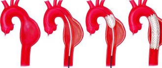
The advantages of endovasal treatment are primarily important in patients in the acute period of hemorrhage, with aneurysms of the paraclinoid part of the ICA and the vertebrobasilar region. Until recently, the main factor determining the possibility of aneurysm occlusion using the endovascular method was considered to be the ratio of the size of the aneurysm body and its neck. It has been established that the larger the size of the aneurysm and the wider its neck, the greater the likelihood of distant recanalization after the initial total occlusion. To solve this problem, stent-assisted techniques have been developed. The essence of the stent-assisted technique in the treatment of aneurysms is to prevent displacement of the coils of the coils into the lumen of the supporting vessel. This is achieved as follows: in the first stage, a stent is installed in the vessel at the level of the aneurysm neck, and then a microcatheter is passed through the stent cell, through which microspirals are delivered. In recent years, a new category of intracranial stents has emerged, the so-called flow divert stents, which reduce blood flow in the aneurysm by directing the main blood flow through the supporting vessel. Thrombosis of the aneurysm after installation of such a stent occurs on average within 4 to 6 months after surgery. This technique is most effective for treating large and giant aneurysms. Currently, the Research Institute of Neurosurgery annually performs about 250 microsurgical and 150 endovascular operations on aneurysms in the “cold” period after SAH (21 days or more). The choice of surgical method depends, first of all, on the anatomical features of the aneurysm. The endovascular method is preferable for ICA aneurysms of the paraclinoid and infraclinoid location. For MCA bifurcation aneurysms and ACA-ASA aneurysms, direct interventions are more often performed, since the structural features of these aneurysms (predominantly wide neck, proximity of the ostia of second-order arteries, variations in the structure of the ACA, etc.) and the impossibility of using stents limit the use of the endovascular method. The decision on the method of operation is influenced by the age of the patient - in young patients direct operations, which are more radical, are preferable. Limitations for the endovascular method are pathological bends and severe atherosclerosis of the brachiocephalic arteries. If a stent-assisted operation is necessary, a history of bleeding of various etiologies should be taken into account, since stent installation requires long-term use of anticoagulants in the postoperative period. In some cases, microsurgical operations are performed as a second stage in cases where only partial occlusion of the aneurysm was achieved during endovascular intervention. The opposite situations are also possible - endovascular intervention after palliative direct surgery (strengthening the aneurysm with gauze, incomplete clipping). The results of microsurgical treatment of aneurysms in the cold period after SAH have remained quite good for many years. The risk of a new neurological deficit is slightly higher with microsurgical exclusion of the aneurysm, and the risk of death is higher with endovasal treatment.
What is the cost of the operation
Due to the fact that clipping an aneurysm is a very complex and dangerous operation, it is not performed in all major cities of Russia. Thus, clinics with appropriate personnel and technical equipment exist in Moscow, Tyumen, Ufa, Kirov, Volgograd, Krasnodar and some other regional centers.
Regarding the cost of the operation, it should be noted that a planned intervention can be performed according to a quota allocated from the funds of the Ministry of Health of the Russian Federation, but for this the patient must contact the regional department of the Ministry of Health and provide all documents confirming the need for the intervention.
In the event that the patient does not intend to wait several weeks or months, he can be operated on in any city at his own discretion, but at his own expense. The cost of operations depends on many factors and ranges from 12 thousand rubles to 180 thousand rubles for endoscopic surgery and from 22 thousand rubles to 170 thousand rubles for clipping an aneurysm.
aneurysm before/after surgery
Treatment of patients in the acute period of the disease
| Comparison of brain appearance in the acute and cold periods after SAH |
Treatment of patients in the acute period after SAH is a complex task that requires the involvement of various specialists. The rationale for operations in the acute period is, first of all, the possibility of repeated hemorrhages, which are accompanied by extremely high mortality. After switching off the aneurysm, it is also possible to begin intensive vascular therapy aimed at eliminating the consequences of hemorrhage. Based on an analysis of the results of surgical treatment of more than 600 patients in the acute period of SAH at the institute, it was shown that prognostically significant criteria for assessing the outcome of the operation are the massiveness of the hemorrhage, the patient’s condition on the Hunt-Hess or WFNS scale, and the duration of surgery after SAH. Taking these factors into account, the basic principles of treating patients have been developed. Indications for surgery in the acute stage of SAH must be strictly individualized. To resolve the issue of patient management tactics, the necessary minimum is to assess the patient’s condition using the Hunt-Hess or WFNS scale, CT, angiography, TC ultrasound and determining the severity of vasospasm. Particular attention should be paid to the period after hemorrhage. The operation is indicated for patients in stages I–II according to Hunt–Hess, regardless of the period after SAH; in stages III–IV according to Hunt–Hess within a period of 14 days after SAH. In stage III–IV patients undergoing surgery on days 0–3 after SAH, it is necessary to install sensors for postoperative ICP monitoring, since these patients are characterized by the development of vasospasm and cerebral edema in the early postoperative period. In stage III–IV patients on days 4–7 after SAH, in the presence of moderate or severe vasospasm, surgery should be abstained. The duration of delay of surgery for vasospasm depends on the TC parameters of the ultrasound scan: the operation can be performed when the blood flow velocity decreases to the level corresponding to a mild spasm, or when it stabilizes at the level of a moderate spasm for several days, but not earlier than 7 days after SAH. In stage V, surgery is indicated only in the presence of large intracerebral hematomas. In these cases, surgery is most effective with the earliest interventions possible. In the acute period of SAH, endovascular exclusion of the aneurysm should be considered the method of choice, especially in severely ill patients. In some cases, if it is impossible to completely occlude the aneurysm, you can limit yourself to partial shutdown, followed by a repeat operation after the condition improves. In addition to anatomical features, a limitation for endovascular surgery in the acute period is severe vasospasm, which prevents the insertion of a catheter. When performing direct intervention, it is necessary to take into account that the brain of a patient in the acute period of SAH is much more sensitive to surgical trauma; therefore, it is necessary to use the most gentle methods of access to the aneurysm with limited traction of the brain, the use of relaxation methods (osmodiuresis, removal of cerebrospinal fluid) and brain protection from ischemia. Throughout the entire period of treatment of patients in the acute stage of SAH, the leading task is the prevention and treatment of complications typical for hemorrhage from an aneurysm, primarily vasospasm, cerebral edema and acute or delayed hydrocephalus. Currently, to combat these complications, the technique of intravascular administration of antispasmodics under the control of TC ultrasound, 3H-therapy under multimodal monitoring, and decompressive craniotomy are used. Over the past 10 years, postoperative mortality during direct operations in the acute period averaged 7.3%, and overall mortality - 12.4%. Among surviving patients, 80% are completely independent in daily life, and about 40% have returned to work and school. The data obtained were used to create a recommendation protocol for the management of patients in the acute period of SAH together with leading neurosurgeons in Russia. The experience of the institute allowed us to take part in several international studies on the treatment of patients in the acute stage of SAH.
Contraindications for surgery
The tactics of surgical treatment in patients with hemorrhage is determined on the basis of a scale developed specifically for neurosurgeons (Hunt and Hess scale (1968); WFNS SAH Scale (1988)). The main criteria here are the presence of clinical manifestations of an aneurysm, motor or speech disorders, as well as vascular spasm in the patient (angiospasm), diagnosed based on the results of an X-ray contrast study. Thus, in a patient with severe vasospasm, one should refrain from surgery, since the risk of postoperative complications outweighs the benefits of surgery.
In addition, the operation is contraindicated if the patient has severe acute somatic diseases or decompensation of chronic diseases - acute infectious process, blood clotting disorder, severe diabetes mellitus, severe exacerbation of bronchial asthma, etc.
Treatment of large and giant aneurysms
| Exclusion of a giant ICA aneurysm using the IAC technique |
Surgical treatment of large and giant aneurysms is one of the most difficult issues in vascular neurosurgery. In the 70s, surgical treatment for such patients was limited to ligation of the internal carotid artery in the neck, while postoperative mortality was 15.2%. With the advent of endovasal technology, it became possible to conduct a balloon occlusion test under EEG control followed by stationary occlusion of the ICA. In some cases, this treatment method was combined with the application of EICMA; however, postoperative mortality in the group of giant aneurysms remained high. Since 1995, the institute has developed and used a technique for intravascular aspiration of blood from an aneurysm. The ICA technique has proven to be very effective in the surgery of large and giant aneurysms of the ICA; it has made it possible to reduce postoperative mortality by 4 times and significantly reduce the group of patients who are denied surgical treatment. Currently, the department has the most experience in the world in applying the HAC technique.
| Shutdown of a large aneurysm using a flow-directing stent |
Along with direct interventions, endovascular methods have been used at the Institute for many years to treat large and giant aneurysms. Before the advent of stent-assistance and flow-directing stents, it was rarely possible to completely shut off such aneurysms with coils. The advent of stents has significantly changed the possibilities for endovascular treatment of large and giant aneurysms. Since the use of flow-directing stents, there has been a significant decrease in the proportion of deconstructive operations. There is no doubt that this technology has great prospects and in the near future will significantly change the structure of endovascular interventions in the treatment of aneurysms in general.
Causes
{banner_banstat9}
As for specific provocateurs of the pathological process:
- Congenital anomalies, mutations. They occur relatively often, but the degree of change is so small that it is very difficult to suspect deviations. Not always, but it happens.
- Complicated anamnesis. If there was at least one elder in the family with a similar problem, the risks automatically increase by 50-70%. It is worth taking a closer look at the people of the side branch: brothers, sisters. The probability increases by a quarter or a little less. It makes sense to undergo regular examinations with a neurologist or therapist; this is an important factor in prevention.
- Encephalitis. A type of inflammation of brain structures.
- Meningitis. The same thing, but of a different origin. Both pathological processes lead not only to an aneurysm, but also to immediate destruction of nerve tissue, deficiency, and focal symptoms. Be it dementia or speech, hearing, or vision disorders.
- Brain injuries. TBI. From concussions to open injuries, penetrating wounds and more. The probability is calculated based on a specific pathological process.
- Smoking. Nicotine and harmful substances affect blood vessels, their strength, and the elasticity of the internal lining.
- Alcohol consumption.
- Drug use.
- Arterial hypertension. Stable increase in pressure in the bloodstream. At any stage, it increases the likelihood of a pathological process.
- Heart diseases. IHD, angina pectoris.
- Tumors. They can compress blood vessels, thereby reducing their regenerative capabilities and adaptive potential.
- Atherosclerosis of cerebral vessels.
- Vasculitis. Autoimmune inflammation of the internal lining of blood vessels. Potentially lethal disorders. They manifest themselves in dozens of ways. Extremely dangerous in themselves. They require treatment under the supervision of a rheumatologist and as quickly as possible.
The causes of aneurysm are vascular, autoimmune, traumatic, cardiogenic pathologies.
If there is at least one of these disorders, and the aneurysm has not yet formed, the patient is classified as a high-risk group and is closely monitored.
Microsurgical treatment of asymptomatic aneurysms
Until recently, asymptomatic aneurysms were detected extremely rarely. In recent years, the widespread availability and availability of noninvasive neuroimaging techniques has led to a significant increase in the number of patients with incidentally discovered aneurysms. Over the past 10 years, the institute has operated on about 400 patients with asymptomatic aneurysms. The results of surgical treatment in this category of patients should be considered satisfactory. Taking into account these data, as well as the extremely high probability of an unfavorable outcome in case of rupture of an aneurysm, it can be argued that unruptured asymptomatic aneurysms in most cases require surgery. It should be noted that the patient must be fully informed about the nature of the disease, the upcoming operation and possible complications.
Possible complications
Complications may include dissection and rupture, thrombosis, stroke, compression of the esophagus, loss of vision or hearing, memory loss, thyroiditis, paralysis of half the body.
Rupture is a common complication of the natural course of the disease . It occurs due to overstretching of the carotid artery, which has undergone irreversible dystrophic changes.
Causes:
- Hypertensive crisis;
- Injuries;
- Detachment of an atherosclerotic plaque.
Risk factors are hypertension, smoking, diabetes, stress, physical overexertion.
Symptoms of a rupture:
- Anxiety;
- Dyspnea;
- Tachycardia;
- Increased pressure;
- Violation of visual, auditory, cognitive functions;
- Paralysis of half the body;
- Lack of response to external stimuli;
- Loss of consciousness.
Treatment in 100% of cases is surgical. The prognosis is unfavorable. Stroke occurs in a third of patients.
Multiple aneurysms
To date, the proportion of patients with multiple aneurysms is 20–25% of all patients with aneurysms operated on at the institute. In the vast majority of cases, no more than two aneurysms are found in a patient with multiple aneurysms, but there are patients with three or more aneurysms. Combinations of aneurysms in localization, size, number, and clinical manifestations are quite diverse. The primary task at the diagnostic stage is to determine the aneurysm that served as the source of hemorrhage. The main principle of treatment for AF is the initial exclusion of a ruptured aneurysm from the bloodstream using microsurgical or endovascular techniques, depending on the anatomical features of the aneurysm. Operations can be performed in one or several stages. In the treatment of multiple aneurysms of various vascular territories, especially in the acute period of SAH, as well as in complex aneurysms, two-stage or multi-stage interventions are preferable.
Treatment of aneurysms in pediatric patients
Cerebral aneurysms in children are rare and can be combined with various connective tissue pathologies (Marfan’s syndrome, Ehlers-Danlos’s syndrome) and various developmental anomalies of the circle of Willis. In children, aneurysms with complex anatomical characteristics are detected three times more often than in adults. Pseudotumorous and ischemic types of course in “children’s” aneurysms occur several times more often than in adults. Incidentally discovered aneurysms in children are rare. The principles of surgical treatment of aneurysms in children are generally the same as in adults. It should be noted that the compensatory capabilities of a child in the event of the development of any complications are greater than those of adults, so children can more easily tolerate the switching off of the arteries carrying the aneurysm. In this regard, deconstructive operations can be considered as the method of choice in cases of complex aneurysms when it is impossible to switch off the aneurysm while maintaining arterial patency.
Indications for surgery
Surgery on a cerebral aneurysm is indicated if the patient has an aneurysmal protrusion larger than 7-10 mm, confirmed by instrumental diagnostic methods:
- Ultrasound examination of cerebral vessels,
- MRI and/or CT scan of the brain, including using a radiopaque contrast agent injected into the patient’s vascular bed,
- Spinal puncture with examination of the cerebrospinal fluid (CSF) for subarachnoid hemorrhage in a ruptured aneurysm.