Cerebellar stroke is still one of the least studied diseases of the central nervous system. The variability of clinical manifestations, their similarity to symptoms of damage to the cerebral hemispheres and other features significantly complicate diagnosis. This pathology occurs relatively rarely: approximately 2% of patients with acute disorders of cerebral circulation have a localization of the lesion in the cerebellum. Men of middle and retirement age suffer more often. What are the characteristic symptoms of the disease and how to treat such a stroke? The answers to these questions are in our article.
Types of cerebellar stroke
According to the standard classification based on pathogenesis, two types of cerebellar stroke are distinguished:
- ischemic;
- hemorrhagic.
According to statistics, the first type significantly predominates. Ischemic lesions of the cerebellum, according to various sources, account for 80 to 90 percent of cases. However, according to expert forecasts, within 50 years the number of diseases with a hemorrhagic nature is expected to double - the result of aging and changes in the racial composition of the planet's population.
Ischemic cerebellar stroke
The development of ischemic damage to the cerebellum is based on partial or complete blockage of blood vessels bringing arterial blood by a thrombus. In this regard, nerve cells stop receiving oxygen and nutrients. As is known, nervous tissue is very sensitive to hypoxia, and if blood flow is not normalized, neurons begin to die.
Hemorrhagic cerebellar stroke
With the hemorrhagic nature of the disease, a sharp disruption of the blood supply to the cerebellum occurs due to rupture of the artery or capillary feeding this structure. Due to the anatomical features of blood circulation, the consequences of a vascular accident are very difficult to predict, since the brain stem is often involved in the pathological process.
Diagnostics
To confirm the diagnosis of cerebellar stroke, an ultrasound is performed. The study allows you to determine the displacement, see the area of edema, and detect a damaged artery.
An informative diagnostic method is magnetic resonance imaging or computed tomography. Both methods provide an accurate picture of the changes, the size of the damage, the depth, and detect the cause of the disease.
A blood test is taken for general formula and biochemistry. The data will allow you to identify the level of sugar, cholesterol, and other substances that are signs of the diseases that provoked the attack.
Symptoms of cerebellar stroke
The clinical picture of both ischemic and hemorrhagic disorders of the blood supply to the cerebellum is varied, but there are characteristic symptoms that make it possible to suspect damage to the blood vessels in this particular area. These include cerebellar ataxia. This syndrome is associated with the functional activity of the cerebellum and includes:
- Balance imbalance. The patient cannot stand steadily and sways in different directions when walking.
- Changes in speech. Disturbance in the flow of arterial blood into the cerebellum is accompanied by scanned speech. The patient loudly tries to pronounce ordinary words.
- Loss of precision in movements. It is difficult for the patient to control fine motor skills, movements become sweeping.
- Tremor. Trembling of the limbs, head, and torso appears.
- Nystagmus. The eyeballs make involuntary oscillatory movements. Their frequency can reach several hundred per minute.
Among the general symptoms characteristic of acute cerebral circulation disorders are headache, dizziness, spontaneous vomiting, fever, loss of consciousness, difficulty swallowing, and muscle paralysis.
Main symptoms
Cerebellar stroke has its own characteristic symptoms:
- The cerebellum is responsible for the coordination of movements. With a cerebellar stroke, the organ can no longer perform its functions. For this reason, the patient experiences motor disturbances. Movements become uncoordinated, although there is no muscle weakness. The trunk and limbs may twitch or shake involuntarily. Doctors call this condition cerebellar ataxia.
- Due to a lack of coordination of movement, a person may experience vomiting; the person seems to be seasick.
- Severe pain in the back of the head.
- There is severe dry mouth. Swallowing is also impaired. This can cause a person's speech to become slurred. It is important to understand that this condition sometimes persists for a long time. In such cases, the person is taught to speak again.
- Partial or complete hearing loss.
- Chaotic movement of the pupils.
- Dulling of tactile sensations.
- A sharp increase in temperature.
- Loss of consciousness.
Causes and factors of cerebellar stroke
The ischemic type of cerebellar stroke is usually considered the outcome of atherosclerotic disease. The thrombus travels through the vertebral or basilar artery through the bloodstream. Embolism can also occur with fresh myocardial infarction or atrial fibrillation. Surgical interventions on the neck are very dangerous, since during their implementation it is possible to traumatize the vertebral arteries with further disruption of blood flow.
The main causes of hemorrhagic stroke of the cerebellum:
- high blood pressure or sudden changes;
- cerebral amyloid angiopathy - deposition of amyloid in small blood vessels through which blood enters the central nervous system;
- taking antithrombotic drugs (“blood thinners”);
- neoplasms;
- inflammatory diseases of the walls of blood vessels.
Wrong lifestyle
In addition to the immediate causes, risk factors play a significant role in the development of cerebellar stroke. One of the key ones is the wrong lifestyle. It includes:
- low mobility during the day;
- food based on foods high in fat and sodium;
- smoking, alcohol abuse, taking narcotic drugs;
- constant nervous tension, chronic stress.
These factors contribute to disruption of normal blood circulation in organs, including the nervous system.
Diseases and medications
Diseases of the internal organs can provoke a cerebellar stroke. The relationship between cerebral circulatory disorders and the following somatic diseases has been proven:
- arterial hypertension;
- metabolic syndrome, accompanied by high levels of cholesterol and low-density lipoproteins in the blood;
- diabetes mellitus and other endocrine pathologies;
- cardiac ischemia;
- diseases in which blood clotting increases.
Uncontrolled use of medications that affect the functioning of the heart, change hormonal levels, and thin the blood can also cause acute impairment of cerebral circulation (including in the cerebellum).
Other reasons
Other causes of vascular accident include genetic predisposition and age factor. A number of genes are currently being studied that are potentially associated with an increased risk of cerebral circulatory disorders.
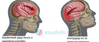
Acute cerebrovascular accidents (ACI) in Russia occupy 2nd place in the structure of overall mortality of the population and are the main cause of permanent disability. Every year, more than 20 million people worldwide suffer a stroke, and in Russia - more than 450,000 [8, 9]. Among all types of stroke, cerebellar strokes are uncommon. Cerebellar infarctions (CI) range from 0.5 to 1.5%, reaching 2.3% of all cerebral infarctions (in autopsies, these observations range from 1.5 to 4.2%), but the mortality rate from them is high and amounts to about 20% [7]. In this regard, early and reliable diagnosis of MI is especially relevant.
One of the reasons for the difficulty of early diagnosis of MI is the atypical course of cerebellar damage, which is observed in almost one third of cases [7], as well as diagnostic difficulties that occur in the most acute phase of MI, when the most characteristic signs of cerebellar disorders may be absent [16]. It is believed that in 28% of cases, stroke in the vertebrobasilar system (VBS) is misdiagnosed. This is due to the fact that CT shows no focal changes both in the acute stage of the disease and subsequently (due to difficulties in visualizing the brain stem), MRI subsequently reveals an infarction in the pons, or there is a widespread infarction (midbrain, cerebellum, thalamus , occipital region). The negative results of a CT study of the brain correspond to the low severity of focal deficits in the acute period, often masked by general cerebral symptoms [3]. If a stroke in the VBS is suspected in patients with dizziness, it is necessary to evaluate vascular lesions of the arteries of the carotid system, since the most common finding detected during duplex scanning (DS) of the carotid arteries is stenosing atherosclerosis in the bifurcation areas of the common carotid arteries with small grade stenoses [5]. In case of stroke in the VBS with DS, a significant decrease in volumetric cerebral blood flow in the arteries of the carotid territory is recorded, while volumetric cerebral blood flow in the arteries of the VBS is reduced only in patients with ischemic stroke in it [6]. A characteristic sign of vertebral artery (VA) stenosis in DS is a change in the pattern of blood flow in the VA distal to the stenosis and a decrease in the linear velocity of blood flow along it, in one or both posterior cerebral arteries. Among patients with transient ischemic attack in the VBS, this fact is confirmed by contrast angiography data on the localization and severity of VA stenoses in 95.7% of cases [15].
Another reason for the difficulty of diagnosis is the masking of the MI clinic with manifestations of brainstem symptoms (in particular, the manifestation of alternating syndromes), since there is a common blood supply to these two structures. Systemic dizziness, nausea and vomiting are early symptoms of ischemia in the anterior inferior cerebellar artery, leading to the development of infarction of the caudal parts of the tegmentum of the pons. Similar symptoms are observed in MI [32]. Such symptoms require a differential diagnosis with peripheral vestibular disorders of various origins. In MI, in contrast to damage to the labyrinth, the fast component of nystagmus is directed towards the lesion [24]. Fixing the gaze on any object does not affect nystagmus and dizziness. In addition, with MI, there is discoordination in the limbs, which is absent with damage to the labyrinth [18, 21, 25]. With central vestibular vertigo, when the position of the eyes changes to the right or left, a change in the direction of nystagmus is observed, and there is also no harmonious deviation of the hands [2, 4].
Differential diagnosis of acute dizziness should be based on identifying concomitant disorders (hearing loss, focal neurological symptoms), analysis of the course of the disease (new or recurrent dizziness), provoking factors, as well as the characteristics of nystagmus accompanying dizziness [11, 14]. Prolonged dizziness, lasting weeks and sometimes months, allows us to make an assumption about its psychogenic nature [21, 26], but repeated dizziness is not typical for patients with stroke [1], especially with MI.
Another diagnostic difficulty may be long-term asymptomatic occlusion of the VA, leading to the development of extensive infarction of the lateral medulla oblongata and the inferior surface of the cerebellar hemisphere [21]. Occlusion of the posterior inferior cerebellar artery leads to lateral medulla oblongata infarctions, causing Wallenberg-Zakharchenko syndrome. Isolated dizziness is increasingly being identified as a manifestation of infarction of the posterior inferior cerebellar artery [22, 24]. Infarctions limited to the medial or lateral branches of the posterior inferior cerebellar artery usually have a favorable outcome [26].
Another difficulty in diagnosing MI may be the presence of clinical symptoms of cerebellar disorders, which are not caused by damage to the cerebellum, but are a consequence of subcortical infarctions of the cerebral hemispheres. Developing crossed cerebellar diaschisis is associated with damage to the corticopontocerebellar pathways and a decrease in cerebral blood flow and metabolism in the cerebellar hemispheres opposite to subcortical strokes [20, 35]. This is confirmed by data [17, 28], indicating that when the striatum is damaged, there may be a decrease in metabolism in the opposite hemisphere of the cerebellum and the cortex of the hemisphere of the same name, mainly in the parietal region. The severity of metabolism correlates with the severity of subcortical aphasia in the case of left-sided localization of lesions and neglect of the paralyzed half of the body in combination with spatial hemiagnosia in pathological foci in the subdominant hemisphere [29], which is also accompanied by corresponding changes in cerebral perfusion according to SPECT data [32]. A moderate decrease in cerebral blood flow in the cerebral hemisphere opposite to MI is clearly recorded with SPECT [19, 20, 31, 33]. The authors explained the presented cerebellar-cortical effect by anterograde deafferentation of the cerebellar-thalamocortical pathway. They did not exclude cases with lesions outside the cerebellum, which complicate the diagnosis of MI [27]. The examined patients with intratentorial infarctions showed a decrease in attention and visuospatial skills [30], and a deficit in working memory [23, 27, 34].
Another difficulty is justifiably considered to be the presence of cognitive dysfunction, which can also occur with damage to the cerebellum, with the occurrence of frontal dysfunction phenomena contralateral to the cerebellar lesion [10].
The next clinical and diagnostic difficulty of MI is speech disorders. Since the cerebellum is involved in processing information coming not only from the motor areas of the cerebral cortex, but also from areas associated with cognitive, including speech functions [12-13], even a small MI can cause isolated cognitive dysfunction in the absence of vestibulocerebellar disorders. In addition, in some cases, especially without neuroimaging studies, cognitive impairment can be interpreted as cerebral circulation disorders in the middle cerebral artery basin [12].
Diagnostic difficulties include the relatively rapid progression of the pathological process compared to its localization in the cerebral hemispheres, which sometimes does not allow a timely multifaceted examination of the patient to specify further therapeutic measures.
Summarizing the literature data on the difficulties of diagnosing MI in its most acute period, taking into account both clinical manifestations and instrumental (USDG, DS, CT, MRI) data, it can be noted that the main difficulties in diagnosing MI lie in the vagueness of neurological manifestations due to the common blood supply VBS of many anatomical structures of the brain and the impossibility in some cases, using intravital neuroimaging methods (for example, CT) of the brain, to recognize a stroke in the cerebellum in its most acute period. The alertness of neurologists should be to suspect MI in cases of sudden headache in the occipital region, dizziness, nausea, hemiataxia in men of working age and elderly women with risk factors - arterial hypertension and atherosclerosis. It is important to emphasize that with a timely diagnosis, MI often has a favorable course.
First aid for stroke
When providing first aid, it is necessary to understand what a cerebellar stroke is, otherwise there is a huge risk of harming the patient. The main goal is to prevent further death of neurons, and inept and incorrect actions can lead to extremely negative consequences. Recommend:
- Lay the patient on the bed, if the circulatory disorder occurred at home, or on the ground, slightly elevate the torso.
- Turn your head to the side to avoid vomit getting into your respiratory tract.
- Seek medical attention immediately.
- Try to calm the victim down and calm down yourself.
- Provide sufficient fresh air and avoid tight clothing.
Is it possible to drink alcohol after a stroke?
After a stroke and rehabilitation, your lifestyle will have to be changed once and for all to prevent its recurrence. In addition to following a diet and constant maintenance therapy, it is necessary to give up alcohol forever. The slightest dose of alcohol can cause a new attack and death. If after an illness a person still experiences a craving for alcohol that he cannot overcome on his own, he should consult a narcologist. Coding after a stroke is prohibited as it can cause complications.
Dr. Shorin’s clinic provides safe treatment of alcohol addiction of any severity with psychotherapy. There are group and individual sessions, as well as a rehabilitation center with sanatorium conditions. Along with the main treatment of alcohol addiction, moderate physical activity, yoga, walking, and physiotherapy are carried out, which also contributes to recovery after a stroke. If necessary, consultations with highly specialized doctors are provided.
It is better to prevent an alcoholic stroke than to undergo long-term rehabilitation therapy or die from an attack. Therefore, in case of binge alcoholism, it is better not to delay treatment and go to a drug treatment clinic to avoid a deadly complication.
Stroke treatment
The volume, tactics and duration of treatment depend on the type of vascular disorder. Common activities include:
- Ensuring the supply of oxygen to brain tissue.
- Correction of blood pressure indicators.
- Prevention of convulsive syndrome.
- Maintaining water-electrolyte and energy balance, normal body temperature.
Drug treatment of cerebellar stroke
The use of medications is possible only after the cause of the vascular accident has been established. For the treatment of ischemic stroke of the cerebellum, the following is used:
- Thrombolytics. Such drugs are able to dissolve blood clots in blood vessels, relieving areas of nervous tissue from ischemia.
- Neuroprotectors. Used to reduce the sensitivity of neurons to oxygen starvation. The use of these drugs allows you to avoid further death of nerve cells and gradually restore the functions of damaged areas.
- Diuretics are medications that help fight swelling of the brain.
In the treatment of acute circulatory disorders in the cerebellum of the hemorrhagic type, neuroprotectors and diuretics are also used, but the key drugs are hemostatic agents.
The amount of drug therapy is selected individually for each patient. It expands in the presence of paralysis, concomitant somatic or endocrine pathology.
Surgery
In parallel with drug treatment, the issue of surgical intervention is being resolved. Its functions:
- Removal of a blood clot or hematoma.
- Redistribution of blood flow to supply affected areas of nervous tissue.
- Stopping bleeding when drug therapy is ineffective.
Traditional surgery is performed openly or using an endoscope.
Rehabilitation
During the recovery period, a set of measures is used: physiotherapy, drug treatment, massage, classes with psychologists and speech therapists.
Depending on the clinical picture and each individual case, rehabilitation courses are selected individually.
Their main goals are:
- restoration of motor functions and motor skills;
- renewal of everyday skills;
- speech restoration;
- memory restoration.
The rehabilitation period depends on the severity of the disease. On average, it takes six months or more for the patient to feel significant improvements.
After a stroke, to minimize the possibility of relapse, you must adhere to the following recommendations:
- monitor blood cholesterol levels;
- undergo MRI periodically;
- control blood pressure levels;
- avoid stressful situations;
- do not give in to bad habits: smoking, drinking alcohol;
- If possible, play sports and lead a healthy lifestyle.
Recovery and rehabilitation after cerebellar stroke
Early initiation of rehabilitation treatment makes it possible to more effectively activate the relationship between neurons, returning lost functions to nerve structures. Rehabilitation measures should be varied and include both medications and physical procedures, special physical exercises, and occupational therapy. The main goals of rehabilitation treatment:
- regaining control over motor activity;
- normalization of speech;
- restoration of lost self-care skills;
- improvement of muscle tone after paralysis;
- prevention of complications (contractures).
The cerebellum and its functions
Below the occipital hemispheres in the lower part of the skull, close to the spinal column, is the cerebellum. It consists of two hemispheres and a worm. The weight of an adult reaches 160 g. At the time of birth, the child is very small, but is distinguished by noticeable development during the first year of life. By the age of 6 it reaches the minimum size for an adult.
On top, the cerebellum is covered with gray matter, inside it consists of white matter - it contains four nuclei.
Nutrition is provided by three cerebellar arteries, originating from the vertebral and main trunks.
The cerebellum receives information about the current position of the body or its parts. At the same time, an adjustment comes to him from the cerebral cortex regarding what next movement needs to be made. This allows him to correct voluntary and involuntary actions.
The main functions of this part of the brain are coordination of movements, regulation of muscle tone, balance, and muscle memory.
- Stroke on the left side: consequences and features of recovery. How long do people live with this diagnosis?
Stroke in the cerebellum: possible consequences, prognosis
Despite its low prevalence, cerebellar stroke has a poor prognosis for the patient and is accompanied by high mortality, reaching thirty percent or more. Common consequences of cerebellar stroke:
- Disability. According to statistics, only 20% of patients who have suffered an acute cerebellar circulatory disorder are able to care for themselves independently by six months from the onset of the disease. The rest of the victims need help from others.
- Paresis and paralysis. Restoration of motor activity in damaged limbs is slow and not always complete.
- Speech impairment. Some patients still have scanned speech.
Often such severe consequences of a cerebellar stroke cause despair in the patient. However, you should not give up: daily rehabilitation activities can partially or even completely restore lost skills.
Causes of hemorrhage
The trigger mechanism for hemorrhage in the cerebellum is most often hypertension. Basically, the reasons for this type of hemorrhage do not differ from the reasons for the occurrence of other types of strokes that affect the brain. Most often these are the following phenomena and conditions:
- A sharp rise in blood pressure;
- Severe anxiety, stress;
- Physical fatigue;
- Intense mental activity;
- Taking inappropriate medications;
- Alcohol consumption;
- Excessive overheating (in the sun, in a bath, a very hot bath, and so on);
- Smoking can cause vascular damage, which, in turn, starts a whole chain of diseases, ultimately leading the patient to stroke, paralysis or death.
Stroke Prevention
To prevent acute cerebral circulatory disorders, WHO specialists have developed simple recommendations:
- Control and correction of blood pressure.
- Prevention of heart rhythm disturbances.
- Minimizing risk factors.
- Annual examination of the condition of the great vessels.
- Monitoring blood parameters.
Cerebellar stroke is an extremely serious disease, but a number of modern diagnostic and treatment methods increase patient survival and improve their quality of life in the future.
First aid
If there is a suspicion that a stroke is developing, then the most important thing is to immediately call an ambulance .
Then you need to check whether the person can breathe, help him by emptying the oral cavity in a position lying on his side. If possible, record blood pressure and pulse readings. If the patient is on the floor, then lay him on his back, placing a towel folded in several layers under his head. It is very important to establish when the attack occurred, this will help in the correct prescription of therapy. You cannot independently lower blood pressure, give water, and especially feed the patient.
First aid for stroke
Basic principles of treatment
Treatment is mostly etiotropic in nature, that is, it initially affects the cause of the stroke in the cerebellum. This therapy is called systemic thrombolysis. Thrombolytic treatment is primarily aimed at restoring blood circulation in the vessels.
Despite its effectiveness, this method is not always used, as it has a number of contraindications.
It is indicated immediately after a stroke has occurred and only if it is ischemic.
If you have time to give the patient the appropriate medications, the consequences of the attack can be prevented or minimized.
The most commonly used means:
- Antiplatelet agents - these include Aspirin or Curantil. They are able to reduce blood density, which means they are preventive agents for blood clots;
- Anticoagulants - in the form of heparin and its low molecular weight fractions. Prevents the development of thrombosis, especially if the patient is immobilized;
- Antihypertensive medications can reduce pressure in the arteries when it increases;
- Statins are medications aimed at reducing cholesterol levels in the blood. These include Atorvastatin, Simvastatin, Rosuvastatin, etc.;
- Neurometabolic drugs such as Glycine, Mexidol, Ceraxmon additionally nourish brain tissue. They are used for a long time.
Surgical intervention is often indicated for hemorrhagic cerebellar stroke or ischemic attack with signs of dislocation.
The two most commonly used surgical methods are:
- Ventriculostomy , in which a shunt is installed;
- Trepanation , characterized by decompression therapy.
Recovery
Recovery from this type is quite possible, but you need to follow a number of recommendations.
Post-stroke recovery involves not only drug therapy; daily and nutritional regimen is also important.
At first, all nutrients will be supplied through the tube.
It will protect the patient from getting food particles into the respiratory system. Otherwise, a complication will begin, when pieces of food penetrate the respiratory tract, the patient will begin to choke, and this can provoke a repeat attack of stroke.
Specific therapy to lower blood pressure is important, which is achieved by administering thrombolytics and anticoagulants. In some cases, the blood clot will have to be removed surgically. During the recovery period, it is important to take neuroprotectors, including Euphyllin, Glycine or Cavinton.
Rehabilitation begins after the patient's condition has stabilized. At this time, massage, breathing and restorative physical exercises, and physiotherapeutic procedures are added to the main treatment. For many patients, psychological professional help is needed; the support of family and loved ones comes first.
About the disease
With this pathology, hemorrhage occurs in the cerebellum.
It is very difficult to predict such a stroke.
It happens, for the most part, suddenly and occurs within 2-3 minutes.
During this time, a person risks getting the following complications:
- Partial damage to motor abilities;
- Possibility of complete paralysis;
- The patient falls into a coma;
- If the case is severe, then the person dies.
Pathology is divided into ischemic and hemorrhagic types.
The cerebellum performs important functions in the body. Despite its small size, only 10% of the volume of the entire brain of the head, it ensures clear coordination of all human processes, controls the musculoskeletal system, and helps maintain muscle tone. Cerebellar stroke is dangerous because all of the above functions are disrupted.