MR angiography of cerebral vessels is a modern diagnostic instrumental study aimed at visualizing blood vessels using a magnetic resonance imaging scanner. MR angiography of the brain allows one to evaluate the anatomical and functional features of blood flow and identify existing pathological conditions. The process may use a contrast agent, which is safe for the human body and is eliminated from it within a few hours after the procedure. MR angiography of the cerebral arteries can easily be called the “gold standard”. It allows you to achieve clear visualization of brain vessels and determine even minimal changes in them and adjacent tissues.
MR angiography of cerebral vessels - 4,000 rubles.
2-5 hours
(duration of procedure)
You can make an appointment and undergo MR angiography of the cerebral arteries at the CELT multidisciplinary clinic. We employ leading domestic diagnostic radiologists, in whose hands our modern equipment turns into a highly effective instrument capable of providing almost one hundred percent diagnostic accuracy!
Indications
- Frequently recurring headaches;
- Clinical manifestations in the form of dizziness and noise in the head;
- The need to exclude/confirm vascular pathologies;
- Postoperative control.
Contraindications
Absolute contraindications:
- The patient has a pacemaker;
- The patient has implants made of metal and metal alloys;
- The patient has an electronic or ferromagnetic middle ear implant;
- The patient has hemostatic clips of cerebral vessels.
Relative contraindications:
- The presence of metal implants;
- The patient has prosthetic heart valves;
- Pregnancy.
How the study is performed
Cerebral angiography is performed in the operating room of the cath lab department. The patient's head is gently fixed to the table with a special tape passing through the frontal area. This is necessary to eliminate head movements during the examination and obtain a high-quality “unblurred” image.
In the department, before transportation to the operating room, premedication is carried out using a mild sedative and an antiallergic drug.
The puncture site in the area of the femoral artery through which the examination is carried out must first be shaved the day before and treated with an antiseptic in the operating room. The examination is performed under local anesthesia. Through a small incision with a diameter of 2.0-3.0 mm, a catheter is inserted into the mouth of the arteries of the brain, through which contrast is injected, and the image is recorded on a computer. If there is pathology of the cerebral vessels, this will be reflected in the resulting image. After completion of the study, a pressure aseptic bandage is applied to the puncture site. For 12 hours after the examination, it is necessary to remain in bed in a position with the lower limb extended on the side where the intervention was performed. After completing the study, it is recommended to drink at least 0.5-0.7 liters of clean water to ensure rapid removal of the contrast from the body.
Angiography of cerebral arteries
Angiography of the arteries of the brain (cerebral angiography) is a study in which a special substance containing iodine (contrast) is introduced into the lumen of the arteries of the brain through a thin, flexible and long tube (catheter), and an image of the vessels of the brain is obtained using X-rays.
How the study is performed
Cerebral angiography is performed in the operating room of the cath lab department. The patient's head is gently fixed to the table with a special tape passing through the frontal area. This is necessary to eliminate head movements during the examination and obtain a high-quality “unblurred” image.
In the department, before transportation to the operating room, premedication is carried out using a mild sedative and an antiallergic drug.
The puncture site in the area of the femoral artery through which the examination is carried out must first be shaved the day before and treated with an antiseptic in the operating room. The examination is performed under local anesthesia. Through a small incision with a diameter of 2.0-3.0 mm, a catheter is inserted into the mouth of the arteries of the brain, through which contrast is injected, and the image is recorded on a computer. If there is pathology of the cerebral vessels, this will be reflected in the resulting image. After completion of the study, a pressure aseptic bandage is applied to the puncture site. For 12 hours after the examination, it is necessary to remain in bed in a position with the lower limb extended on the side where the intervention was performed. After completing the study, it is recommended to drink at least 0.5-0.7 liters of clean water to ensure rapid removal of the contrast from the body.
How to prepare for the test
Before the study, be sure to inform your doctor about the presence of:
- allergies to strawberries, shellfish, seafood or iodine-containing substances;
- history of bleeding of unknown origin;
- an allergic reaction to a contrast agent in the past;
- pregnancy.
Before the study, it is necessary to exclude food and liquid intake for 4-8 hours. Before the examination, you need to remove all jewelry, decorations and costume jewelry to prevent their contours from interfering with the resulting image.
How will you feel during the study?
When contrast is administered, you may feel a short-term burning sensation in the groin, heat in the face and head. After the examination, small hematomas (hemorrhages) may remain in the area of the puncture site, which resolve within a few days.
Why is the research being done?
Cerebral angiography is used to detect or exclude pathologies of the blood vessels of the brain, such as:
- abnormal blood vessels (vascular malformation);
- aneurysms;
- narrowing of the arteries of the brain;
- vasculitis.
The study is also used for:
— confirmation of the presence and location of a brain tumor;
— assessment of the artery of the head and neck before surgery;
- determining the presence of blood clots in the vessels of the brain, which can cause a stroke.
In some cases, this procedure is used to clarify the diagnosis after abnormalities have been identified according to MRI or CT. Contrast that extends beyond the outline of a blood vessel may be a sign of internal bleeding. Narrowing of the arteries may indicate cholesterol deposits, spasms, or hereditary diseases. Abnormally developed blood arteries may be associated with brain tumors, intracranial bleeding, aneurysm (bulging of the artery wall), or arteriovenous malignancy.
Cerebral angiography is also performed to prepare for x-ray surgery to eliminate malformation, aneurysm or thrombosis of cerebral vessels.
Possible complications:
- development of an allergic reaction to the contrast agent;
- subcutaneous hemorrhage (hematoma) at the puncture site;
- damage to the artery or artery wall.
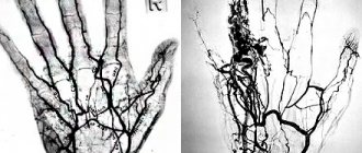
| Angiography of cerebral arteries (normal) | Pathological formation in the parietal region on the right (arteriovenous malformation) |
How to prepare for the test
Before the study, be sure to inform your doctor about the presence of:
- allergies to strawberries, shellfish, seafood or iodine-containing substances;
- history of bleeding of unknown origin;
- an allergic reaction to a contrast agent in the past;
- pregnancy.
Before the study, it is necessary to exclude food and liquid intake for 4-8 hours. Before the examination, you need to remove all jewelry, decorations and costume jewelry to prevent their contours from interfering with the resulting image.
3. Risks of analysis and what can affect the result?
Risks of angiography of the brain and neck vessels
After the dye is injected, you may feel nauseous and nervous. These symptoms go away quickly. Some people are allergic to the dye. Tell your doctor if you experience dizziness, nausea, or itching after the dye is injected.
The dye can worsen the condition of the kidneys if they are sick. The dye may also cause fetal harm in pregnant women. Consult your doctor before the procedure.
What can affect angiography?
Pregnancy, curvature of blood vessels as a result of atherosclerosis or with age, and the inability to remain in a quiet position may interfere with angiography.
Why is the research being done?
Cerebral angiography is used to detect or exclude pathologies of the blood vessels of the brain, such as:
- abnormal blood vessels (vascular malformation);
- aneurysms;
- narrowing of the arteries of the brain;
- vasculitis;
The study is also used for:
- confirmation of the presence and location of a brain tumor;
- assessment of the head and neck artery before surgery;
- determining the presence of blood clots in the vessels of the brain, which may cause a stroke.
In some cases, this procedure is used to clarify the diagnosis after abnormalities have been identified according to MRI or CT. Contrast that extends beyond the outline of a blood vessel may be a sign of internal bleeding. Narrowing of the arteries may indicate cholesterol deposits, spasms, or hereditary diseases. Abnormally developed blood arteries may be associated with brain tumors, intracranial bleeding, aneurysm (bulging of the artery wall), or arteriovenous malignancy.
Cerebral angiography is also performed to prepare for x-ray surgery to eliminate malformation, aneurysm or thrombosis of cerebral vessels.
How is MR angiography done?
The procedure takes 40-45 minutes.
Scanning steps:
- The patient is placed on the tomograph conveyor, the limbs and body are fixed with special rollers. Random movements during an MRI session lead to distortion of MR angiography results. The device may produce noise when operating; use headphones for protection.
- After briefing, medical personnel are located in an adjacent room. Communication with the patient is maintained using an intercom. For emergency communication, the patient can use the panic button.
MRI of the neck with MR angiography on a closed tomograph
- For more accurate scanning, a gradient coil is placed above the area of interest, creating an alternating electromagnetic pulse. The table with the subject is moved into the tube of the apparatus, where the constant force field generator is located.
- After a series of native images, the patient is injected with a gadolinium solution. Scanning continues after filling the vascular bed with a contrast agent.
- During an MRI session, angiography of veins and arteries may require holding your breath for 10-20 seconds.
The shooting is carried out in axial, sagittal and frontal projections. Based on the results of MR angiography, a three-dimensional image of the arteriovenous network can be reconstructed. The 3D model helps to clarify the localization of the pathologically changed area. The thickness of the scanned section is from 1 mm.
MRI angiography of vessels is performed using open and closed type devices. The first ones do not have a tunnel module, the power of the tomograph is a maximum of 0.8 Tesla. Such devices are recommended for MRI in patients with claustrophobia and overweight patients.
Closed tomographs provide a magnetic field strength of 1.5 Tesla.
MRI of the neck with contrast MR angiography
High power allows you to make high-quality images of the studied area.
Methodology and preliminary preparation
Vascular angiography is an invasive procedure that requires medical supervision of the patient’s condition before and after the diagnostic procedure. Because of these features, as a rule, it is necessary to hospitalize the patient in a hospital and conduct a certain clinical set of laboratory tests: general blood test, urine test, biochemical blood test, determination of blood group and Rh factor and a number of other tests as indicated. The person is advised to stop taking certain medications that affect the blood clotting system (for example, aspirin) several days before the procedure.
Modern angiography department
Before angiography procedures, the doctor examines the patient and obtains consent to perform the procedure.
The patient is advised to refrain from eating 6-8 hours before the start of the diagnostic procedure. If hair grows at the intended site of needle insertion, it is shaved off and then a hygienic shower is taken.
The procedure itself is carried out using local anesthetics, and the person is usually prescribed sedatives (calming) drugs on the eve of the test.
The angiographic examination technique itself involves introducing an X-ray contrast agent into the vascular bed and taking several x-rays. Depending on the pathology and the goals of the procedure, the injection site for the contrast agent may vary.
Before angiography is performed, each patient is tested for an allergic reaction to the drugs used in contrast. To do this, a certain amount of the substance is injected subcutaneously and the body’s reaction is observed. If side effects occur (rash, nausea, itching, etc.), the angiographic study is canceled. Then, instead, MR angiography (magnetic resonance angiography) is performed, for which the use of contrast agents is not mandatory.
Catheter insertion
After pre-treatment with antiseptic solutions and local anesthesia, a small skin incision is made and the required artery is found. It is pierced with a special needle and a metal conductor is inserted through this needle to the desired level. A special catheter is inserted along this conductor to a given point, and the conductor along with the needle is removed. All manipulations taking place inside the vessel occur strictly under the control of X-ray television. A radiopaque substance is injected into the vessel through a catheter and at the same moment a series of X-rays are taken, changing the patient’s position if necessary.
After the procedure is completed, the catheter is removed, and a very tight sterile bandage is applied to the puncture area. The substance introduced into the vessel leaves the body through the kidneys within 24 hours. The procedure itself lasts about 40 minutes.
Video: animation of the angiography process
Where can you get angiography with contrast in Moscow?
You can make an appointment with the specialists of JSC "Medicine" (clinic of Academician Roitberg) on the website - the interactive form allows you to select a doctor by specialization or search for an employee of any department by name and surname.
Each doctor’s schedule contains information about visiting days and hours available for patient visits. Clinic administrators are ready to accept requests for an appointment or call a doctor at home by calling +7 (495) 775-73-60.
Convenient location on the territory of the central administrative district of Moscow (CAO) - 2nd Tverskoy-Yamskaya lane, building 10 - allows you to quickly reach the clinic from the Mayakovskaya, Novoslobodskaya, Tverskaya, Chekhovskaya and Belorusskaya metro stations .
Application of vascular angiography
Vascular angiography is widely used to determine a variety of vascular pathologies, such as stenosis (narrowing) of a vessel, aneurysm (dilation) of a vessel, etc., as well as to identify pathological conditions of the heart, diagnose kidney function, to identify malformations and damage to various organs, and to diagnose tumors , cysts and many other pathological conditions.
Angiographic image
This type of research can visualize vessels of any size (from the aorta to the smallest capillaries) and all systems and organs of the human body. In addition, vascular angiography is often used before surgical interventions for preoperative preparation and diagnosis.