Brain tumors include all neoplasms localized in the central spinal canal and inside the skull. The development of brain tumors is associated with uncontrolled cell division, their classification depends on factors such as the primary factor and cellular composition.
A brain tumor can be benign or malignant. In addition, all brain tumors are classified according to certain criteria:
- by occurrence;
- by histological type;
- by localization;
- according to Smirnov.
Oncologists, neurosurgeons and radiologists at the Yusupov Hospital Oncology Center, when choosing an effective method for treating brain tumors, take into account not only the location and type of tumor, its features, the general condition and age of the patient, but also the likelihood of developing intraoperative and postoperative complications. The most advanced methods are used to treat brain cancer, providing maximum results.
Classification of brain tumors by occurrence
There are different types of brain tumors in adults, which are classified according to certain criteria.
First of all, all brain tumors are divided into primary and secondary.
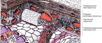
A primary tumor (for example, primary CNS lymphoma) is formed by the brain tissue itself and adjacent to it: tissue of the cranial nerves, meninges, pineal gland, lymphoid tissue or pituitary tissue. The development of these neoplasms is associated with mutations that lead to the appearance of abnormalities in the DNA of brain cells.
The occurrence of secondary brain tumors is most often associated with a metastatic process from other organs affected by cancer.
Symptoms
When brain cancer appears, symptoms in the early stages are local and depend on the location of the tumor. The disease manifests itself:
- impaired sensitivity in certain parts of the body, deterioration of orientation in space;
- memory impairment;
- decreased muscle activity up to complete paralysis;
- convulsive seizures of an epileptic nature;
- hearing damage and decreased speech recognition function;
- partial or complete visual dysfunction;
- partial or complete loss of the ability to write and speak;
- weakness, fatigue;
- pathological changes in the production of hormones from the pituitary gland or hypothalamus;
- changes in character, emotional imbalance;
- decrease in intellectual level;
- the appearance of hallucinations.
The first signs of brain cancer appear as minor changes in one or more of these areas, but the deterioration progresses over time. With a significant increase in pressure inside the skull, general cerebral symptoms appear:
- persistent headaches of high intensity that cannot be relieved with analgesics;
- dizziness caused by compression of the cerebellum or decreased blood supply to brain structures;
- vomiting independent of food intake.
Benign and malignant brain tumor
A benign brain tumor (for example, a brain lipoma) is distinguished by the relatively normal appearance of its constituent cells, slow growth, and lack of spread to other organs and penetration into the tissue of the brain itself. Despite this, a benign brain tumor can be quite dangerous and in some cases threatens the patient’s life - when the tumor is localized in a vital area of the brain, a benign tumor in the head is accompanied by compression of sensitive nerve tissue, as well as an increase in intracranial pressure.
A malignant brain tumor is a pathological process characterized by uncontrolled and unrestrained cell proliferation. Malignant neoplasms exhibit rapid growth and the ability to metastasize; the tumor often grows into nearby organs and tissues.
Malignancy and benignity are rather relative concepts. Some malignant tumors, in fact, have a benign course, which can last quite a long time.
How is the operation performed?
Neurosurgical operations to remove malignant tumors are complex and high-tech. It is important for the doctor to remove the tumor completely, without damaging functionally significant areas of the brain.
The procedures that determine the success of the operation often begin before it even begins. Doctors conduct a thorough examination of the patient, especially if the tumor is deep. They determine the boundaries of the tumor using PET or functional MRI. Then it is necessary to identify functionally significant areas in order to bypass them by dissecting the brain tissue. For this purpose, functional mapping and electrophysiological studies are used. If necessary, studies can be performed directly during surgery.
The operation is performed under stereotactic control. Stereotaxis allows the doctor to perfectly navigate inside the patient's skull. The classic version involves the use of a rigid frame, which is fixed with screws to the periosteum. In recent years, frameless stereotaxis systems have become widespread.
The most important task of the neurosurgeon, which determines the success of the operation, is to completely remove the tumor. But to do this, it must be distinguished from healthy tissue. They look almost the same. To find the boundaries of the tumor, fluorescent diagnostics and laser spectroscopy are used. The doctor uses a substance that is absorbed by abnormal cells and causes them to glow.
A German study involving 350 patients showed that without fluorescent diagnostics, glioblastoma can be completely removed in only 1 out of 3 patients, and in 2/3 of cases with its use. The probability of no tumor recurrence within 6 months after surgery increases by 2 times.
Our expert in this field:
Malakhov Igor Yurievich
Neurosurgeon
Doctor of the highest qualification category
Experience: More than 27 years
Call the doctor
Call the doctor
Types of brain cancer by location
The localization of the tumor in the brain is an important factor, which determines the method of surgical access to it.
Depending on the location of the brain tumor, there may be:
- intracerebral: their growth begins in the gray or white matter of the brain. Supratentorial affects the frontal, parietal and temporal lobes of the brain, subtentorial - the brain stem, cerebellum, bottom of the 4th ventricle (a tumor of the medulla oblongata develops, a tumor of the brain stem, a tumor of the brain stem, a tumor of the 4th ventricle of the brain, a tumor of the cerebellum of the brain, a tumor of the subcortical nodes and midbrain);
- extracerebral: develop in the soft or hard shell of the brain, blood vessels, bones of the skull (meningioma, cerebral neuroma occurs).
Treatment methods
Brain tumors are not only difficult to diagnose, but also difficult to treat. The surgical operation is technically complex, and the risk of complications after such an intervention is quite high. Treatment with chemotherapy and targeted drugs is also not always effective.
Thanks to the advent of modern intelligent equipment, neoplasms, including inoperable ones, are very effectively treated using radiosurgery and other innovative radiation therapy technologies.
Detailed information about modern treatment methods.
Classification of brain tumors by histological type
The tissue from which the tumor develops is of great importance for predicting the further “behavior” of a tumor. According to histological classification, tumors are divided into the following types:
- glial tumor of the brain;
- tumor of nervous tissue;
- vascular tumor (eg, brainstem cavernoma);
- tumor of the meninges;
- teratoma.
Glial brain tumors include:
- astrocytoma - a benign tumor of glial tissue localized in the cerebral hemispheres and cerebellum;
- oligodendroglioma - a tumor of glial tissue that is not prone to grow into other tissues and is localized in the cerebral hemispheres;
- ependymoma - brain ependymoma is a benign tumor localized in the lateral or 4th ventricle;
- glioblastoma multiforme - this type of tumor is diagnosed, as a rule, in elderly patients;
- medulloblastoma - a tumor affecting the cerebellum, most often found in children;
- pinealoma is a neoplasm that affects the pineal gland.
Tumors formed from nerve tissue include:
- Brain neuroma (schwannoma): the tumor is formed by Schwann cells or nerve sheath cells. Most often, this type of disease is benign, but sometimes it can be malignant;
- neurofibroma of the brain - is, as a rule, a benign neoplasm consisting of Schwann cells, mast cells or fibroblasts;
- tumors developing in the area of the sella turcica, for example, from the pituitary gland.
A tumor of the meninges is, for example, a meningioma of the brain. This tumor is characterized by slow growth and benign nature.
Teratomas are cancerous tumors that develop from cells with a morphological structure close to stem cells. Most often diagnosed in patients of childhood and young age.
What is the brain and why do we need it?
Thanks to the brain, we think, feel, remember, talk, hear, see, move and breathe. With the help of nerves, it exchanges signals with other tissues, transmits and receives information from them, and coordinates the work of internal organs.
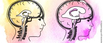
It has several important departments, each of which performs special tasks:
- Cerebrum
: Surrounded by cerebrospinal fluid and divided into left and right hemispheres, which control reasoning, thoughts, emotions, language, human planned muscle movements such as walking or chewing. In addition, this is where the awareness of the received information about everything seen, taste, smell, touch and pain occurs. - Cerebellum
: Helps us coordinate movements. - Brainstem
: Contains bundles of very long nerve fibers that transmit signals between the brain and other tissues that control muscles and sensations. The centers located in it control breathing and heartbeat. - The hypothalamus
and the small gland
pituitary gland
: thanks to their joint work, hormones are produced that regulate the production of hormones. Hormones are substances that are created by our glands, enter various tissues through the bloodstream and tell them how to act - to work or rest, to secrete something or absorb .. - Blood-brain barrier:
the inner lining of capillaries - small blood vessels - creates a reliable barrier that prevents harmful substances from reaching the brain. Unfortunately, it also delays most chemotherapy drugs that can destroy cancer cells. - Choroid Plexus:
The area that produces cerebrospinal fluid, which protects our major organ.
Classification of brain tumors according to Smirnov
This classification involves the division of brain tumors into several types in accordance with cell maturity and morphological characteristics.
According to the degree of maturity, brain tumors can be:
- mature (for example, cerebral ependymoma);
- immature (for example, ganglioblastoma of the brain);
- immature (for example, medulloblastoma of the brain).
In addition to the degree of maturity, this classification is also based on morphological characteristics, combining classification by localization and histological classification.
Treatment of brain tumors at the Yusupov Hospital
Treatment of brain tumors at the Yusupov Hospital Oncology Center is carried out using the most advanced non-surgical techniques:
- stereotactic radiosurgery – which allows you to remove small tumors in one session;
- radiation therapy using the latest generation installations, the latest linear accelerators and systems that ensure maximum safety and effectiveness of therapy, etc.
If surgical treatment is unavoidable, it is performed by the best specialists who masterfully master all the technologies and methods of interventional neuroradiology and modern neurosurgery. Doctors at the Yusupov Hospital Oncology Center make every effort to use gentle, minimally invasive methods and operations with transnasal access, which do not require an incision in the skull.
Thanks to the innovative equipment of the oncology center, doctors have the opportunity to perform high-quality diagnostics and carry out effective treatment of brain tumors using non-surgical or minimally invasive methods.
In parallel with neurosurgeons, neurologists, oncologists, and radiologists take part in the treatment of patients with brain tumors. The patient is provided with supportive therapy and qualified psychological support.
You can make an appointment with an oncologist at the oncology clinic and find out the conditions of hospitalization by calling the Yusupov Hospital or online on the website from the coordinating doctor.
Diagnostic methods
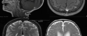
To make a diagnosis, doctors prescribe various types of scans (CT, MRI), laboratory tests, and functional tests.
The use of the latest generation computerized complexes allows specialists to take tumor tissue for examination under a microscope without resorting to a full-scale operation. At the same time, the risk of complications is reduced to a minimum even when the tumor is located in a hard-to-reach place.
Detailed information about modern diagnostic methods and capabilities.