General information
Arachnoiditis is an infectious disease of the central nervous system and is a serous inflammation of the structures of the arachnoid membrane of the brain or spinal cord. The arachnoid membranes do not have their own vascular system, therefore the lesions are not isolated, and infectious processes spread from the dura or soft meninges, therefore the symptoms of arachnoiditis are conclusively attributed to the serous type of meningitis .
The pathology was described in most detail by the German doctor Benninghaus, and the term was first used in the dissertation of A.T. Tarasenkov, who studied the signs of cerebral inflammation and arachnoiditis in particular. Some scientists call this disease serous meningitis, but according to ICD-10 it is assigned the code G00 and the name bacterial arachnoiditis , G03 - which includes meningitis due to other or unspecified causes, including arachnoiditis , meningitis , leptomeningitis , pachymeningitis , as well as G03 .9 – for unspecified meningitis – spinal arachnoiditis NOS (no additional instructions).
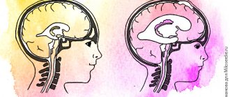
The brain has three membranes: hard, arachnoid and soft. Thanks to the hard one, sinuses are formed for the outflow of venous blood, the soft one provides trophism, and the arachnoid one is necessary for the circulation of cerebrospinal fluid . It is located above the gyri, but does not penetrate the sulci of the brain and separates the subarachnoid and subdural space. Its structure contains arachnoidendothelial cells, as well as bundles of collagen fibrils of varying thickness and quantity.
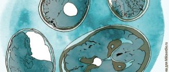
Histology of the meninges
Prognosis and prevention for patients
The best prevention is a healthy lifestyle, strengthening the immune system, minimizing stress, and regular medical examinations for timely detection and treatment of diseases. This is especially true for expectant mothers.
But if a cyst is still detected, there is no need to panic. It is necessary to see a doctor and follow his recommendations, and get more rest. In some patients, a recurrence of the cyst occurs after treatment. In case of relapse of the disease, regular monitoring of cholesterol levels, blood pressure, and treatment of existing diseases is necessary.
With timely treatment, however, the prognosis is good. Many patients recover completely and say goodbye to the cyst forever.
Pathogenesis
Arachnoiditis causes morphological changes in the form of clouding and thickening of the arachnoid membrane, which may be complicated by fibrinoid deposits. Most often they are diffuse, but in some cases they can be limited, that is, we are talking about more severe local disorders initiated by an extensive process of arachnoiditis. Macroscopic changes in this case are:
- clouding and thickening (hyperplasia of the arachnoid endothelium) of the arachnoid membrane, its fusion with the choroid and dura mater of the brain;
- diffuse infiltration;
- expansion of subarachnoid slit-like formations and cisterns at the base of the brain, development of their hydrops (overflow of cerebrospinal fluid).
The further course of the pathology leads to fibrosis and the formation of adhesions between the choroid and arachnoid membrane, impaired circulation of the cerebrospinal fluid (cerebrospinal fluid) and the formation of one or more arachnoid cysts. In this case, the normal circulation of the liquor fluid is disrupted and, as a result, hydrocephalus , the mechanism of which is based on two development paths:
- occlusive - resulting from a violation of the outflow of fluid from the ventricular system, for example, the closure of the Luschka or Magendie holes by the resulting adhesions or cysts;
- aresorptive – in which the processes of fluid absorption through the structures of the dura mater are disrupted, as a result of a diffuse “adhesive” process.
How to treat a cyst?
If the arachnoid cyst does not grow or develop, then treatment is not required. A person will need to register with a neurologist and undergo an MRI examination every year. This will make it possible to monitor the growth and development of the cyst, if it occurs.
When the cyst progresses and causes neurological symptoms, it requires treatment. Drug correction comes down to taking medications that normalize cerebral circulation (nootropics, vasotropics, antioxidants). Sometimes the patient is prescribed drugs that dissolve adhesions (Karipain, Longidaza). The help of a surgeon is needed when taking medications does not solve the problem.
Surgical intervention is designed to reduce intracranial pressure; it is implemented in three ways:
- Shunting. The result of this procedure is the formation of pathways for the outflow of cerebrospinal fluid from the cyst cavity into the peritoneal cavity.
- Fenestration. This method involves aspiration of the contents of the cyst and further creation of holes. They connect the cystic cavity with the cerebral ventricle or subarachnoid space.
- Drainage of the tumor using needle aspiration.
Modern neurosurgeons prefer to use endoscopic interventions, as they have minimal brain trauma.
Surgery is prescribed only if other treatment methods are ineffective. If there is hemorrhage in the area of the cyst, or if it ruptures, removal of the tumor is required. The operation is performed by trephination, which is associated with a number of complications. The rehabilitation period is quite complex and time-consuming.
Classification
There are several classifications of arachnoiditis. Based on the established cause, arachnoiditis can be post-traumatic, infectious (rheumatic, post-influenza, tonsillitis) and toxic, depending on the type of changes - cystic, adhesive-cystic, limited and diffuse, single-focal and multifocal.
Depending on the clinical picture and course, acute, subacute and chronic arachnoiditis are distinguished, but for diagnosis it is most important to determine the location of arachnoiditis and predict the pattern of exposure and consequences of membrane lesions.
Depending on the predominant location and structures involved in the pathology, arachnoiditis can be of various types: cerebral, basal, opto-chiasmal, pontocerebellar, precerebellar, spinal, etc.
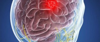
Cerebral arachnoiditis
The cerebral type of arachnoiditis usually covers the membranes of the brain of the anterior parts of the cerebral hemispheres and areas of the central gyri, affecting not only the archnoid endothelium, but also the structures of the soft medulla with the formation of adhesions between them. As a result of the adhesive process, cysts with liquor-like contents are formed. Thickening and compaction of cysts can lead to xanthochromic tumor-like formations containing large amounts of protein, which can manifest as the development of status epilepticus .
Arachnoid cyst of the brain
Optico-chiasmatic arachnoiditis
It is most often localized in the chiasmal region and affects the base of the brain, involving the optic nerves and their chiasm in the pathology. This is facilitated by traumatic brain injuries (concussion or contusion), infectious processes in the paranasal sinuses, as well as diseases such as tonsillitis, syphilis or malaria . Its result can be irreversible vision loss, which begins with pain behind the eyeballs and visual impairment, which can lead to unilateral and bilateral temporal hemianopia, central scotoma , concentric narrowing of the visual fields.
The development of pathology is slow and not strictly local; it can spread to areas distant from the chiasm, usually accompanied by the formation of multiple adhesions, cysts, and even the formation of a scar membrane in the chiasm area. A negative effect on the optic nerves causes their atrophy - complete or partial, which is ensured by mechanical compression by adhesions, the formation of congestive nipples and blood circulation disorders ( ischemia ). In this case, initially one of the eyes suffers more, and after a few months the second one is also involved.
Spinal arachnoiditis
In addition to these well-known causes, spinal arachnoiditis can be caused by furunculosis and purulent abscesses of various locations. In this case, cystic limited formations cause symptoms similar to an extramedullary tumor, symptoms of compression of spinal cord structures, as well as radicular syndrome and conduction disorders, both motor and sensory.
Chronic inflammatory processes cause protein-cell dissociation of the cerebrospinal fluid and most often affect the posterior surface of the thoracic, lumbar or cauda equina spinal cord. They can spread to several roots or, in case of diffuse lesions, to a large number, changing the lower limit of the sensitivity disorder.
Spinal archnoiditis can be expressed as:
- in the form of tingling, numbness, weakness in the legs, unusual sensations in the limbs;
- the occurrence of leg cramps, muscle spasms, spontaneous twitching;
- in the form of a disorder (increase, loss) of such reflexes as knee, heel;
- attacks of severe shooting pain like electric shocks or, on the contrary, aching pain in the lower back;
- disruption of the pelvic organs, including decreased potency .
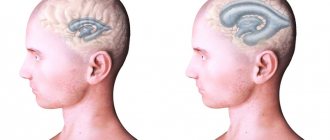
Irritation and compression of the cortex and nearby parts of the brain during arachnoiditis can be complicated by the formation of cysts of various types - retrocerebellar, cerebrospinal fluid, left or right temporal region.
Retrocerebellar arachnoid cyst
A retrocerebellar cyst is formed when the choroid plexus of the fourth ventricle is displaced upward and backward from the intact vermiform part of the cerebellum. To identify this type of cyst, CT and MRI are approximately equally informative.
Arachnoid cerebrospinal fluid cyst
It is customary to distinguish between intracerebral and subarachnoid cerebrospinal fluid cysts, the former are more common in adults, and the latter are more typical for pediatric patients, which is very dangerous and causes mental retardation.
Liqueur cysts are formed by arachnoid endothelium or scar collagen and filled with cerebrospinal fluid. They can be congenital or formed during the resorption of intracerebral hemorrhages, foci of bruises and crush injuries of the brain, in the zone of ischemic softening after injuries. They are characterized by a long-term remitting course, initiating epileptic seizures of different structure, duration and frequency.
A cerebrospinal fluid cyst can also occur as a result of subarachnoid hemorrhage or reactive adhesive leptomeningitis.
Arachnoid cyst of the right temporal region
A cyst in the right temporal region can cause headaches, a sensation of pulsation, squeezing of the head, noise in the ear, attacks of nausea, convulsions , and uncoordination of movements.
Arachnoid cysts are frozen, stable and most often do not cause discomfort or cerebral disorders. An asymptomatic course can lead to the formation being detected only during a brain tomography if arachnoiditis is suspected.
Arachnoid cyst of the left temporal lobe
If the left temporal lobe cyst is progressive, then it can gradually increase focal symptoms due to pressure on the brain. It is usually located in the left temporal lobe and looks like an extension of the external cerebrospinal fluid space.
When a patient learns about a cyst in the left temporal region, it often turns out that it is not fatal and may not cause negative symptoms. However, in some cases there is a risk of developing speech disorders (sensory aphasia ), loss of visual fields, sudden spasms of the limbs or the whole body.
Features of treatment
After an accurate diagnosis of an arachnoid cyst lesion, treatment options depend primarily on the size of the arachnoid cyst and the dynamics of its progression. If a small, slow-growing tumor is found, you will be monitored regularly and treated for the underlying cause. Once the triggers are eliminated, the cysts will resolve. A rapidly growing large cyst poses a serious threat to the health and life of the patient, so its treatment can be carried out using a combination of drug therapy and surgical intervention.
Conservative therapy
If a medium-sized tumor is detected at the time of diagnosis, it can be treated without surgery, but only pharmacologically. In this case, treatment is determined individually for each patient, and the following drugs can be used.
- Antiviral drugs (Amiksin, Pyrogenal);
- metabolic stimulants (Gliatilin, Actovegin);
- drugs that reduce adhesion (Karipatin, Longidaza);
- Immunomodulators (Timogen, Viferon).
During drug therapy, it is important to strictly follow all recommendations and recommendations of the attending physician and not deviate from the prescribed regimen of taking the drug.
Surgical treatment
When a fast-growing cyst is diagnosed, patients are prescribed urgent surgery to remove the tumor. The operation can be performed using the following methods:
- puncture - the contents of the cyst are removed through special punctures;
- Bypass - a drainage tube is inserted into the cavity of the cyst to expel its contents;
- Endoscopic method - this method involves removing the cyst using special equipment (endoscope);
- Craniotomy is a radical surgical procedure during which the skull is opened and the cystic tumor is removed.
Surgery for an arachnoid cyst is the most effective way to remove the tumor, but there is a risk of infection or damage to adjacent brain tissue during surgery.
Causes
There are several ways of developing inflammation of the arachnoid membranes and it has been established that arachnoiditis is polyetiological and can occur as a result of such factors as:
- past acute and chronic infectious processes (including influenza , rheumatism, measles , fever , sepsis , pneumonia , syphilis , tuberculosis , brucellosis , toxoplasmosis , osteomyelitis of the skull bones);
- inflammatory diseases of the nasal paranasal sinuses;
- acute or more often chronic suppurative otitis media , especially caused by low-virulent microorganisms or toxins;
- complication of suppurative otitis media, for example, labyrinthitis , petrositis , sinus thrombosis ;
- complication of cured purulent meningitis or brain abscesses;
- chronic intoxication with alcohol, lead, arsenic ;
- various injuries - craniocerebral and spinal cord (mainly as residual effects);
- reactive inflammation caused by slowly growing tumors or encephalitis, most often non-purulent otogenic.
Standard size and purpose of operation
The absence of a cyst is considered normal, but if there is an arachnoid cyst, regardless of its size, this is already an anomaly. An arachnoid cyst can reach different sizes, but cannot grow significantly - due to the pressure of the cerebral fluid, it resists pressure.
If a small cystic tumor (1-2 mm) is diagnosed, changes in volume are monitored to see if the arachnoid cyst increases in size. This is usually in the early stages of development. The average cyst is about 0.9-1 cm, and more serious, severe cysts exceed 1 cm and can reach 12 cm.
In a situation where the arachnoid cyst has a constant size and does not increase in adults, there is no need to perform surgery to remove it. Your doctor will make some recommendations and perform regular yearly exams to monitor changes in the cyst.
A large arachnoid tumor is congenital and occurs in the fetus along with the central nervous system. Characteristic are small arachnoid cysts acquired in adults, enlarged due to the accumulation of fluid in them.
If during observation we notice changes in size, it grows, and its symptoms begin to make themselves felt, then surgical intervention is simply necessary urgently. The need for surgery is also due to the negative impact of such factors as: increased pressure inside the skull, cerebral hemorrhages, convulsions, rupture of the cyst itself.
Symptoms of brain arachnoiditis
Symptoms of arachnoiditis are usually caused by intracranial hypertension , in more rare cases by liquor hypotension , as well as manifestations reflecting localization affecting the meningeal processes. Moreover, general or local symptoms may predominate, depending on this, the first symptoms and the clinical picture changes.
The initial subacute course of the disease can eventually turn into a chronic form and manifest itself in the form of general cerebral disorders:
- local headaches , the most intense - in the first half of the day can cause nausea and vomiting ;
- development of the jumping symptom , when pain occurs locally during jumping or an awkward, unabsorbed movement with landing on the heels;
- dizziness of a non-systemic nature;
- sleep disorders;
- memory impairment;
- mental disorders;
- the occurrence of causeless irritability, general weakness and increased fatigue.
Focal disorders primarily depend on the location of the development of the pathology and can manifest themselves in the form of symptoms of damage to the trigeminal, abducens, auditory and facial nerves. Besides:
- With convexital (convex) arachnoiditis, inflammatory processes affect areas of the central gyri and anterior parts of the cerebral hemispheres, while the phenomena of irritation of brain structures predominate over manifestations of loss of functions, which are expressed in the form of anisoreflexia , central paresis , generalized and Jacksonian epileptic seizures , impaired circulation of cerebrospinal fluid, disorders of sensitivity and movement ( mono- or hemiparesis ).
- With inflammation of the basal sections (optic-chiasmatic, cerebellopontine and in the region of the postcranial fossa), general cerebral symptoms most often occur and the functions of the nerves of the base of the skull are disrupted.
- Optico-chiasmatic arachnoiditis is manifested by a decrease in visual acuity and changes in fields, resembling optic neuritis and combined with autonomic dysfunction - severe dermographism , increased pilomotor reflex, profuse sweating, acrocyanosis , sometimes thirst , increased urination, hyperglycemia , adiposogenital obesity , decreased sense of smell.
- Pathology affecting the region of the cerebral peduncles causes pyramidal symptoms, as well as signs of damage to the oculomotor nerves and meningeal signs.
- Arachnoiditis of the cerebellopontine angle causes headaches in the occipital region, tinnitus, neuralgia , paroxysmal dizziness, sometimes with vomiting, unilateral cerebellar disorders - when the patient staggers or supports weight on one leg, the fall occurs on the affected side; a thorough examination can reveal an ataxic gait , horizontal nystagmus , pyramidal symptoms, dilatation of the veins of the fundus, provoked by disturbances of the venous outflow.
- If the large (occipital) cistern is affected, the disease develops acutely with fever, obsessive vomiting, pain in the back of the head and cervical region, which intensifies when coughing, trying to turn the head or make a sudden movement.
- Localization of inflammatory processes in the area of IX, X, XII pairs of cranial nerves leads to nystagmus , increased tendon reflexes, pyramidal and meningeal symptoms.
- Arachnoiditis of the posterior cranial fossa can affect the V, VI, VII, VIII pair of cranial nerves and cause intracranial hypertension with meningeal symptoms, cerebellar and pyramidal disorders, for example, ataxia , asynergia , nystagmus , adiadochokinesis , headache becomes a constant symptom, one of the earliest .
- Diffuse damage is provoked by general cerebral phenomena and uneven expansion of the ventricles, which is expressed in the occurrence of frontal, hypothalamic, temporal, midbrain and cortical syndrome; the pathology initiates a disruption of the normal exchange of cerebrospinal fluid, vague pyramidal symptoms, and can affect individual cranial nerves.
Possible consequences and complications
After a radical intervention with excision of the cyst, some neurological consequences may remain. This depends on the condition of the patient’s body, concomitant diseases, and compliance with medical recommendations.
The most severe consequences (disability, blindness, deafness, intractable headaches, rupture with effusion of cerebrospinal fluid, death) are possible in case of late detection of a large cyst.
An arachnoid cyst is a type of benign tumor. Practice shows that it can degenerate into cancer, but in fact there are not many such cases.
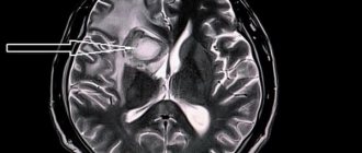
Tests and diagnostics
When making a diagnosis, a differential diagnosis must be made with abscesses and neoplasms in the posterior cranial fossa or other parts of the brain. To determine arachnoiditis, it is important to conduct a comprehensive and detailed examination of the patient.
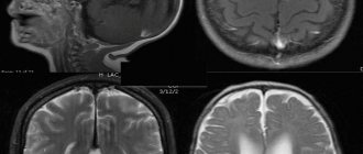
Electroencephalography , angiography , pneumoencephalogram , scintigraphy , survey craniograms , skull x-ray, myelography , CT, MRI are indicative These studies make it possible to identify intracranial hypertension, local changes in biopotentials, expansion of the subarachnoid space, cisterns and ventricles of the brain, cystic formations and focal changes in the brain substance. Only if there is no congestion in the fundus, then a lumbar puncture to detect moderate lymphocytic pleocytosis and slight protein-cell dissociation. In addition, it may be necessary to perform an index and finger-nose test.
How to detect?
An examination by a neurologist allows one to suspect the presence of a tumor in the brain only if the cyst reaches a significant size. Only in this case does it give certain neurological symptoms.
Therefore, if the doctor suspects the presence of a cyst, he will refer the patient for the following studies:
- EEG.
- REG.
- Echo-EG.
- MRI or CT have the maximum information content and allow you to clarify the diagnosis. The use of MRI with contrast is especially important. This method makes it possible to distinguish a cyst from a brain tumor.
Diet for arachnoiditis
Diet for the nervous system
- Efficacy: therapeutic effect after 2 months
- Timing: constantly
- Cost of food: 1700-1800 rubles per week
Treatment of arachnoiditis is best combined with a diet based on the principles of healthy eating:
- limited consumption of seasonings and salt;
- products containing refined sugar, butter, flour are excluded;
- meat should be of dietary grades - rabbit, turkey, chicken, etc.;
- no sausages or smoked meats;
- there should be fresh vegetables and fruits on the table;
- You need to enrich your diet with baked potatoes, cabbage and carrot salads, spicy and leafy greens;
- you need to add currants, sea buckthorn, persimmons, grapes, and raisins to dishes and teas.
Symptom complex
The manifestation of symptoms primarily depends on the location and volume of the cystic neoplasm. When pressure from a cyst associated with its increased size is applied to any area of the brain, the patient experiences:
- sharp pain in the head;
- dizziness that suddenly appears;
- severe attacks of nausea and vomiting reactions;
- state of constant fatigue;
- drowsiness, hallucinations;
- movement coordination disorder;
- visual and hearing impairments;
- the presence of convulsions and seizures.
Among the primary symptoms are convulsions, hallucinations, partial paralysis of the body, disorganization of mental functioning. If any, you should immediately seek specialized help and undergo a full examination to prescribe further treatment.
Why is the disease dangerous?
A cerebrospinal fluid cyst, if not properly treated, can become a dangerous disease with serious complications and tragic consequences. It entails disability, and in isolated cases even death.
With timely assistance, the prognosis is more than favorable. The risk of worsening the condition is reduced. Otherwise, a number of conditions may develop:
- Neurological disorders. These include disturbances in motor activity, loss of sensitivity, and disruptions in the functioning of the nervous system.
- Epilepsy, frequent convulsions, involuntary movements of arms and legs, loss of consciousness.
Strict adherence to all doctor’s prescriptions will help prevent their development.
Arachnoid cysts do not pose a serious danger for the time being. They do not manifest themselves in any symptoms and are detected only during a routine examination. But sometimes, due to an increase in the amount of fluid, they begin to grow. In such situations, the person’s condition noticeably worsens and requires medical intervention.