MRI expansion of convexital liquor spaces
The cerebrospinal fluid spaces are filled with cerebrospinal fluid, which transports nutrients and regulates the level of intracranial pressure.
The distribution of cerebrospinal fluid within the skull is ensured by the subarachnoid space, the third and fourth ventricles.
The movement of cerebrospinal fluid is ensured by cardiac activity, body position, the respiratory cycle, and the movement of epithelial cilia. Normal indicators of intracranial pressure are ensured by the optimal amount of cerebrospinal fluid - 140 ml.
The expansion of convexital spaces is detected by ultrasound and MRI in newborns. Acquired forms of nosology in infants occur after traumatic brain injuries. Doppler ultrasound (USDG) allows for timely diagnosis of pathology before the fontanelles of the skull close. In other cases, magnetic resonance imaging reveals the disease. The procedure is harmless to the baby's health. For high-quality results, a motionless position is required during the scan, so the procedure is performed for the child under anesthesia.
MRI of expanded cerebrospinal fluid spaces
What types of external hydrocephalus of the brain are there?
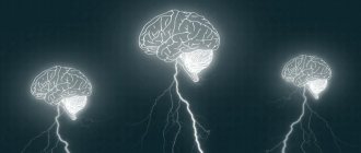
External hydrocephalus of the brain refers to the accumulation of cerebrospinal fluid (cerebrospinal or cerebrospinal fluid) outside the cerebral hemispheres - in the subarachnoid space. Due to the large accumulation of fluid, the subarachnoid fissures widen, which causes increased pressure on the cerebral cortex and the resulting negative consequences.
The nature and level of complexity of the disease directly depend on the specific type of dropsy. Several criteria are used for classification. The most common ones are:
- intensity of manifestation (pronounced - accumulation of a large amount of cerebrospinal fluid, causing neurological symptoms; moderate - minimal amount of fluid, no signs);
- degree of impact on brain structures (compensated - cerebrospinal fluid does not affect the brain; decompensated - there is a deterioration in the functioning of the nervous system and brain);
- causes of occurrence (replacement - more often diagnosed in older people and is accompanied by the death of brain cells; acquired - occurs due to the spread of infections and mechanical traumatic brain injuries);
- nature of the course (chronic form - a gradual increase in neurological disorders; acute form - a sharp deterioration in the patient’s well-being).
Employees of the Department of Neurosurgery of the City Clinical Hospital named after. Eramishantsev will first determine the type of external hydrocephalus and only then begin treatment. Particular attention is paid to diagnostic data and a thorough study of the symptoms of the identified disease.
Reasons for the expansion of convexital liquor spaces in children
Etiology of congenital forms:
- Benign and malignant neoplasms;
- Inflammatory processes (meningitis, meningoencephalitis);
- Traumatic brain injuries;
- Cerebral origin when passing through the birth canal;
- Hydrocephalus (dropsy);
- Increased intracranial pressure.
The first signs of pathology can be detected by parents by the protrusion of the skull in the area of the sutures. The second specific manifestation is the divergence of the gap between the hemispheres. The changes lead to limited head elevation. Initially, neuropathologists make a diagnosis of perinatal encephalopathy. Diagnostic methods allow you to more accurately identify cerebral changes.
Secondary symptoms:
- Decrease in reflex activity of the limbs;
- Loss of appetite, weight loss;
- “Moonlight gaze” - lower drooping of the eyelids, visualization of the iris and pupil.
The expansion of the convexital grooves of the brain in the subarachnoid spaces can result in atrophy of the parietal, frontal, temporal, and occipital regions. Against the background of pathology, deformation of the ventricular system occurs.
Atrophic cerebral changes provoke post-ischemic conditions, meningococcal, tuberculous encephalitis, and cancer processes. Expansion of the subarachnoid space is a consequence of the destruction of certain areas of the cerebral parenchyma. A decrease in the amount of white and gray matter leads to the filling of free spaces with cerebrospinal fluid and blood.
Symptoms of external hydrocephalus
The clinical picture in each specific case will be different, and the nature of the manifestation of the disease depends on the severity of the pathological process and the state of the central nervous system. Common symptoms are frequent headaches, blurred vision, nausea, vomiting, and weakness. By the way, pain is more localized in the frontoparietal region and in the area of the eyeballs. A person with dropsy experiences pain in the first half of the day, with sudden movements, coughing, sneezing, or severe physical exertion.
Symptoms may vary depending on the severity of the disease. Scientists distinguish 3 stages, and each has its own characteristics:
Mild external hydrocephalus. With a minimal amount of dropsy, the human body will try on its own to cope with such a problem as impaired circulation of the cerebrospinal fluid. In this case, you will feel a slight malaise, periodic dizziness, short-term darkening in the eyes, and a tolerable headache.
The middle stage of development of the disease. At this stage of the spread of the disease, symptoms appear intensely and are more pronounced. Due to an increase in intracranial pressure, severe headaches occur during physical activity, swelling of the optic nerve and facial tissues, increased fatigue, nervousness, depression, and surges in blood pressure.
Severe form of the disease. Signs of pathology in severe forms of external hydrocephalus are reduced to convulsive seizures, frequent fainting, a state of apathy, loss of intellectual abilities, memory loss and inability to care for oneself. Progressive dropsy can even lead to death, so there is no need to delay going to the doctor. It is better to do this at the first suspicion and a slight deterioration in health.
With chronic accumulation of cerebrospinal fluid, symptoms such as unsteady gait, paralysis of the upper and lower extremities, urinary incontinence, nighttime insomnia and daytime sleepiness, depressed mood, and a complex of psychoneurological disorders can be observed.
Causes and symptoms
Hydrocephalus in adults is an acquired pathology and is divided into forms and degrees. Clinical manifestations depend on the speed of the pathological process.
Root causes of pathology:
- The secretion of cerebrospinal fluid and its absorption by the villi of the arachnoid membrane are impaired. The circulation of secretions is not impaired;
- Violation in the cerebrospinal fluid spaces;
- The disorder is caused by changes in the brain parenchyma (senile age, central nervous system pathologies);
- Hypersecretion of cerebrospinal fluid.
The liquor system in the brain fills all free cavities (ventricles, cisterns, gaps between the membranes). A closed system allows the liquor fluid to circulate, as a continuous cycle of secretion formation and absorption occurs.
Impaired cerebrospinal fluid dynamics may be caused by concomitant diseases.
Secondary causes:
- Malignant or benign neoplasm, cyst;
- Inflammation of the meninges;
- Vascular pathology;
- Traumatic brain injuries;
- Intracranial hemorrhage;
- Intoxication due to poisoning (food, heavy metals, poisons).
Acquired external hydrocephalus is a fairly rare pathology in adults. A small accumulation of cerebrospinal fluid in the subarachnoid space has a blurred clinical picture. The patient experiences only minor headaches and does not seek medical help.
Symptoms increase as the subarachnoid cavity expands (circulation in the ventricles of the brain is not impaired). An increase in intracranial pressure leads to damage or death of nerve cells, which can have serious consequences.
Common clinical manifestations:
- Jet lag;
- Fatigue and drowsiness immediately after rest;
- Continuous headaches;
- Memory disorder;
- Disorientation in space;
- Change in gait (elementary purposeful actions are impossible, movements are uncertain and shaky);
- Development of strabismus;
- Attacks of vomiting, followed by a decrease in the intensity of the headache;
- Meteosensitivity.
The increase in fluid leads to pressure from the membranes on vital centers in the brain. The chronic form is manifested by additional symptoms:
- Disturbance in the respiratory center (from difficulty breathing to complete stop);
- Enuresis (urinary incontinence);
- Convulsive syndrome;
- Paresis or paralysis (lower limbs);
- Development of dementia;
- Heart rhythm failure.
For children, this symptomatology is characteristic of the expansion of the subarachnoid convexital spaces (the area of the central sulcus). This pathology is not typical for adults.
Why does dropsy of the brain occur?
In adult patients, acquired hydrocephalus is often encountered, which develops either due to any mechanical damage to the head, or as a result of the development of pathological processes. Why does cerebrospinal fluid accumulate outside the cerebral hemispheres? The explanation is simple: brain structures are disrupted, adhesions appear in the veins, arachnoid villi are destroyed, and as a result, the cerebrospinal fluid does not circulate as it should.
If we delve deeper into the question of the causes of such a disease as external dropsy of the brain, we can identify some factors:
- infectious diseases (tuberculosis, meningitis, encephalitis);
- post-stroke condition, development of sepsis, extensive hemorrhage;
- concussion, head or cervical spine injury;
- malignant tumors that develop in the stem region.
Frequent intoxication of the body leads to the appearance of external hydrocephalus. For example, alcohol abuse, which damages neurons and leads to tissue death. Those patients who suffer from metabolic disorders, diabetes mellitus, multiple sclerosis, encephalopathy, and atherosclerosis are also at risk. Another reason that deserves due attention is irreversible age-related changes that cause aging of blood vessels and brain tissue.
Main services of Dr. Zavalishin’s clinic:
- consultation with a neurosurgeon
- treatment of spinal hernia
- brain surgery
- spine surgery
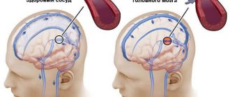
Diagnosis of external hydrocephalus in adult patients
Studying the symptoms and visual examination of the patient is not a sufficient condition for determining external hydrocephalus of the brain. Indirect signs, of course, are important, but you can’t do this without professional diagnostics. Today, 6 methods for detecting dropsy are used:
- Ultrasound examination (US) of the neck and head to assess the condition of blood vessels;
- Magnetic resonance imaging (MRI) helps to identify changes in soft tissues and most accurately determine the type of hydrocephalus and the stage of development of the pathology;
- computed tomography (CT) is intended to determine the degree of damage to brain tissue, the size of subarachnoid fissures, and the presence of neoplasms;
- X-rays with the introduction of a contrast agent are aimed at identifying disturbances in the outflow of venous blood and damage to the vascular bed;
- a spinal puncture is prescribed if there is a suspicion of the development of dropsy after encephalitis or meningitis and you need to find out what level of cerebrospinal fluid pressure;
- ophthalmological examination - an opportunity to determine whether the patient has swelling of the optic nerve and atrophy of the tissues of the ocular apparatus.
IMPORTANT! If the diagnosis of “chronic external hydrocephalus of the brain” is confirmed, it is advisable to carry out an additional diagnostic examination after 6 months. The intensity of further visits to the doctor depends on the data obtained and is determined individually.
Clinical symptoms of increased liquor spaces
Signs of dilation of the ventricles and subarachnoid cavities in newborns:
- Constant irritability;
- Negative reaction to strong noises, flashes of light;
- Restless sleep;
- Frequent regurgitation;
- Strabismus;
- Different pupil sizes;
- Moodiness when weather changes;
- Slow overgrowth of the fontanel;
- Twitching of the chin.
Deviations must be detected immediately after their occurrence. Correction of changes should be carried out immediately after detection.
Clinical symptoms of subarachnoid enlargement in adults:
- Dyspeptic disorders (vomiting and nausea);
- Headache;
- Drowsiness;
- Constant dizziness;
- Increased intracranial pressure;
- Dementia;
- Memory problems;
- Disorders of spatial orientation;
- Unsteady gait.
Determining clinical signs in a child and an adult requires contacting a neurologist. The specialist will detect the pathology and give a referral for an MRI of the convexital cerebrospinal fluid cavities.
At the initial stages there are no manifestations of pathology. Symptoms depend mainly on the severity of the deformity and the causes of the pathology. In small children, the disease is provoked by arachnoiditis, birth trauma, and meningitis. Tumors are a common etiological factor in the occurrence of pathology in adults. Compression of the cerebral parenchyma causes defects in the destruction of white and gray matter. The formed cavities are filled with liquor.
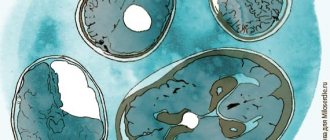
Analysis of brain tomograms in people with enlarged cerebrospinal fluid cavities: atrophic changes in the cortex of the frontal, occipital, and temporal regions. The number of convolutions is smoothed out, and blood accumulations are visible.
The presence of concomitant complications in a child is dangerous, so invasive diagnostic measures are postponed to the second year of life. A reliable diagnosis can be made after taking a biopsy of the enlarged cavities. Microscopic assessment of the properties of cerebrospinal fluid makes it possible to differentiate inflammatory, tumor, and ischemic changes.
Magnetic resonance imaging reveals accompanying pathological changes:
- Leukomalacia is a pathology of impulse signal transmission due to softening of the meninges;
- Inflammatory foci;
- Collections of blood.
An MRI picture of a moderate expansion of the external cerebrospinal fluid spaces shows morphological changes, but a whole range of diagnostic methods is required to make a correct diagnosis.
Tomograms of hypertensive hydrocephalus
Treatment of external hydrocele in adults
Treatment methods are selected at a consultation with a neurosurgeon or neurologist after diagnosing the disease. Intervention must be timely, otherwise the risk of various neurological complications increases. It is important to take into account both the type of pathology and the characteristics of the patient’s body.
In the Department of Neurosurgery of the City Clinical Hospital named after. Eramishantsev practice only effective methods of treating external hydrocele of the brain. Methods are divided into two large groups: conservative (medicinal) and surgical (operative), each of which has its own characteristics and advantages.
CONSERVATIVE TREATMENT
Drug treatment is only relevant at a mild stage of the disease. Special medications accelerate the outflow of fluid from the brain, increase urination, relieve inflammation and swelling, strengthen blood vessels, and normalize the functioning of the cardiovascular system. To combat severe headaches, your doctor may prescribe non-steroidal anti-inflammatory and painkillers.
Common groups of medications are vascular, neurotropic, venotonics, diuretics. But in acute illness they will be ineffective. Mixed hydrocephalus is poorly corrected. In this case, conservative treatment will not get rid of the disease, but will only restore or improve the functioning of individual systems and functions of the human body. Often surgical intervention is not possible.
SURGERY
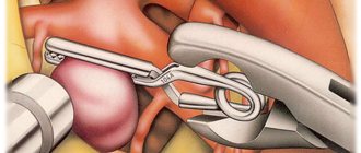
If acute external dropsy is diagnosed, in most cases drainage of the cerebral ventricles is prescribed. The main technologies are endoscopy and open surgery.
In the first case, we are talking about manipulations that are characterized by minimal trauma, a very low risk of complications, and fairly rapid postoperative recovery. Endoscopy methods allow, with minor intervention, not only to remove excess cerebrospinal fluid, but also to eliminate vein defects, hematomas, and blood clots.
Currently, open surgery is chosen only in exceptional cases. Why? It is difficult to imagine performing open surgery without craniotomy. And trepanation always means increased risks and a long postoperative recovery period.
Another way to get rid of external dropsy is bypass surgery. Doctors use a system of valves and silicone tubes to remove excess cerebrospinal fluid from the skull. The fluid is redirected to other cavities of the body, in particular to the abdominal cavity, right atrium, and superior vena cava. According to statistics, the effectiveness of this technique is 85%.
Is it possible to protect against the occurrence of external hydrocephalus of the brain? This is a very difficult question. But, if you completely give up bad habits and avoid traumatic brain injuries, there is a high probability that trouble will bypass you. Another important point is the timely and professional treatment of such serious diseases as encephalitis, polio, meningitis, as well as other infectious diseases.