Causes of the disease
Hydrocephalus of the brain in adults and children is accompanied by obstructed outflow of cerebrospinal fluid, which serves to protect the brain and spinal cord, normalize blood pressure and metabolic processes. Causes of hydrocephalus include:
- mechanical damage to the skull;
- spread of acute and chronic infections;
- the occurrence of inflammatory processes in different parts of the brain;
- the presence of intracerebral tumor neoplasms;
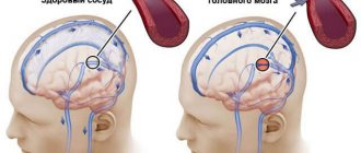
- problems with the vascular system;
- hypertension;
- encephalopathy, etc.
Hydrocephalus syndrome can appear as a result of meningitis, stroke, diabetes, or tuberculosis. Disruption of cerebrospinal fluid circulation can be complete or partial, and problems with the movement of cerebrospinal fluid arise due to blockage of the lumens or adhesions.
Treatment of hydrocephalus in children
Treatment of hydrocephalus in children
HYDROCEPHALUS IS THE MOST COMMON CAUSE OF SURGICAL INTERVENTIONS ON THE CENTRAL NERVOUS SYSTEM ORGANS IN CHILDREN
Hydrocephalus
, hydrocele - a disease characterized by excessive accumulation of cerebrospinal fluid in the ventricular system of the brain as a result of difficulty in moving it from the place of formation (cerebral ventricles) to the place of absorption into the circulatory system (subarachnoid space) -
occlusive hydrocephalus
, or as a result of malabsorption -
aresorptive hydrocephalus
.
The frequency of occurrence is 1 case in 2000-4000 newborns, more common in boys.
Causes of occurrence.
Hydrocephalus occurs at any age, but most often in young children, due to various reasons: tumors, infectious and inflammatory diseases, traumatic brain injury, congenital anomalies.
Hydrocephalus in a newborn can be caused by a birth craniocerebral injury, infectious diseases suffered by the mother during pregnancy (cytomegalovirus infection), leading to disruption of the ventricular system of the fetal brain. This, in turn, leads to difficulty in the circulation of cerebrospinal fluid and/or its excessive formation. In addition to congenital hydrocephalus, acquired hydrocephalus can also develop (most often in the first months of a newborn’s life) after meningitis, meningoencephalitis, head injuries, intoxication, intraventricular hemorrhages, etc.
Impaired circulation of cerebrospinal fluid (CSF) leads to increased pressure in the cranial cavity and the occurrence of the so-called hypertensive-hydrocephalic syndrome. As a result of the pressure exerted on areas of the brain, vision begins to decrease, convulsions occur, compression of the brain stem is manifested by oculomotor disorders (strabismus, limited upward gaze (the “setting sun” symptom)), weakness in the upper and lower extremities. This can lead to death, severe neurological disorders, and decreased intellectual abilities.
Manifestations
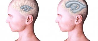
. The most characteristic sign of hydrocephalus in newborns is an accelerated growth of head circumference, leading to a visually well-defined hydrocephalic shape of the skull, greatly increased in volume. Signs of hydrocephalus include a bulging, tense fontanelle, frequent tilting of the head, and a downward displacement of the eyeballs. In places where normal fusion of the skull bones has not occurred, rounded pulsating protrusions may form. Strabismus and nystagmus (involuntary eye movements with high frequency) often occur. There is high excitability due to headaches, the child does not eat well, often cries, vomits, and is lethargic. Sometimes you may notice a decrease in vision and hearing.
Hydrocephalus in older age is characterized by headaches, especially in the morning, nausea and vomiting at the height of headache, dizziness; the size of the head is not increased.
These graphs reflect normal head circumference depending on the age of the child in boys and girls.
Diagnostics.
The most informative are magnetic resonance and computed tomography, which reveal sharply enlarged ventricles of the brain. In young children with an open large fontanelle, neurosonography (ultrasound examination of the brain) is also very informative.
Normal ventricles of the brain.
CT picture of hydrocephalus (dilatation of the lateral ventricles).
IT IS IMPORTANT TO UNDERSTAND THAT THERE IS NO EFFECTIVE DRUG FOR TREATING HYDROCEPHALUS.
MEDICATION TREATMENT OF HYDROCEPHALUS IS MAINLY APPLIED IN THE FORM OF TEMPORARY MEASURES, ESPECIALLY IN CASES WHEN THE DECISION ABOUT SURGICAL TREATMENT HAS NOT BEEN MADE AND MAY BE DELAYED.
ANNUALLY, AT THE NEUROSURGICAL DEPARTMENT OF THE ROSTOV REGIONAL CHILDREN'S CLINICAL HOSPITAL, ABOUT 150 OPERATIONS FOR SURGICAL TREATMENT OF HYDROCEPHALUS ARE CARRIED OUT FOR CHILDREN IN THE ROSTOV REGION AND IN NEIGHBORING REGIONS OF THE SOUTH RUSSIA
Surgical treatment of hydrocephalus in children.
The main method of treating hydrocephalus is endoscopic ventriculostomy of the third ventricle, which consists in creating a connection between the third ventricle and the interpeduncular cistern by dissecting the bottom of the cistern. If endoscopic triventriculostomy is ineffective, a cerebrospinal fluid shunt operation is used - this is a surgical intervention during which a special shunt is installed that drains fluid from the cerebrospinal fluid spaces of the brain into the circulatory system. As a result of the installation of a shunt, fluid does not accumulate in the cranial cavity, and hydrocephalus no longer develops, and a person’s life completely depends on the functioning of this device (shunt).
The entire range of operations performed to treat this pathology is divided into two groups:
1. Operations with drainage of cerebrospinal fluid outside the central nervous system:
-Installation of a ventriculoperitoneal shunt (shunt between the brain and peritoneum);
-Installation of a ventriculoatrial shunt (between the brain and heart);
-Installation of a ventriculovenous shunt (between the brain and veins).
1. “Internal shunting” with the creation of normal channels for the movement of cerebrospinal fluid through the central nervous system systems:
-Torkildsen operation (ventriculocisternostomy). It consists of creating a communication between the lateral ventricle and the occipital cistern by installing a silicone catheter passed under the skin on the back of the head;
-Endoscopic ventriculostomy of the third ventricle. It consists of creating a communication between the third ventricle and the interpeduncular cistern by dissecting the bottom of the cistern in the area of the gray tubercle;
-Implantation of internal stents. It consists of installing stents that expand the Magendie and Luschka holes to normal;
-Plasty of the cerebral aqueduct. It consists of expanding the lumen of the water supply system to ensure normal circulation of cerebrospinal fluid;
-Fenestration of the interventricular septum. It consists of creating an opening between the ventricles through which cerebrospinal fluid can circulate freely.
At the moment, in modern pediatric neurosurgery, only 3 methods of surgical treatment of hydrocephalus are widely used and recognized:
Endoscopic triventriculostomy.
The essence of the method is to create, using a neuroendoscope, a hole in the bottom of the third ventricle for the outflow of fluid into the extracerebral cisterns. As a result, the cerebrospinal fluid flows freely and the person is cured, without shunt dependence. The neuroendoscope is a complex optical system of Hopkins lenses, an illumination channel, a video channel, as well as channels for inlet and outlet of fluid, built into a tube with a diameter of only 6 mm. The operation is performed through a small hole in the skull. The image from the endoscope camera is transmitted to the screen and the doctor sees where the instrument needs to be inserted to restore the outflow of fluid. The method is less traumatic and reliable. Unlike bypass surgery, which takes about an hour, the operation takes only 10-20 minutes.
TO CARRY OUT TRIVENTRICULOSTOMY, THE NEUROSURGICAL OPERATING ROOM OF THE CSTO ROOM IS EQUIPPED WITH MODERN ENDOSCOPIC EQUIPMENT FROM THE LEADING MANUFACTURER
KARL STORZ.
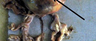
This is what the perforated bottom of the 3rd ventricle looks like.
Endoscopic ventriculostomy of the third ventricle is an effective method for the treatment of obstructive triventricular hydrocephalus; with the correct choice of indications, it allows achieving a lasting cure in 90% of such patients and, therefore, is the preferred method. The risk of serious complications after endoscopic ventriculostomy of the third ventricle is low and amounts to 6.6%. The key to the safety of the operation is: its planning on the basis of complete and high-quality clinical and radiological data and strict adherence to the methodological standard of the operation at all its stages, which together makes it possible to reduce the risk of serious complications by three times. The peculiarity of this surgical intervention is that it is highly effective only for obstructive forms of hydrocephalus.
Ventriculoperitoneal and ventriculoatrial shunting.
Ventriculoperitoneal shunt surgery has been in practice for more than fifty years, being the main standard method for getting rid of almost any form of hydrocephalus. Regardless of the technical nuances of the operation, the final result of VPS is the creation of an artificial outflow of cerebrospinal fluid from the dilated ventricles into the abdominal cavity. In some cases, in order to reduce the risk of trauma to the abdominal organs, in the presence of adhesions after previous abdominal operations, it is possible to use laparoscopic techniques. The shunt system consists of 3 main elements:
— ventricular catheter
, which is installed into the ventricular system through a small burr hole at one of the standard points;
— shunt pumps
regulating the outflow of cerebrospinal fluid from the ventricles; All models of pumps equipped with valves can be divided into three types depending on their throughput, or, in other words, on the level of cerebrospinal fluid pressure (low, medium and high pressure). Shunt systems with valves of a given pressure pose a certain complexity for the surgeon, since an error in selecting the parameters of a shunt system can lead to both excessive and insufficient levels of cerebrospinal fluid drainage; this problem can be solved by shunt systems that have appeared not so long ago, equipped with programmable valves, the “highlight” of a programmable valve is that it is equipped with a device designed for the possibility of remotely changing the level of cerebrospinal fluid drainage;
— peritoneal or atrial catheter
, immersed in the abdominal cavity or atrium to ensure the outflow of cerebrospinal fluid.
This is what a shunt system looks like, consisting of a ventricular catheter, a shunt pump and a peritoneal/atrial catheter
ü IN THE NEUROSURGICAL DEPARTMENT OF GBU RO "CSCH", FOR SURGICAL CORRECTION OF HYDROCEPHALUS ONLY MODERN SHUNT SYSTEMS FROM LEADING FOREIGN MANUFACTURERS, SUCH AS
INTEGRA, AESCULAP, ETC.
Complications.
The key point in the development of complications after liquor shunt operations using shunt systems is the presence of a foreign shunt system in the child’s body. According to various authors, in children in the first year of life, the risk of developing dysfunction of the shunt system (that is, its failure), even with a favorable course, is up to 50% and subsequently decreases by 10% with each subsequent year of the child’s life. The essence of the dysfunction is the impossibility of ensuring the shunt system of normal outflow of cerebrospinal fluid for one reason or another, due to the failure of the main elements of the shunt system, in addition to purely technical issues associated with the breakdown of the valve apparatus of the pump, frequent causes of shunt dysfunction are the development of shunt infection, the formation of pseudoperitoneal cysts in the abdominal cavity (a limited cavity that does not allow cerebrospinal fluid to be normally absorbed into the peritoneum). Clinically, the development of dysfunction is manifested by the development of general cerebral (the child becomes lethargic, adynamic, refuses to eat, nausea, vomiting appears) and focal neurological symptoms (Graefe’s “setting sun” symptom, meningeal symptoms, etc.).
It is important to remember that dysfunction of the shunt system is a serious complication in which repeated surgical intervention is indicated to eliminate it; If there is a suspicion of dysfunction of the shunt system, it is necessary to urgently seek specialized neurosurgical help to examine the child and decide on further treatment tactics!!!
ü Neurosurgical Department of the State Budgetary Institution of the RO “CSTO” operates around the clock and has the ability to conduct emergency surgery for hydrocephalus, including in cases of the development of displacement of shunting systems
Unfortunately, even a successfully performed operation is not a guarantee of hydrocephalus cured for life, since the anatomical dimensions of organs can change, the head can grow (especially in children), the holes in the shunt systems become clogged, etc. Children who have undergone such operations need to be constantly monitored by a neurologist and neurosurgeon in order to promptly identify emerging disorders that require correction. Thus, due to changes in the position of organs or the growth of the head, repeated operations have to be performed to replace the shunt with a more suitable one in terms of parameters.
How does hydrocephalus of the brain manifest?
The main signs of hydrocephalus are directly related to the deterioration of the patient’s general health. Symptoms can be pronounced or unexpressed. Common consequences of cerebral hydrocephalus are headaches, nausea, and vision problems. Secondary symptoms: the occurrence of convulsive syndrome, tinnitus, impaired coordination of movement and fear of light.
Even moderately severe hydrocephalus, located in the cerebral ventricles, can lead to impaired sensitivity of the limbs and even paralysis. Convulsive seizures also appear, which in turn can be symmetrical or asymmetrical. Ventricular hydrocephalus is a rather dangerous disease. In newborns, the consequences of hydrocephalus are retardation in physical and mental development.
The main types of hydrocephalus
At the initial consultation, the neurosurgeon will listen to the patient’s complaints, but will be able to make an accurate diagnosis only as a result of a comprehensive diagnosis. Special studies make it possible to determine what stage of the disease we are talking about - advanced or mild hydrocephalus.
There are quite a large number of varieties of dropsy:
- by origin, acquired and congenital diseases are distinguished;
- according to the characteristics of the course, hydrocephalus can be closed (occlusive), open (non-occlusive) and mixed;
- by location - internal and external hydrocephalus;
- according to clinical signs - acute, subacute, chronic degrees of hydrocephalus.
The dynamics of pathology development is also important. The most dangerous is considered progressive hydrocephalus, in which the number of symptoms constantly increases. The mild type is considered a regressive disease. With stable hydrocephalus, significant changes in the human body, as a rule, do not occur.
Material and methods
This single-center retrospective study included 59 children with shunt system dysfunction aged 1 to 14 years who were treated in our department from 2014 to 2021. Inclusion criteria: 1) age at the time of examination over 1 year; 2) implantation of a shunt system in the first 12 months of life. Medical histories, pre- and postoperative CT scans of the brain with 3D reconstruction in the bone window were analyzed to assess the condition of cranial sutures. Of the 60 children meeting the inclusion criteria, 1 patient was excluded due to the lack of CT images and the resulting inability to assess bone structures. In the remaining 59 cases, medical records were analyzed to determine the causes of hydrocephalus, the age of primary implantation and the type of shunt system. The condition of the bone sutures was assessed using 3D reconstruction of the patient's pre- or postoperative computed tomography in the Jemys: RIS+PACS program. Cases of primary craniosynostosis were excluded based on images taken before the initial implantation of the shunt system or in the first months after it.
In determining the indications for surgical intervention, the most significant was the combination of clinical manifestations of intracranial hypertension, radiologically confirmed closure of cranial sutures, narrowing of the subarachnoid spaces and ventricles of the brain. Additional diagnostic criteria may include thickening of the calvarial bones, severe digital indentations, acquired Chiari malformation, and the results of invasive monitoring of intracranial pressure (ICP).
Diagnosis and treatment methods for dropsy
Diagnosis of hydrocephalus is carried out in several ways. For example, using multislice computed tomography. As a result, the location of the pathology and its nature are determined - closed or open. The second common research method is MRI (magnetic resonance imaging), which provides accurate data on the condition of the brain vessels, the presence of structural changes and inflammatory processes.
In the early stages of identifying hydrocephalus, conservative treatment is prescribed - for example, medication. If the results are unfavorable, surgery is indicated:
- shunting for hydrocephalus involves draining excess fluid through a tube;
- external ventricular drainage - organization of a special drainage that is inserted into the cavity with cerebrospinal fluid;
- surgical removal of a septum, tumor, or blood clot that obstructs the movement of fluid.
Contraindications to surgery to eliminate symmetrical or asymmetrical hydrocephalus are severe cardiac pathologies, inflammatory processes in the nervous tissue, and serious problems with the blood vessels or respiratory system.
Neurosurgeons of the city clinical hospital named after. A.K. Eramishantsev will help cope with problems such as replacement, normotensive, triventricular, vicarious hydrocephalus. Sign up for a consultation to get more accurate answers to your questions. With timely intervention, the rehabilitation period after surgery occurs with minimal discomfort, and within a few days after surgery, adult patients can return to work, and children can return to school and a full-fledged active lifestyle.
Hydrocephalus: symptoms
If you notice the following symptoms in your little child:
– rapid increase in head circumference, – enlarged and swollen fontanel, – pronounced venous pattern on the forehead and face, – oculomotor disorders (strabismus, setting sun symptom, congestion of the optic nerves), – if the child does not hold his head up, throws it back, – eats poorly, sleeps poorly, cries often -
Contact a neurologist immediately! Time in this case can work against you!
Your actions:
STEP 1. Get a referral from a neurologist for an examination (MRI or CT). All examinations are carried out within the framework of compulsory medical insurance! MRI is performed free of charge for young children; the examination is performed under general anesthesia in a day hospital.
STEP 2. Contact a neurosurgeon with the results of the examination. If there is no neurosurgeon in the region (or to get a “second opinion”), send the results of the examination (MRI image) for an online consultation at a specialized clinic (the consultation is free, the contacts of medical organizations are in the memo) or to the post office of the municipal non-profit organization “He Needs You” marked “for consultation”
STEP 3. The hospital conducts an absentee medical consultation based on the medical documents you provided and, if hospitalization is necessary, prepares a voucher for you, which will indicate the date of hospitalization, a list of tests for the child and accompanying person, doctor’s contacts, hospital address and other important information.
STEP 4. When a diagnosis of Hydrocephalus is made based on MRI results, surgical treatment is performed. The method of surgical intervention is determined by the neurosurgeon depending on the type of disease.