© Author: Sazykina Oksana Yuryevna, cardiologist, especially for SosudInfo.ru (about the authors)
Transesophageal electrophysiological study (TEPE) is a technique for studying cardiac contractility and heart rhythm by applying physiological doses of electric current to the area of the heart most closely adjacent to the esophagus.
For the first time, the PPE technique began to be widely introduced into practice by arrhythmologists in the 70-80s of the last century. The study is otherwise called a non-invasive electrophysiological study, as opposed to an invasive one, in which electrodes are usually inserted through large arteries directly into the heart cavity from the inside. A little later, TEE began to be actively used in pediatrics, significantly expanding diagnostic capabilities in arrhythmology.
The essence of the method is based on the anatomical proximity of the esophagus and the heart. Often, in many patients, heart rhythm disturbances (arrhythmias) cannot be detected using a standard ECG or even with 24-hour blood pressure and ECG monitoring. Then stimulation of the heart muscle is applied, and if the patient has an arrhythmia, this will manifest itself during TEE. In other words, in some cases the myocardium must be “provoked” in order to understand whether electrical impulses are carried out correctly or whether there is a rhythm disturbance.
Using an electrode inserted through the esophagus, impulses are sent to the heart muscle, and electrodes placed on the chest record the rhythm during the study.
In addition, the rapid heart rate that occurs due to electrical stimulation can cause episodes of myocardial ischemia in patients suffering from coronary heart disease that is not detected on a standard ECG.
In addition to transesophageal EPI, there are invasive methods:
insertion of electrodes into the heart during invasive catheter EPI
- With endocardial EPI, a catheter with an electrode is delivered through the femoral vein into the ventricle or atrium, and the diagnostic procedure is often combined with subsequent RFA of pathological areas of the myocardium.
- EPI is also performed through open access (during cardiac surgery). Stimulation occurs epicardially - from the “outer” surface of the heart.
The essence of invasive procedures is generally similar to transesophageal procedures. Naturally, such interventions are correspondingly traumatic, but can be more effective.
The essence of the method, advantages and disadvantages
The essence of an EFI examination of the heart is as follows:
- Typically, various heart rhythm disturbances or coronary heart disease can be determined based on a standard electrocardiogram.
- If arrhythmia or myocardial ischemia cannot be detected using a single ECG, the doctor prescribes 24-hour monitoring of blood pressure and Holter ECG. Under conditions of normal household activity, the indicated diseases can be registered in most cases during the day.
- If it is not possible to track them on the monitor, the patient performs exercise tests. As a rule, based on such tests (bicycle, treadmill, 6-minute walk test), an accurate diagnosis is established, since the heart is under conditions of increased stress, but increased naturally (walking, running).
- When the above methods do not reliably establish the diagnosis of arrhythmia or ischemia, and the patient still has cardiac complaints, he is prescribed EPI (electrophysiological study of the heart)
.
With EPI, the load on the heart also increases, but not as a result of physical activity, but as a result of electrical stimulation of the myocardium
.
Such stimulation is carried out using electrodes, which begin to supply electrical currents of physiological power to the heart muscle, but at a high frequency. As a result, the myocardium contracts faster, causing a provoked increase in heart rate. And at a high heart rate, either arrhythmia or ischemia occurs if a person already has pathological processes in the myocardium, which are prerequisites for the development of these diseases. In other words, EPI allows you to provoke the desired diseases and register them on an ECG for the purpose of further treatment of the patient.
But depending on how the electrodes are applied to the heart muscle, there are three types of techniques:
insertion of an electrode during transesophageal EPI
- Transesophageal EPI (TEPI)
.
The electrodes are supplied using a probe inserted into the lumen of the esophagus. It is a non-invasive technique, and its technique is similar to conventional fibrogastroscopy. Performed more often than the next two types of EFI. (We will not dwell on the technique of conducting PPE in this article in too much detail; there is a separate material about it). - Endocardial EPI (endoEPI)
. It is an invasive technique; electrodes are inserted into large vessels using a sterile probe and advanced under the control of X-ray equipment. Refers to high-tech types of medical care (HTMP). Despite the complexity of implementation, as well as the need to use high-quality personnel and expensive technical equipment, it is a very informative diagnostic method, and it identifies cardiac diseases better than TEE. - Epicardial EPI (epi-EPI)
. It is also an invasive technique when myocardial stimulation is performed during open-heart surgery with an incision into the chest (thoracotomy). In terms of information content, it is not inferior to endoEFI. Due to such a disadvantage as the need for thoracotomy, it is performed mainly during heart surgery for other diseases.
insertion of a catheter into the heart during invasive endoESI
Classification
The heterogeneity of the disease causes serious disagreement among specialists regarding the definition of an accurate classification. Based on the anatomical characteristics, the following types are distinguished: atrial, ventricular, atrioventricular and sinus arrhythmias. Many experts use the frequency and rhythm of heart contractions to create a classification.
However, despite such contradictions, there is the most complete classification. It is based primarily on electrophysiological parameters of cardiac arrhythmia. Based on this classification, we can distinguish the following types of arrhythmia:
1. Diseases that are the result of a serious disturbance in the formation of an electrical impulse.
In turn, this group includes two types of heart rhythm disturbances: nomotopic and heterotopic. In the first case, we are talking about damage to the sinus node, which provokes bradycardia or tachycardia.
Heterotopic arrhythmia is divided into two types:
- Passive. An ectopic impulse occurs. This occurs due to the fact that the main impulse slows down (or disturbances in its conduction are noted).
- Active. In this case, the ectopic impulse excites the myocardium even earlier than the impulse that is formed in the main pacemaker. Thus, the resulting ectopic contractions seem to “interrupt” the normal sinus rhythm. When it comes to active heterotopic arrhythmia, we distinguish between extrasystole, ventricular fibrillation and other negative factors.
2. Diseases that are provoked by dysfunction of intracardiac conduction.
Treatment of arrhythmia is impossible without accurate identification of the disease. Those ailments that are provoked by a violation of the conduction function inside the organ appear due to a noticeable decrease in the propagation of the impulse or its complete cessation. In this case, there are syndromes of early excitation of the ventricles or blockade within them.
3. Combined arrhythmias. It is not difficult to guess that the disease includes the symptoms of the first two options.
When is EPI indicated?
Any type of EPI is carried out if the patient has certain complaints that the doctor cannot associate with disorders identified by the ECG or which arise in the patient with satisfactory examination results, or when certain diseases are suspected.
Thus, invasive cardiac EPI is performed when symptoms of the following nature occur:
- Paroxysmal interruptions in the work of the heart, especially short-term, but causing significant subjective discomfort,
- Interruptions in the heart, accompanied by severe general poor health, as well as shortness of breath and wheezing in the chest at rest, blue discoloration of the nasolabial triangle or the skin of other parts of the body (cyanosis), severe pallor of the skin, very high or low blood pressure, intense pain in the chest or in the chest on the left,
- Loss of consciousness and presyncope, with the exception of pathology of the central nervous system or other diseases (in the case of cardiac causes, loss of consciousness is called an attack or the equivalent of a Morgagni-Adams-Stokes attack, an attack of MES),
- Episodes of cardiac arrest (asystole) leading to clinical death with successful resuscitation of the patient.
Among the diseases that require invasive cardiac electrophysiology to clarify the diagnosis, the following can be noted:
- Paroxysmal types of arrhythmias - atrial fibrillation-flutter, supraventricular tachyarrhythmia, frequent ventricular extrasystole with transition to ventricular tachycardia,
- Sick sinus syndrome (SSNS), accompanied by alternating tachy- and bradycardia (tachy-brady syndrome), as well as bradycardia with the above-mentioned attacks of MES,
- Ventricular fibrillation with transition to asystole,
- Cardiac ischemia.
In the case where TEE does not help to reliably establish or exclude a diagnosis, that is, in diagnostically unclear cases, the patient undergoes endo- or epi-EPI.
In addition, endoEPI is carried out as part of an intraoperative examination when performing an intravascular RFA (radiofrequency ablation) operation, in which the pathological paths of the impulse that are the cause of one or another type of arrhythmia are destroyed by an intracardiac probe.
Indications
- Attacks of palpitations that are not recorded during daily monitoring of the electrocardiogram;
- episodes of rare pulse followed by attacks of palpitations;
- constant low pulse;
- fainting and dizziness, especially in young people;
- WPW syndrome;
- study of various characteristics of supraventricular paroxysmal rhythm disturbances with an established diagnosis;
- assessment of the effectiveness of antiarrhythmic treatment, including surgery;
- in some cases - diagnosis of coronary heart disease.
In what cases is EPI contraindicated?
Any type of cardiac EPI has a number of contraindications. These include the following:
- The patient develops an acute heart attack or stroke,
- The occurrence of fever, acute infectious disease,
- Unstable angina (new or progressive),
- Suspicion of pulmonary embolism (PE),
- Acute surgical pathology,
- Severe decompensation of chronic diseases (diabetes mellitus, bronchial asthma),
- Development of acute heart failure (cardiac asthma, pulmonary edema), or severe decompensation of chronic heart failure,
- Decompensated heart defects,
- Stage III of chronic heart failure,
- Severe dilated cardiomyopathy with low ejection fraction (less than 20=30%).
How to prepare for the procedure?
All nuances of preparation for the study must be carefully explained by the doctor to the patient. Firstly, the patient (under the supervision and as directed by a doctor!) must stop taking any antiarrhythmic drugs, as they can distort the results of the study. Secondly, before the TPE procedure, a patient experiencing even slight discomfort from the stomach must undergo fibrogastroscopy in order to exclude acute gastroesophageal pathology.
Before the endoEPI procedure for attacks of loss of consciousness, a neurologist must exclude brain pathology that could cause fainting, and this may require a CT scan or MRI of the skull.
Due to the fact that endo- or epi-EPI requires hospitalization in a hospital, a patient undergoing a routine examination must provide the doctor with the results of tests for HIV, syphilis, hepatitis and blood clotting no later than two weeks ago (different institutions have their own deadlines) .
The study is carried out strictly on an empty stomach. The need for epi-EPI on an empty stomach is due to the fact that during general anesthesia, vomiting of eaten food or liquid and aspiration of vomit may occur.
After the necessary preparation, the patient is hospitalized in a hospital. He must have the results of the examination (ultrasound of the heart, 24-hour monitor), as well as an extract from the outpatient card or discharge summary from the institution where he received examination and treatment before. The extract must indicate the rationale for the need for EFI with a detailed clinical diagnosis.
Carrying out EPI of the heart
Due to the fact that the essence of electrical stimulation of the myocardium is the same for all three methods, and TPEPI is similar in technique to FEGDS, it makes sense to dwell in more detail on invasive EPI methods.
So, invasive endoEPI
is carried out in the department of x-ray and surgical diagnostic methods, while the patient is undergoing inpatient treatment in the cardiology, cardioarrhythmology or cardiac surgery department.
After a little preparation in the form of intravenous administration of sedatives, the patient is transported on a recumbent gurney to the X-ray surgery. The doctor conducting the examination, under conditions of complete sterility, accesses the femoral (less often the subclavian) vein under local anesthesia. A small incision is made in the vein in the most favorable place for the technique (called venesection).
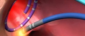
Next, through the resulting incision, a thin plastic or metal conductor called an introducer is inserted into the patient's vein. A probe with electrodes at the end, which has X-ray contrast properties and is therefore visible on the screen, is passed along it. After the probe gradually moves through the vein to the right atrium, controlled on the screen, and the probe reaches the chamber of the heart (atrium or ventricle) necessary for examination, the myocardium is stimulated in a physiological mode.
The probe typically has three to five miniature electrodes that are connected to a device that can switch their operation from stimulation mode to recording mode, and vice versa. The resulting cardiograms are recorded using a computer device.
example of electrode placement for EndoEPI
The duration of the procedure is half an hour or more, without causing significant pain. The patient remains conscious throughout the operation. After removing the probe, a pressure aseptic bandage will be applied to the skin in the venesection area.
EpiEPI
carried out in the department of cardiac surgery. After putting the patient into a medicated sleep (general anesthesia), the chest is dissected with access to the pericardial cavity. The issue of using a heart-lung machine (ACB) is decided strictly individually. After exposing the inner layer of the outer layer of the heart (epicardium), electrodes are applied to it, and stimulation begins with simultaneous recording of the response received from the heart muscle. The study takes more than an hour. After all the necessary manipulations have been carried out, the wound is sutured layer by layer, and drainage remains in the pleural cavity and is removed on the 2-3rd day.
After any of the invasive EPI methods, the patient remains under observation in the intensive care unit for a period of one day or more, depending on the severity of the patient’s condition.
Are there any complications?
As with any invasive research method, complications of endo- and epi-EPI are possible, but they occur in extremely rare cases. The main types of adverse consequences are cardiac acute conditions provoked by artificially created tachycardia. These include:
- Angina attack
- Development of acute myocardial infarction,
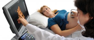
- Thromboembolic complications caused by the entry of a blood clot that has broken away from the heart cavity, if the latter was not identified before the procedure using echocardiography (ultrasound of the heart).
Prevention of this kind of complications is a thorough examination of the patient before surgery, as well as competent determination of indications for examination.
In the postoperative period, there is an extremely low probability of developing inflammatory and thromboembolic complications, as well as the occurrence of life-threatening arrhythmias.
What it is?
TEE is a functional diagnostic method used to determine the state of the cardiac conduction system. It allows you to determine whether this system is working normally, and also helps in diagnosing its violations. TEE identifies arrhythmias and helps clarify their characteristics necessary for proper treatment. Thus, TEE is a method for non-invasive diagnosis of heart rhythm disorders.
Decoding the results
Interpretation of the results is carried out by the doctor performing the study and the attending physician who referred the patient for the procedure.
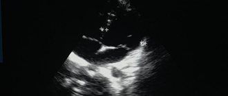
Normally, the electrogram obtained during EPI reveals sinus tachycardia with a heart rate of 100 to 120 per minute or more. Such tachycardia is transient and is not dangerous for the patient.
example of EFI results
If the study protocol contains the phrase that no rhythm disturbances were achieved using all types of stimulation, it means that the patient does not have a suspected type of arrhythmia, and the EPS results are regarded as normal. Also, normally there should be no depression or elevation of the ST segment and negative T waves, indicating myocardial ischemia.
If such changes are detected, their location is indicated, as well as the type of electrical stimulation in which they occurred.
When an arrhythmia is detected, its type is indicated (atrial fibrillation, ventricular tachycardia, frequent ventricular extrasystole, etc.), and the stimulation parameters that caused the rhythm disturbance.
Any of the disorders recorded on the electrogram requires careful medical supervision due to the need to prescribe certain antiarrhythmic drugs or perform RFA.
Carrying out, based on the results of EFI, RFA – “cauterization” of the area of pathological electrical activity of the heart
Features of disease prediction
In terms of prognosis, arrhythmia is a rather complex disease. Still, those ailments that are not associated with organic pathology of the organ do not pose a particular threat to human life.
But atrial fibrillation, on the contrary, can lead to serious consequences. Often, advanced disease leads to problems such as stroke or the development of life-threatening heart disease.
The most dangerous arrhythmia is considered to be ventricular fibrillation, as well as their flutter. If the disease is diagnosed in a patient, immediate resuscitation measures are necessary.
The success of treatment largely depends on when a person turns to a specialist. If there are even subtle symptoms, you should visit a doctor. Normal dizziness may be a consequence of the rapid development of the disease. Therefore, the matter should not be put off for a long time, especially since modern clinics are ready to offer inexpensive, but at the same time qualified treatment.
Approximate cost of EFI
Cardiac EPI can be performed in any large medical institution that has the appropriate personnel and technical equipment. Typically, EFI is carried out in regional or district centers, as well as in city hospitals in large cities (Moscow, St. Petersburg, Tyumen, Chelyabinsk, etc.).
Typically, EPI of the heart is carried out according to a quota from the Ministry of Health using funds from the federal budget. However, if the patient can pay for the procedure on his own, then there is no need to wait several weeks, since EFI can be performed for paid services.
Prices for cardiac electrophysiology testing vary widely. Thus, the cost of PPE is from 2000 to 4000 rubles, depending on the institution and equipment. The cost of endoEPI is much higher and amounts to 60-180 thousand rubles, depending on the payment for the probe and catheters, as well as the payment for the subsequent stay in the clinic.