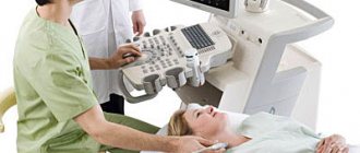
Echocardiography is considered one of the safest research methods, therefore it is often prescribed to children. Usually, a child is sent for an ultrasound of the heart after an ECG
, the results of which remained mixed. Modern ultrasound examination of the heart has many advantages over other types of diagnostics.
What is cardiac ultrasound?
Echocardiograms, also known as cardiac ultrasounds, are used to study a baby's or fetus's heart as it functions. This is a simple and painless procedure, performed according to the same principles as the ultrasound examination of the fetus in the womb of a pregnant woman, which is familiar to all mothers. Echocardiography uses sound waves (ultrasound) to create images of the heart. The Doppler test uses sound waves to measure the speed and direction of blood flow. By combining these tests, the pediatric cardiologist obtains useful information about the anatomy and function of the heart. Echocardiography is the most common test used in children to diagnose or rule out heart disease, and to monitor children who have already been diagnosed with heart problems. This test can be done on children of all ages, even newborns or unborn children.
Contraindications to cardiac ECHO
Echocardiography has absolutely no contraindications. It does not have any harmful effects on the human body. Therefore, it is used even for a child in the womb.
There are no harmful radiations or unpleasant sensations. The procedure itself does not take much time and is carried out in a comfortable environment.
Despite the absence of contraindications, there may be individual characteristics of the patient’s body that complicate cardiac echocardiography. These are bronchial asthma and a long history of smoking, a woman’s large breast size, significant obesity, and a large deformation of the chest.
Answers to frequently asked questions on the topic cardiologist:
- Call a cardiologist to your home
- ECG
- Chest pain
- Heart disease in adults
- Prevention of heart disease
- Holter monitoring
- Treatment of heart diseases
- Blood chemistry
- Ultrasound at home
- Ultrasound of arteries
- Ultrasound of blood vessels and heart
- Coronary angiography
- Chest pain
Why is heart ultrasound done in children?
A cardiologist, pediatrician, or obstetrician may order an echocardiogram of the baby or fetus to see how well the heart muscle is working and to check for heart abnormalities. An echocardiogram can evaluate a heart murmur or a number of other conditions. This test allows doctors to very accurately diagnose disorders of the structure and function of the heart and assess blood flow in the heart and blood vessels. This is very informative and one of the most common types of studies in cardiology.
Ultrasound can help identify the causes of many heart problems, including:
- Noises
- Chest pain
- Aneurysm
- Cardiomyopathy
- Congenital heart defect
- Chronic heart failure
- Pericarditis
- Acquired heart defects
How to prepare for a cardiac ECHO
The standard procedure does not require special preparation. The patient simply comes on the appointed day, takes off his clothes to the waist and lies down on the couch. The doctor conducts the examination within half an hour.
However, there are some general recommendations:
- the day before the procedure, avoid heavy physical activity, including canceling sports;
- stop using sedatives and stimulants;
- do not drink coffee and drinks containing caffeine;
- do not overeat;
- do not be nervous, avoid stressful situations.
If the patient suffers from an increased heart rate (over 90 beats per minute) or has high blood pressure (approximately 170/99 mm Hg), then before the study you should consult a cardiologist about whether you need to take medications to lower your pulse and blood pressure.
Transesophageal ECHO CG requires preparation:
- within six hours before the procedure, refuse food, 4 hours – from water;
- give up alcohol and cigarettes, as well as fatty foods in a day.
This method of examining the heart is carried out in order to study the organ as closely as possible. The patient's throat is lubricated with an anesthetic and a probe is passed into the stomach through the esophagus. It takes no more than 12 minutes to receive data.
You need to prepare and stress - Echo KG. This examination takes about 45 minutes.
Necessary:
- for 3 hours do not perform any work that requires physical strength and stress;
- do not eat 3-4 hours before the procedure;
- Stop drinking 2 hours in advance, and food should consist of a small snack.
Clothing on the day of the appointment should be loose and not restrict movement.
What are the types of cardiac ultrasound?
There are four special types of echocardiography:
- Echocardiography in M-mode
This is the simplest type of echocardiography and produces an image that looks like a slice rather than the actual structure of the heart. M-mode echo is useful for measuring structures of the heart, such as the chambers of the heart, the size of the heart itself, and the thickness of the heart walls.
- Doppler echocardiography
The Doppler method is used to measure and evaluate blood flow through the chambers and valves of the heart. The amount of blood pumped out with each beat is an indicator of the heart's performance. In addition, Doppler ultrasound can detect abnormal blood flow in the heart, which may indicate problems such as a hole between the chambers of the heart, a problem with one or more of the four heart valves, or a problem with the walls of the heart.
- Color Doppler
Color Doppler is an advanced form of Doppler echocardiography. Color Doppler uses different colors to indicate the direction of blood flow. This simplifies the interpretation of the Doppler method.
- 3-D (3-dimensional) echocardiography
This method is used to see the actual structures and movement of the heart structures. The 3D echo image appears cone-shaped on the monitor, and the movement of heart structures can be observed in real time. This allows the doctor to see the functioning of various structures of the heart and evaluate them.
Who needs a cardiac ECHO?
If a person suspects a pathology of the cardiovascular system, then he should definitely undergo a cardiac ECHO. The heart is an important organ located in the chest and is a hollow muscle with a dense structure. The heart supplies blood flow throughout the body. Divided into two atria and two ventricles.
The heart must receive enough oxygen and nutrients to function normally. With any, even minor, changes in the functioning of the heart, a person begins to feel certain ailments. The consequences are very serious for the body.
Echocardiography is recommended:
- people who periodically feel pain of unknown etiology in the chest area;
- those who suffer from shortness of breath often feel a lack of air;
- those suffering from frequent headaches;
- if there is swelling, especially in the face and legs;
- if there is a disturbance in the heart rhythm, the heart beats too quickly (tachycardia);
- suffered a heart attack;
- hypertensives – people prone to high blood pressure;
- with a prolonged increase in body temperature;
- if there are fainting, dizziness;
- with established diagnoses of heart disease - heart defects, coronary artery disease, angina pectoris, and so on.
It is also worth undergoing regular examinations for those who play sports, who have suffered a severe form of influenza or pneumonia, with frequent bronchitis, or with stress. The heart reacts to physical and emotional stress, so echocardiography serves as a preventive monitoring of the condition of the organ.
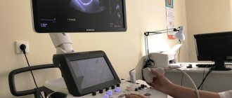
What happens during an ultrasound of the heart?
This is a harmless and painless test, the same as an ultrasound for a pregnant woman. Mom and baby enter a darkened exam room where your baby can lie down on a regular bed. It is important that your child remains silent and still during the test, so an entertaining video may be shown to help keep him occupied and calm. Young children may be bottle fed during the study.
An ultrasound technician with special training in working with babies and children will apply warm ultrasound gel to the baby's chest and heart area and then place a small probe on the chest to obtain images from multiple vantage points. A small amount of clear gel is applied between the sensor and the chest to ensure proper contact. The gel does not leave stains and is wiped off at the end of the test. Several stickers called electrodes will also be placed on your chest to track the ECG during the exam. The echocardiogram is recorded digitally, so the doctor can review it and compare it with echocardiograms done in the past or ones that will be done in the future. The doctor can select and show many images on the monitor screen. You will be able to see an image of a beating heart, and you will also be able to see and hear the flow of blood. The picture usually changes as the sensor moves. Loud sounds of blood flow may be heard. If your child has too much trouble relaxing for a routine echocardiogram, he or she may need to be sedated before the test. If sedation is required, the child will be asked not to eat or drink for several hours before the test. Before giving the medicine, the doctor or nurse will make sure there is no reason why the sedatives should not be given (such as a bad cold), and then explain the procedure to the parent.
Children usually fall asleep within 30 minutes and sleep for about an hour. Although sedated echocardiograms are very safe, careful monitoring of heart rate, blood pressure and oxygen is performed while the child is sleeping. When the child wakes up, he or she may have poor balance (may last more than an hour) and should be monitored closely. The doctor and nurse will continue to monitor you closely until your baby is fully awake and drinking juice or milk.
Older children and teenagers usually do not need sedatives and may enjoy watching a monitor. To avoid anxiety, make sure your child knows that the exploration is fun, that it won't hurt, and that mom will be there.
Except in cases of sedation, no special preparations need to be made. It will help if your child wears a shirt or blouse with buttons down the front. The child may also eat normally before the examination. Bring a bottle of milk for a baby or a smartphone with a cartoon for an older child.
Heart ECHO for pregnant women
An echocardiogram is performed for pregnant women according to indications. The indications are:
- there is a hereditary predisposition to heart defects;
- the previous pregnancy ended in spontaneous miscarriage;
- if the expectant mother has diabetes;
- if the expectant mother suffers from epilepsy and took special medications for this disease in the first and second trimesters of pregnancy;
- if a pregnant woman is sick with rubella or antibodies to rubella are found in the blood (a dangerous disease for the fetus).
Pregnancy and childbirth itself increase the load on the heart. Therefore, all expectant mothers are recommended to have their heart examined - this vital organ to eliminate all risks. It is advisable to undergo a cardiac ECHO while planning a pregnancy.
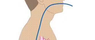
What is a transesophageal echocardiogram?
Sometimes a regular echocardiogram does not provide all the images needed. Your doctor may recommend a test that uses special echocardiography probes to take pictures from inside the esophagus. This procedure is called a transesophageal echocardiogram. In this test, a tube with an ultrasound probe is passed down the throat into the esophagus. The esophagus is located directly behind the heart, and images taken there can give a very clear picture of the heart and its structures. This procedure requires anesthesia and takes about 20 minutes.
Normal cardiac ECHO readings
During the procedure, the specialist compares normal heart parameters with those observed online. For the right ventricle, the requirements are:
- normal wall thickness is 5 mm;
- size parameters from 0.75 to 1.25 cm/m2;
- the size of the ventricle at rest is from 0.75 to 1.1 cm.
For the left ventricle, 8 indicators are important. This:
- myocardial mass – for women the norm is 95-141 g, for men – 135-182 g;
- myocardial mass index – from 71 to 80 g/m2 for female patients and from 71 to 94 g/m2 for female patients;
- the volume at rest is from 65 to 193 ml in men and from 59 to 136 ml in women;
- dimensions at rest from 4.6 to 5.7 cm;
- size during contraction from 3.1 to 4.3 cm;
- Normal wall thickness without contractile movements should be 1.1 cm;
- the volume of blood ejected during contraction should be within 55-60 percent;
- stroke volume within 60-100 ml.
Only an experienced cardiologist can understand all these designations and compare the data. Self-interpretation may give an erroneous idea of health.
What is a stress echocardiogram?
Stress echocardiography is performed while the heart is beating normally and then again after exercise. In this test, the child is asked to run on a treadmill or pedal an exercise bike to increase their heart rate and the amount of blood and oxygen the heart needs to function.
If your child is too young or unable to run on a treadmill or ride an exercise bike, the doctor may use a medicine called dobutamine to increase the heart rate. A stress echocardiogram may be a more effective way to evaluate arterial stenosis or thrombosis.
Symptoms for which it is necessary to do an ECHO of the heart
The following manifestations of trouble in the functioning of the heart should alert you:
- frequent dizziness and headaches;
- tendency to faint;
- shortness of breath even with slight physical exertion;
- swelling of the limbs;
- blue discoloration of the nasolabial area;
- cold hands and feet;
- the pulse is rapid, sometimes the heart “freezes” or there is a noticeable pounding of the heart in the chest;
- increased sweating;
- pain in the sternum and behind the sternum.
If you have at least two of the listed symptoms, you should not postpone a visit to a cardiologist.
Cost of services
| Name of service | Price |
| Ultrasound of the heart (Echo-CG) with Doppler | 2,600 rub. |
Ultrasound diagnostics of the heart is an informative, fast and safe method of studying the condition of a child’s heart, which allows you to accurately establish a diagnosis and carry out the required treatment with maximum efficiency, or get rid of worries and doubts about the state of your baby’s health.
You can get additional information about cardiac ultrasound and make an appointment for diagnostics at a medical center by phone.
ULTRASOUND SCREENING OF NEWBORNS AND CHILDREN UP TO 1 YEAR OF AGE
at the EVO Specialized Medical Center.
WHAT IS A BABY ULTRASOUND SCREENING?
Ultrasound screening of newborns and children in the first year of life includes:
- examination of the abdominal organs and kidneys (deviations from the norm in shape, size, structure and location of organs) ;
- examination of the hip joints for orthopedic deformities (dislocation, dysplasia) ;
- examination of the brain for developmental defects.
WHY DO AN ULTRASOUND OF YOUR BABY?
For every mother and every father of a newborn son or daughter, the main issue is the health of the child. And the first year of a child’s life is the most critical stage for future health, during which a number of diseases and problems can be excluded. Modern parents have probably heard a lot about screening studies. For example, all expectant mothers in our country must undergo prenatal (antenatal) screening diagnostics during pregnancy. These studies are aimed at timely detection of abnormalities in the fetus. And the earlier the disease is detected, the greater the chance of a positive pregnancy outcome and the health of the newborn. Screening to identify hidden defects (congenital and acquired), hereditary pathologies, deviations from the norm in the structure and location of the internal organs of a newborn child allows you to calm the worried hearts of young parents or, if necessary, not to miss precious time to begin treatment for the baby.
WHAT DIAGNOSTICS DOES A BABY NEED?
Various disorders in the child’s body can occur both during intrauterine development and during childbirth. Diseases of newborns can be caused by hereditary chromosomal abnormalities, the influence of a number of negative factors during pregnancy, and birth injuries.
Diagnostics includes collecting patient complaints, assessing the presence of disease symptoms identified during an external examination + results of laboratory and instrumental studies. However, newborn children do not complain. Symptoms of the disease are often mild and sometimes completely absent. Laboratory tests and instrumental studies remain in our arsenal.
Currently, there is practically no problem with laboratory tests; in well-equipped child care institutions, any indicator in the blood or urine can be determined. Instrumental studies have some limitations, because screening of the baby must be carried out in the first 3 months of life, and it is during this period that the child’s body is extremely sensitive to any external influences.
The slightest carelessness during diagnosis can lead to serious and even irreparable consequences. The screening method for newborns should not only be highly informative, but also convenient and safe. Today, ultrasound examination (ultrasound) is the best diagnostic method for newborns and children under 1 year of age.
BENEFITS OF ULTRASOUND FOR CHILDREN UNDER 1 YEAR OF AGE:
- There are no harmful external influences on the child.
- Ultrasound is absolutely harmless even for a child’s body.
- Possibility of repeated research.
- Harmlessness will remain with repeated diagnostic procedures.
- Non-invasiveness of the method (ultrasound is not associated with damage to the skin, mucous membranes, or penetration into the internal environment of the body).
- The short duration of the study.
- There is no preliminary preparation for the study, in particular, ultrasound can be performed immediately after feeding, and not just on an empty stomach; there is no need for a cleansing enema, intravenous administration, ingestion of a contrast agent, etc.
- The method is completely painless. The child experiences absolutely no pain or discomfort, so there is no need for anesthesia.
IS ULTRASOUND HARMFUL FOR A NEWBORN?
Ultrasound waves, unlike X-rays, are completely safe - for both adults and children, even for the smallest inhabitants of the planet and the most precious people for you personally. Over several decades of ultrasound use, no side effects or negative effects on the body have been recorded. During an ultrasound examination, a special sensor comes into contact with parts of the child’s body. The ultrasonic wave emanating from it passes through the tissues without causing any harm or pain to the child’s body. The reflected signal is transmitted to the computer monitor in the form of an image.
WHY SHOULD CHILDREN BE PERFORMED IN EVO?
- Specialists. Diagnosis is carried out only by experienced and qualified doctors who can easily detect even the slightest deviation in the initial stage.
- Equipment. Our ultrasound machines meet all modern requirements and maximum resolution.
- Medical consultations. Upon completion of the diagnosis, if certain deviations from the norm are detected, you can immediately obtain advice from an appropriate specialist and recommendations for treatment and/or prevention. Find out more here.
- Time. Ultrasound is performed only at a time convenient for you, which can be agreed upon in advance with the administrators; queues and unwanted contacts are excluded.
NEURSONOGRAPHY in EVO.
Ultrasound of the child's brain through the large fontanel. It is performed on children in the 2nd month of life, whose head bones have not yet fused, and between them there are “fontanelles” - canals with softer tissue. The presence of this natural bone defect will allow ultrasonic waves to easily penetrate deep into the brain structures, which means to qualitatively examine the internal structure of the brain, identify congenital anomalies, as well as pathology acquired during the prenatal period or during labor. As the “fontanelles” harden, this becomes difficult. Brain ultrasound can detect:
- brain tumors, cystic formations of the meninges, areas of hemorrhage in the medulla (intracranial hemorrhage);
- increased intracranial pressure;
- hydrocephalus due to the accumulation of cerebrospinal fluid (CSF) in the ventricles of the brain.
Untimely detection of circulatory pathology and brain disorders will not allow timely initiation of treatment and can lead to irreversible consequences such as brain dysfunction and neurological disorders.
Ultrasound of internal organs in EVO.
Allows you to identify congenital diseases, anomalies in the structure and location of organs, congenital neoplasms (cysts, tumors) in the abdominal cavity.
- Ultrasound of the kidneys and urinary tract is used to diagnose changes in the shape and size of the kidneys, underdevelopment of the kidneys, as well as various anomalies in the structure of the bladder and muscle valves (sphincters).
- Ultrasound of the liver, pancreas and gall bladder is performed to diagnose various structural and degenerative changes in the liver, pancreas and biliary tract.
Ultrasound of the hip joints at EVO.
This study will allow timely recognition of congenital hip dysplasia and its complication – congenital hip dislocation. Screening is carried out on infants aged 1 to 2 months and makes it possible to detect abnormalities that were not previously noticeable, which gives a high chance of complete normalization of the child’s musculoskeletal system. Early detection of orthopedic pathology allows immediate correction to begin, thanks to which the baby can be completely healthy within six months.
How much does ultrasound screening of a newborn and a child up to one year cost at EVO?
Currently, the EVO Specialized Medical Center is running a promotion: the total cost of systemic screening ultrasound examinations is 3,000 rubles instead of 5,050 rubles. Your savings are 2,050 rubles!!! And this is much lower than in other clinics!
FAQ:
My baby was born premature. Will an ultrasound harm him?
Ultrasound is harmless even for premature babies. Moreover, prematurity often entails an increased risk of a number of congenital diseases. Therefore, screening ultrasound is not only desirable for premature babies, but also indicated.
I did all the tests for the child. Everything is fine there. Why do I need an ultrasound?
Normal clinical indicators of a newborn’s tests do not at all exclude abnormalities in the internal organs, musculoskeletal system, and brain. Therefore, during screening studies, laboratory diagnostics are carried out only in combination with ultrasound.
Is it possible to limit yourself to one study (to look at the head or kidneys) and at the same time save additional money, and postpone the rest for later?
Yes, you can. But it's not necessary. The essence of screening diagnostics lies, firstly, in its timeliness, and secondly, in its complexity. “The miser pays twice” and, unfortunately, your savings today may cost you dearly in the future.
CT is a more informative method than ultrasound. Why don't you do it?
In terms of its diagnostic value, ultrasound is not inferior to computed tomography, and in some cases even surpasses it. It is worth noting that CT scanning is not safe for a child, since the baby is exposed to x-ray radiation and must lie still for a certain time, which entails anesthesia. In our opinion, without any specific evidence for this, such risks for a newborn and a child in the first year of life are unjustifiably unnecessary.
You can get answers to other questions, as well as sign up for an ultrasound scan, by calling the EVO Medical Center (8) 812 701 03 30.
Remember that “nature knows no stop in its movement and punishes all inactivity” (Johann Wolfgang von Goethe).
Patient reviews about ultrasound diagnostics in medical
If you want to know where ultrasounds are performed for children, then call Art Medic. We see good children's doctors - ultrasound specialists, as evidenced by the reviews of our patients.
I thank all the employees of the Art Medic MC for their warm attitude and professionalism! It is thanks to you that your son happily goes to “aunt doctor”, be it for an injection or a pediatrician’s examination. The nurses in the treatment room are wonderful, they always smile and talk to my son to take his mind off the injections. Thank them very much for their gentle hands and humane attitude. They work with soul and know how to find an approach to anyone, even the smallest patient! And I have never met pediatricians like Elena Nikolaevna Nemurova - always (!!!) correct diagnoses, effective treatment and sensitive attitude, willingness to help. The administrators are always friendly and attentive. And management that exceeds all expectations!!! You don't just make people healthier, you do it from the heart! Thank you for being! We treat only with you!!!
June 13, 2021, Svetlana
Is it possible to do an ultrasound on a baby?
On various forums you can often find messages about the supposed potential harm of ultrasound for a child. Medical specialists are ready to dispel this unscientific myth. For several decades, doctors around the world have declared with all responsibility that ultrasound is the safest, and in some cases, the most effective diagnostic tool. It can be taken without fear even during pregnancy at any stage and any medically reasonable number of times.
Moreover, there are some studies that need to be done at an early age. This is due to the peculiarities of the anatomy and physiology of newborns.
The interpretation of the child’s ultrasound is carried out on site, by specialists from the Art Medic clinic.
At what age do children have their first ultrasound?
If there are no medical indications, then the first examination is recommended when the baby reaches 3 months of age. For the procedure, you should choose a clinic that specializes in examining newborns. Firstly, its specialists know how to establish contact with the baby, and secondly, the possibility of contact with potentially infectious patients is minimized.
If the baby was born premature, has developmental defects or birth injuries, then an ultrasound of the newborn baby is performed at 1 month or even earlier. It is worth noting that ultrasound standards for a monthly toddler vary widely, so instead of searching the Internet for transcripts and getting nervous, it is better to immediately go with the conclusion to a qualified pediatrician, who will make a diagnosis, taking into account the nuances of the birth and development of your baby.
Do children need an ultrasound after one year?
After routine screening, examination is carried out only according to indications until a comprehensive diagnosis is carried out before kindergarten. For example, if during screening a non-closed foramen ovale was detected, then an additional ultrasound of the heart is indicated for a child aged 2 years.