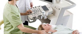
Today, one of the main methods for diagnosing heart disease is echocardiography (EchoCG). This is a non-invasive study that does not have a negative effect on the body, and therefore can be performed on patients of all age categories, including children from birth.
This article will discuss what echocardiography is, why it is performed, what types of this procedure exist, what may be a contraindication to its implementation, and how to prepare for it.
The essence and purpose of echocardiography
Echocardiography or echocardiography is a non-invasive method of examining the heart using ultrasound. The echocardiograph sensor emits a special high-frequency sound that passes through the tissues of the heart, is reflected from them, and is then recorded by the same sensor. The information is transmitted to a computer, which processes the received data and displays it on the monitor in the form of an image.
Content:
- The essence and purpose of echocardiography
- Benefits of EchoCG
- Indications and contraindications for Echo-CG
- Types of echocardiography
- Preparation for echo-CG
- Research methodology
- Decoding the results
- Where to get an echocardiogram
- Echocardiography in children
- Preparing and performing the procedure for children
- Fetal echocardiography
Echocardiography is considered a highly informative research method, since it makes it possible to assess the morphological and functional state of the heart. Using this procedure, it is possible to determine the size of the heart and the thickness of the myocardium, check their integrity and structure, determine the size of the cavities of the ventricles and atria, find out whether the contractility of the heart muscle is normal, find out about the condition of the valvular apparatus of the heart, examine the aorta and pulmonary artery. This procedure also allows you to check the level of pressure in the structures of the heart, find out the direction and speed of blood movement in the heart chambers and find out the condition of the outer lining of the heart muscle.
This cardiological examination allows you to diagnose both congenital and acquired heart defects, find out about the presence of free fluid in the heart sac, identify blood clots, changes in the size of the chambers, thickening or thinning of their walls, detect tumors and any disturbances in the direction and speed of blood flow.
How is cardiac ultrasound performed?
Like preparation for the study, the results shown by ultrasound of the heart depend on the type of technique. There are also technical parameters of ultrasound that expand diagnostic capabilities.
One-dimensional ultrasound in M-mode
One-dimensional echocardiography shows the heart in one plane in real time. Interpretation of the results of a one-dimensional ultrasound of the heart will show the size of the chambers and the lumen of the heart valves.
2D ultrasound
This research method allows you to examine the heart in two planes to see the areas of the valve openings and the lumens of the great vessels.
Three-dimensional ultrasonic scanning
This type of study is also called Doppler. Three-dimensional ultrasound of the heart, which simultaneously shows the movement of blood flow and the anatomy of the organ, is the most advanced method of ultrasound diagnostics.
Benefits of EchoCG
Echocardiography has a number of advantages over other types of cardiac examination.
First of all, this is an absolutely painless and non-invasive procedure that does not cause any discomfort to the patient. It is performed like a regular ultrasound. No injections or any other similar manipulations are performed before the procedure.
In addition, the procedure is completely safe for patients of any age group. It can be performed on children, adolescents, and pregnant women, since ultrasound does not have any negative effect on the fetus.
EchoCG is accessible, since the equipment for its implementation is available in almost any medical institution. The cost of echocardiography is much lower compared to MRI.
And the most important advantage of this type of examination is its excellent information content, which will allow the doctor to obtain the maximum necessary information and choose the right therapy.
Indications and contraindications for EchoCG
Echocardiography may be recommended to patients both if the doctor suspects they have any cardiovascular pathology, and during therapy to assess the effectiveness of the drugs used.
Indications for echocardiography are:
- Hypertension.
- Suspicion of the presence of congenital or acquired heart disease, including with a hereditary predisposition to this disease.
- Frequent dizziness, fainting, shortness of breath and swelling.
- Complaints about a “fading” heart, about “interruptions” in its work.
- Pain behind the sternum, especially if it radiates to the area of the left shoulder blade or the left half of the neck.
- Myocardial infarction, diagnosis of angina and cardiomyopathy, suspected heart tumor.
- Preventive examination of patients who often experience emotional and physical overload.
- Changes on the ECG and chest x-ray that require clarification of morphological changes in the heart.
It is worth mentioning separately in what cases echocardiography is recommended for expectant mothers. Pregnant women should undergo echocardiography if:
- The expectant mother has pain in the precordial area.
- The patient has congenital or acquired heart defects.
- Weight gain has stopped or sudden weight loss has occurred.
- Unreasonable swelling of the lower extremities and shortness of breath appeared with a slight antiepileptic load.
- Hemodynamic disturbances during pregnancy.
It should be noted that there are practically no absolute contraindications to echocardiography. However, certain types of this research are not recommended in certain situations, which will be discussed below.
ECG is the first test on the diagnostic path
A patient who complains of chest pain to a therapist is almost immediately given an ECG, that is, an electrocardiogram. This is a test that can be performed very quickly in any clinic, hospital, or even at the patient’s home. An ECG is usually the first test to diagnose cardiovascular disease. An ECG should be performed on every patient visiting a cardiologist or with cardiac disease or chest pain. It is also recommended to perform an ECG for healthy people involved in sports professionally and as amateurs. To perform this test, you will need an electrocardiograph that records the functional currents of the heart muscle. Each muscle, including the heart muscle, produces electrical impulses that are recorded by the machine and transferred to paper. A special printout of electrical impulses is an electrocardiogram. An ECG test can detect dangerous cardiac arrhythmias (atrial fibrillation, conduction block, tachycardia), as well as a recent heart attack. However, the diagnostic value of the test is limited, because even a correct ECG recording does not clearly exclude serious heart disease, and then a more accurate diagnosis is necessary. An at-home ECG is a safe and quick test. The patient, lying on his back, has electrodes glued or attached to his chest, arms and legs. The test itself takes up to 5 minutes and does not require special preparation. The patient receives the test result immediately after it is performed.
Types of echocardiography
Today, there are several types of echocardiography. What specific type of study to conduct is decided in each specific case by a cardiologist.
One-dimensional
At the moment, this type of echocardiography is rarely used independently, because it is considered less informative than others. No image of the heart is generated during the procedure. The data is displayed on the screen in the form of a graph. Using M-echocardiography, the doctor can determine the volume of the heart cavities and evaluate their functional activity.
B-echocardiography (two-dimensional)
During B-echocardiography, data from all structures of the heart enters the computer and is displayed on the monitor in the form of a black and white image. The doctor is able to determine the size of the heart, find out the volume of each of its chambers, the thickness of the walls, assess the mobility of the valve leaflets and how the ventricles contract.
Doppler echocardiography
As a rule, this study is performed simultaneously with B-echocardiography. It allows you to track blood flow in large vessels and on the heart valves, identify reverse blood flow and its degree, which may indicate the formation of pathological processes.
Contrast echocardiography
This test makes it possible to more clearly visualize the internal structures of the heart. The patient is injected intravenously with a special contrast agent, after which the procedure is carried out as usual. This procedure allows you to examine the inner surface of the chambers of the heart. Contraindications for this study are individual intolerance to contrast and chronic renal failure.
Stress echocardiography
To diagnose hidden heart pathologies that manifest themselves exclusively during physical activity, a special type of study is used - stress echocardiography. It makes it possible to identify diseases in the early stages, which do not remind of themselves if the patient is at rest. Stress echocardiography is recommended to assess the condition of blood vessels and their patency, to find out how great the risk of complications is before performing surgical interventions on the heart and blood vessels. The procedure is also carried out in order to determine how effective the therapy for coronary heart disease is and to determine the further prognosis for this disease.
There are several contraindications to stress echocardiography. It should not be performed on patients suffering from severe respiratory, renal, hepatic or heart failure. It is also contraindicated in case of myocardial infarction, aortic aneurysm and a history of thromboembolism.
Transesophageal echocardiography
This is a special type of study, during which an ultrasound-generating sensor is lowered through the oropharynx through the esophagus to the required depth. Since the sensor has very small dimensions, it passes through the esophagus without problems. However, such a study is considered quite complex and is carried out exclusively in specialized medical centers. In addition, there are special indications for it. In particular, a transesophageal examination is performed in cases where a standard transthoracic examination does not allow assessing the condition of the heart and its structures. In particular, when doubts arise about the correct functioning of a previously prosthetic heart valve, if an aortic aneurysm and atrial septal defect are suspected, as well as if the patient has been diagnosed with infectious endocarditis and the doctor suspects an aortic root abscess.
At the same time, this type of study has contraindications from the upper digestive tract, namely, any tumor formations of the esophagus, bleeding from the upper gastrointestinal tract, the presence of a large diaphragmatic hernia or dilation of the esophageal veins. Transesophageal examination should not be performed in patients with severe osteochondrosis of the cervical spine, instability of the cervical vertebrae, or a history of esophageal perforation. Diagnosis may be complicated in patients with thyroid diseases.
What indicators are considered normal?
Having received the results in hand, the cardiologist can interpret them. In this case, the specialist takes into account anamnesis, medical history, the effect of medications taken and many other related factors. To evaluate the results of cardiac ultrasound, the norm of indicators is prescribed in its standard version. Deviations from the norm do not always indicate the presence of serious pathologies. If you consult a doctor in a timely manner and start a correctly selected treatment, there is every chance of bringing the abnormal indicators back to normal.
| Indicator name | Women | Men |
| Left ventricular myocardial mass | From 95 to 142 g | From 135 to 180 g |
| Left ventricular mass index | From 71 to 88 g/m2 | From 71 to 92 g/m2 |
| Left ventricular end-systolic size | From 31 to 42 mm | |
| End diastolic size | From 45 to 58 mm | From 46 to 58 mm |
| Left ventricular wall thickness in diastole | Max 10mm | Max 11mm |
| Volume of blood ejection during left ventricular systole | From 60 to 100 ml | |
| Right ventricular wall thickness | 4.8 to 5 mm | 5 mm |
| Left atrium size | From 17.5 to 33 mm | From 18.5 to 33 mm |
| Left atrial end-diastolic volume | From 38 to 57 ml | From 50 to 82 ml |
| Right atrial end-diastolic volume | From 20 to 100 ml | |
| Thickness of the interventricular septum in systole | 5 to 9.0 mm | From 5 to 9.5 mm |
| Thickness of the interventricular septum in diastole | From 7.5 to 11 mm | |
| Aortic opening area | From 20 to 35 mm2 | |
| Thickness of the outer membrane of the pericardium | 1.2 to 1.7 mm | |
| Volume of fluid in the pericardial cavity | From 10 to 30 ml | |
Ultrasound parameters of the heart, normal for children from birth to 14 years
| Index | 0-1 m | 1-3 m | 3-6 m | 6-12 m | 1-3 g | 3-6 years | 6-10 years | 11-14 years old |
| LV EDC | 13-23 | 16-26 | 19-29 | 20-32 | 23-34 | 25-36 | 29-44 | 34-51 |
| TZS LV | 2-5 | 2-5 | 3-6 | 3-6 | 3-7 | 3-8 | 4-8 | 5-9 |
| LV ESD | 8-16 | 9-18 | 11-20 | 12-22 | 13-22 | 14-25 | 15-29 | 21-35 |
| Diameter AO | 7-13 | 9-15 | 10-16 | 10-17 | 11-18 | 13-21 | 13-26 | 15-30 |
| TM ZhPd | 2-6 | 2-6 | 2-6 | 2-6 | 2-6 | 3-7 | 4-8 | 5-8 |
| LA diameter | 9-17 | 10-19 | 12-21 | 14-24 | 14-26 | 15-27 | 16-31 | 19-32 |
| TSS PZhd | 1-3 | 1-3 | 1-3 | 1-4 | 1-4 | 1-4 | 1-4 | 1-4 |
| Pancreas diameter | 2-13 | 2-13 | 2-14 | 3-14 | 3-14 | 4-15 | 5-16 | 7-18 |
Preparation for echo-CG
As a rule, when performing one- and two-dimensional echocardiography, as well as Doppler echocardiography, there is no need for any special preparation. If a transesophageal study is prescribed, there are a number of limitations.
So, the last meal should be no later than six hours before the procedure. Drinking is also not recommended. Immediately before the procedure, dentures should be removed.
On the eve of a transesophageal examination, persons with a labile nervous system are recommended to take a mild sedative. After the procedure, the patient will need some time to recover, so you should not overload yourself with work until the end of the day. It is also necessary to refrain from driving.
Research methodology
For transthoracic echocardiography, the patient is placed in the left lateral position. When a person lies in this position, the apex of the heart and the left side of the chest are brought closer together. This makes it possible to provide the most accurate visualization of the heart - as a result, all four of its chambers are visible on the monitor at once.
The doctor applies a gel to the sensor, which improves contact between the electrode and the body. After this, the sensor is alternately installed first in the jugular fossa, then in the area of the fifth intercostal space, where the apex beat of the heart can be monitored as clearly as possible, and then under the xiphoid process.
Of course, every doctor strives to ensure that the results of the study are as accurate as possible. It should be noted that how informative the procedure will be depends on three main factors.
First of all, the anatomical features of the patient should be taken into account. Serious obstacles to ultrasound are obesity, chest deformation and other similar factors. As a result, the resulting image may not be clear and cannot be interpreted properly. In order to clarify the diagnosis, doctors in such cases offer a transesophageal examination or MRI.
The quality of the equipment should also be taken into account. Of course, more modern equipment will provide the doctor with more opportunities to obtain sufficient information about the patient's heart.
Finally, the competence of the person conducting the examination should be taken into account. In this case, not only his technical skills are important (the ability to position the patient in the correct position and place the sensor at the right point), but also the ability to analyze the data obtained.
When performing stress echocardiography, the patient first undergoes a regular echocardiography, and then special sensors are applied that record parameters during physical activity. For this purpose, bicycle ergometers, treadmill test, transesophageal electrical stimulation or medications are used. In this case, the initial load is minimal, and then it is gradually increased, monitoring blood pressure and pulse indicators. If the patient's health worsens, the examination is stopped.
All this time, an electrocardiogram is continuously performed, which makes it possible to quickly respond if any extreme situations arise. During exercise, the patient may feel dizziness, increased heart rate, and discomfort in the heart area. After stopping the exercise, the heart rate slows down. Sometimes, in order for the heart to function completely back to normal, it is necessary to administer other medications. In this case, the patient's condition is carefully monitored until complete recovery.
Typically, the entire procedure lasts about an hour.
Transesophageal echocardiography begins with irrigating the patient's mouth and pharynx with lidocaine solution. This is intended to reduce the gag reflex during insertion of the endoscope. After this, the patient is asked to lie on his left side, a mouthpiece is inserted into his mouth and an endoscope is inserted through which ultrasound will be received and delivered.
Ultrasound standards
During the session, the specialist analyzes such indicators as myocardial contractility, the presence and intensity of reverse blood flow, and valve activity. The doctor can see if there are scars, tumors, blood clots, or aneurysms in the heart area.
The results are deciphered exclusively by the doctor.
The indicator standards for an adult are as follows:
- right ventricle at the end of diastole – 90–255 mm;
- posterior left wall of the ventricle – 60–110 mm;
- thickness of the septum separating the atria – 60–110 mm;
- valve dysfunction and reverse blood flow are not observed;
- the speed of blood movement through the carotid artery is 22 cm/s.
Norms for children are calculated depending on body area. For this purpose, a special index is used in medical practice. Pathological conditions are recorded by a sonologist using special tables with the specified index. More details about deciphering the results can be found in this article.
Decoding the results
The doctor who conducted the study interprets the echocardiography results. He either transfers the received data to the attending physician, or gives it directly to the patient.
It should be borne in mind that a diagnosis cannot be made based solely on the result of echocardiography. The data obtained is compared with other information available to the attending physician: data from tests and other laboratory tests, as well as the patient’s existing clinical symptoms. Echocardiography cannot be considered as a completely independent diagnostic method.
Where to get an echocardiogram
Standard echocardiography is carried out both in public medical institutions (clinics and hospitals) and in private medical centers. To register for an examination, you must provide a referral from your attending physician or cardiologist.
More specific types of echocardiography - transesophageal examination or stress echocardiography - can only be performed in specialized medical institutions, since they require special equipment and personnel who have undergone special training.
Description of the method
Many patients who have been referred for electrocardiography are interested in what it is and what the essence of the method is. Echo CG is performed in a hospital setting or at home using special equipment. For this, an ultrasound-emitting device, a special sensor and a transducer are used that transmits an image of the area under study to the screen.
Passing through the heart, ultrasound waves are absorbed and reflected by its tissues. Thanks to this, the device displays an image on the screen, from which a specialist can make a conclusion about the main parameters of the organ’s functioning.
Cardiac echocardiography is considered one of the most informative and health-safe methods for identifying pathologies.
Echocardiography in children
As noted above, the undeniable advantages of EchoCG are the non-invasiveness, painlessness and complete safety of this method of cardiac research. The manipulation is not associated with radiation exposure and does not provoke any complications. Therefore, if there are appropriate indications, the study can be recommended not only for adults, but also for children.
Diagnostics will help to timely detect congenital pathology in young children, which, in turn, will make it possible to select the most effective treatment. As a result, the child will be able to lead an absolutely fulfilling life in the future.
Indications for echocardiography in a child are:
- Heart murmurs.
- The appearance of shortness of breath either during physical activity or at rest.
- Blueness of the lips, nasolabial triangle, fingertips.
- Decreased or complete lack of appetite, too slow weight gain.
- Complaints of constant weakness and fatigue, sudden fainting.
- Complaints of frequent headaches.
- Discomfort behind the sternum.
- Decrease or increase in blood pressure.
- The appearance of swelling in the extremities.
Taking into account the fact that the method is safe, echocardiography can be performed on children more than once in order to track the development of the disease or assess how effective the treatment is. If any pathological changes have been identified, a study is carried out at least once every twelve months.
Best materials of the month
- Coronaviruses: SARS-CoV-2 (COVID-19)
- Antibiotics for the prevention and treatment of COVID-19: how effective are they?
- The most common "office" diseases
- Does vodka kill coronavirus?
- How to stay alive on our roads?
Preparing and performing the procedure for children
Like adult patients, children do not need any preliminary preparation. It is advisable that the child does not eat anything for three hours before the test, because with a full stomach, the diaphragm is high, which can distort the result.
Parents should take with them the results of the electrocardiogram performed the day before, as well as the results of studies that were carried out previously. Without fail, the baby should be psychologically prepared for the procedure, explaining that no one is going to hurt him.
In order to carry out the procedure, the baby is undressed to the waist and placed on the left side on the couch. After moving the sensor across the chest, the doctor examines the resulting image.
Progress of the procedure
How is echocardiography done? The session takes no more than 30–40 minutes, and the patient does not experience discomfort or pain. No anesthesia is required. During the examination, the patient must remove clothes to the waist and lie down on the couch. The sensor is applied to several areas. This is the area of the jugular fossa, the area of the intercostal space to the left of the chest, the place where the sternum ends.
To ensure good contact of the sensor with the skin, the medical worker treats it with a special gel, which must be wiped with a napkin after the procedure.