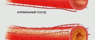
A popliteal artery aneurysm is a pathological expansion of the main artery running from the lower third of the thigh to the upper third of the leg. It is located quite deep in the leg under the knee. The popliteal artery is a continuation of the superficial femoral artery and below the knee it divides into the anterior, posterior tibial arteries and peroneal artery. These arteries supply blood to the leg and foot, so blocking blood flow in the popliteal artery leads to severe circulatory failure in the leg below the knee. The normal diameter of the vessel is about 6-10 mm.
Popliteal aneurysm is a risk factor for sudden acute limb ischemia and subsequent amputation. Unoperated aneurysms lead to leg amputation in 50% of all cases within 3 years.
Popliteal artery aneurysm must be operated on as soon as possible after diagnosis. There is no need to hope that it will “resolve” on its own. The high risk of acute ischemia and good results of planned operations should encourage the patient to consent to surgery. The results of planned interventions are very good.
Treatment technologies at the Innovative Vascular Center
The vascular surgeons of our clinic have significant experience in diagnosing and treating both planned and complicated lesions of the popliteal artery. The main method of treatment in our clinic is autovenous replacement of the popliteal aneurysm. This technology shows better immediate and long-term results. For complicated aneurysms, open surgery allows you to restore the patency of not only the popliteal artery, but also the vessels of the leg. Endovascular interventions for extensions of this localization have very poor results due to the high mobility of the knee joint.
Diagnostics
Source: MART PRODUCTION: Pexels
Asymptomatic course and imitation of the clinical picture of other diseases can hide the fact of the presence of an aneurysm until complications develop. Often an aneurysm becomes an incidental diagnostic finding. It is easiest to diagnose a peripheral artery aneurysm symptomatically, since its symptoms are quite obvious.
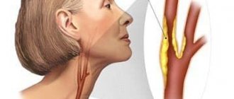
The doctor can detect large aneurysms of the aorta and peripheral arteries during examination using palpation (feeling) and auscultation (listening). Damage to the arteries of the brain can manifest itself as symptoms of transient ischemic attack, sudden headaches, eye pain, fainting and other symptoms.
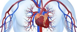
A cardiac aneurysm can manifest itself as symptoms of heart failure: shortness of breath, swelling, bluishness of the skin.
The following instrumental diagnostic methods are the most informative for aneurysmal vascular lesions:
- Ultrasound is a fairly universal technique used to diagnose aneurysms of various locations (except for cerebral vessels in adults). There are several options for ultrasound, used depending on the part of the body being examined and the pathology being sought.
- X-ray examination - allows you to identify aortic aneurysms, as well as the displacement of neighboring organs caused by them.
- CT and MRI are the most accurate diagnostic methods and allow you to visualize aneurysms of any location and size, determine the presence of thrombotic masses in them, and assess the condition of the walls.
- Angiography is a highly informative method that can be used in the diagnosis of any aneurysms. Unlike the above, this procedure is invasive, as it is necessary to deliver a contrast agent through a catheter into the vessel of interest. It is difficult to perform, and therefore is used to clarify cases of questionable diagnosis and controversial indications for surgical intervention.
Causes
Popliteal artery aneurysms account for about 1% of all surgical vascular diseases and often occur in both legs. The main reason is the congenital weakness of the artery wall, which contributes to their pathological expansion. The majority of patients (95%) are elderly men with an average age of approximately 71 years. The exact reasons for the development of dilation in the popliteal artery are unknown, but there is a clear connection with atherosclerotic changes in the vessel wall. Sometimes the pathology develops as a result of injuries to the popliteal region, dislocations or fractures. Patients with multiple aneurysms in different arteries should have general tissue weakness. The exact nature of this is still unclear. The tendency of the popliteal artery to pathological expansion is associated with frequent flexion and extension of the vessel due to movements in the knee joint.
Experience in surgical treatment of false aneurysm of the deep femoral artery (clinical case)
A.V. MAKSIMOV, E.A. GAISINA, A.K. FEISKHANOV, V.V. GLINKIN
Republican Clinical Hospital of the Ministry of Health of the Republic of Tatarstan, Kazan
Kazan State Medical Academy
Maksimov Alexander Vladimirovich
— Candidate of Medical Sciences, Head of the Department of Vascular Surgery, Associate Professor of the Department of Cardiology, Endovascular and Cardiovascular Surgery
420064, Kazan, Orenburgsky tract, no. 138, tel. 2-686-987, e-mail
False aneurysms of the great arteries in the structure of reconstructive operations on the great vessels range from 0.5% to
8-9%.
Their surgical treatment with the modern development of vascular surgery has been developed quite well. Therefore, the observation of large aneurysms in these localizations today is casuistry. The article presents a clinical case of successful surgical treatment of a giant false aneurysm of the femoral artery. Key words: giant false aneurysm, deep femoral artery replacement.
AV MAKSIMOV, EA GAYSINA, A K. PHEYSKHANOV, VV GLINKIN
Republican Clinical Hospital of the Ministry of Health of the Republic of Tatarstan, Kazan
Kazan State Medical Academy
Experience of surgical treatment of pseudoaneurysm of the deep femoral artery (medical case)
Pseudoaneurysm of the magistral arteries within corrective surgery on great vessels is from 0.5% to 9.8%. Their operative treatment in course of the current development of vascular surgery is worked out good enough. Therefore, the observation of large aneurysms of these locations today is casuistry. The article presents a medical case of successful surgical treatment of a giant pseudoaneurysm of femoral artery.
Key words:
giant pseudoaneurysm, prosthesis of the deep femoral artery.
False aneurysms of the great arteries account for from 0.5% to 8-9% in the structure of reconstructive operations on the great vessels [1, 2]. The most well-known causes of their occurrence are catheterization of the femoral artery during diagnostic and therapeutic procedures, trauma to the vessel, failure of the vascular anastomosis, or arrosive bleeding due to a purulent-inflammatory process.
Diagnosis of aneurysms of superficial arteries (femoral, brachial, carotid) is not difficult. Their surgical treatment with the modern development of vascular surgery has been developed quite well. Therefore, observations of large aneurysms in these localizations today are casuistry. In this regard, we present a clinical case of successful surgical treatment of a giant false aneurysm of the femoral artery.
Patient Z., 69 years old, was admitted to the department of vascular surgery of the Republican Clinical Hospital of the Ministry of Health of the Republic of Tatarstan with complaints of the presence of a painful dense formation in the left groin area and the upper third of the thigh. According to the patient, in 1988 (24 years ago) reconstructive surgery was performed on the left femoral artery. After discharge, he notes a slow growth of formation in the area of the postoperative wound. The patient did not seek medical help regarding this matter. The reason for this patient's visit was the appearance of pain.
Laboratory data on admission: Hb - 127 g/l, Er - 3.09 x 10¹²/l, L - 6.7 x 10⁹/l, total protein - 64 g/l; creatinine - 64 umol/l; potassium - 0.6 mmol/l; sodium -137 mmol/l, AST - 26 U/l, ALT - 2-2 U/l, PTI - 73%; APTT - 32.5 s.
On examination: the left lower limb is pale, cool on palpation, the veins are empty. In the groin area, along the anterior surface of the thigh, in the projection of the femoral artery, a dense, painful, weakly pulsating formation with a diameter of 18 cm, rising above the skin by 20 cm, is determined. The skin above the formation is thinned, tense, with areas of cyanosis and developing necrosis. Tactile sensitivity and active movements in the limb are preserved. There is no pulsation in the popliteal artery on both sides (Fig. 1).
Rice. 1.
Giant aneurysm of the left femoral artery
Distal arteriography and CT angiography were performed (Fig. 2), and the diagnosis was made: Atherosclerosis. Obliterating atherosclerosis of the arteries of the lower extremities.
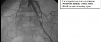
Rice. 2.
Arteriography and CT angiography of patient Z
Occlusion of the superficial femoral artery on both sides. Condition after reconstructive surgery on the left femoral artery (1988). Giant false aneurysm of the left femoral artery. Chronic arterial insufficiency of both lower extremities, degree 2B (according to Pokrovsky A.V.). IHD. (PEAKS?). Hypertension of the 1st degree. Common psoriasis, acute stage.
On December 18, 2012, an operation was performed: resection of a giant false aneurysm of the femoral artery on the left. External-iliac-deep-femoral extra-anatomical prosthetics on the left.
Progress of the operation: under epidural anesthesia, from the retroperitoneal approach according to Pirogov, the external iliac artery was isolated on the left and clamped. A longitudinal incision was made to open a false aneurysm in the groin area. The aneurysm cavity is filled with parietal thrombotic masses and tissue detritus from muscles and surrounding tissues with a volume of up to 2-2.5 liters. At the bottom of the aneurysm, the orifice of the deep femoral artery is visualized. The femoral artery, as an anatomical structure, is absent. From a separate access, lateral to the opened cavity of the aneurysm, the unchanged distal segment of the deep femoral artery was isolated. Extra-anatomical ilio-deep femoral prosthesis was performed using an Intergard-Silver 8 mm prosthesis, which was placed extra-anatomically outside the aneurysm cavity through the lacuna musculorum. Thrombotic masses and tissue detritus from the aneurysm cavity were removed, altered tissue of the aneurysm bottom and excess fasciocutaneous flap were excised. The aneurysm cavity was actively drained and sutured in layers. Blood loss was 250 ml.
In the postoperative period - the formation of marginal necrosis of the wound. Healing by secondary intention. Blood flow in the lower limb is compensated. The ankle-brachial index at discharge was 0.7. Discharged with recovery.
LITERATURE
1. Webber GW, Jang J., Gustavson S., Olin JW Contemporary management of postcatheterization pseudoaneurysms // Circulation. - 2007. - Vol. 115. - P. 2666-2674.
2. Righini M. Post-catheterization false femoral aneurysms // Rev. Med. Suisse. - 2007. - Vol. 3 (97). - P. 341-345.; Tisi PV, Callam MJ Treatment for femoral pseudoaneurysms // Cochrane Database Syst. Rev. - 2009; (2): CD004981.
Complaints and symptoms
Patients with an aneurysm complain of a feeling of heaviness in the popliteal region, swelling of the foot of the affected limb, and sometimes shooting pains. Most often, such complaints are vague, and the patient may not be aware that he has such a dangerous disease.
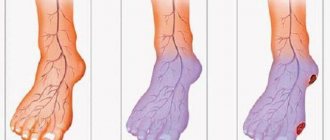
With aneurysm thrombosis, a clinical picture of acute ischemia develops: severe pain in the affected limb, changes in color and skin temperature of the foot. Subsequently, disturbances in sensitivity and movement develop. With advanced acute ischemia, rigor of the leg and foot develops; active and passive movements are impossible due to muscle death.
Types of aneurysm by location
Cerebral artery aneurysm
The most common form of this disease. It is characterized by local dilation of the arteries of the brain. In the case of hemorrhage due to a rupture of an aneurysm, a sharp unbearable headache, loss of consciousness, nausea, vomiting, and convulsions are most often noted. In half of the cases, patients die; many of those who survive risk remaining disabled. However, only about 25 percent of patients with an aneurysm experience a headache similar to a migraine before the critical moment. This disease is often misdiagnosed as a brain tumor.
Aortic aneurysm
This type of aneurysm can develop in different areas of this large blood vessel. According to research results, aortic aneurysm is found in 7 percent of those who die for another reason. In the later stages of the disease, patients complain of pressing pain in one or another part of the body.
There are aneurysms of the thoracic aorta and aneurysms of the aortic arch, the peculiarity of which is that it can develop even within 20 years after a chest injury. There is also an abdominal aortic aneurysm, which is often asymptomatic. However, very thin patients may feel throbbing and pain when they place their hand on their abdomen.
Aneurysm of peripheral vessels (blood vessels of the extremities)
Source: tirachardz / ru.freepik.com
Aneurysms of peripheral vessels are less dangerous in comparison with other types. Most often they develop in the vessels of the lower extremities - the popliteal and femoral arteries. Aneurysms of the vessels of the upper extremities are much less common. The clinical course is often asymptomatic, but sometimes patients note coldness and pallor of the limb, pulsation and pain when palpated.
Such aneurysms are subject to rupture much less frequently, but are often a source of thromboembolic complications.
Heart aneurysm
Characterized by a saccular protrusion of the heart wall. Acquired cardiac aneurysm occurs at the site of a myocardial infarction and is detected in 5-20 percent of patients who have suffered it. Over time, a scar is found at the site of the lesion, which gradually protrudes. An aneurysm can develop either immediately after a heart attack or several months after.
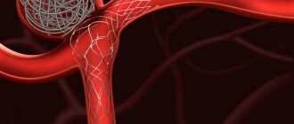
When any form of aneurysm is detected, surgical intervention is most often required. The essence of the operation is to exclude the aneurysm from the bloodstream to prevent life-threatening hemorrhages - endovascularly, by clipping, excision of the damaged section of the vessel and replacing it with a plastic prosthesis or a fragment of a blood vessel from another part of the body.
It is not always possible to prevent the development of an aneurysm, but you can reduce your risk of developing one by quitting smoking and reducing high blood pressure. To do this you need:
- Eat a healthy diet - in particular, eat more fruits and vegetables and less salt;
- drink alcohol in moderation or avoid it altogether;
- maintain a healthy weight;
- devote time to regular physical activity - approximately 150-200 minutes of moderate activity per week;
- reduce caffeine consumption to approximately 400 mg per day, which is approximately 2-3 cups of 250 ml.
Course and complications
The main risk from a popliteal aneurysm is associated with embolization - blockage of the underlying arteries with pieces of blood clots or occlusion of the aneurysm cavity. Both of these complications can lead to acute ischemia and gangrene of the legs (sudden loss of blood supply). Blood clots (thrombi) gradually form in the cavity of the vessel. When this clot remains attached to the wall of the vessel, it does not pose any danger. If a clot fragment breaks off, it can travel far from the aneurysm and cause blockages in small arteries, preventing blood flow to downstream tissues. A popliteal aneurysm can rupture (rupture), but this is much less common than embolization. In this case, a pulsating hematoma occurs behind the knee. Simultaneously with the rupture, the next stage is thrombosis of the popliteal artery with the development of signs of acute circulatory failure of the limb. Most people develop severe ischemic changes and death of the leg. Only surgery performed within the next 6-12 hours after the complication will help avoid amputation.
What is an aneurysm?
An aneurysm is a protrusion of the wall of an artery (less commonly, a vein) or heart due to its thinning, stretching or damage.
As a result, a so-called aneurysmal sac appears, which can compress nearby tissue. An aneurysm may be congenital. Moreover, when a child is born, this defect is invisible, and the baby develops completely normally. Aneurysms are also caused by diseases that affect blood vessels: hypertension, atherosclerosis, syphilis (at a late stage), sepsis. The risk of developing this disease increases when a blood vessel is injured or injured and when infected blood clots form. You can live with an aneurysm for years, go about your daily activities, and not experience any symptoms. Meanwhile, the aneurysm can quietly increase in size, threatening to explode at any moment - like a time bomb.
Sources
- S. Z. Amrakhov, A. S. Vischipanov. Surgical tactics and principles of performing operations in patients with coronary heart disease with post-infarction aneurysm of the left ventricle of posterobasal localization. Bulletin of the Scientific Center for Agricultural Sciences named after. A.N. Bakulev RAMS. 2013; 14 (1): 26-33.
- Koon K. Teo, MBBCh, PhD. Aneurysms of peripheral arteries. // MSD Disease Directory - 2021.
- Mark A. Farber, MD; Thaniyyah S. Ahmad, MD. Aneurysms of the thoracic aorta. // MSD Disease Directory - 2021.
- Brain aneurysms - Research Institute of Emergency Medicine named after. N.V. Sklifosovsky.