Heart in the life of the fetus
During fetal development, there must be structures in the heart that disappear after birth. Basically we are talking about the oval hole in the interatrial septum, which connects the atria of the heart during pregnancy and the ductus arteriosus of Botalla. Its presence is a physiological phenomenon that ensures the proper functioning of the circulatory system in the fetus.
Both openings close only after childbirth. Premature closure of the foramen ovale or duct of Botalla can lead to impaired cardiac function and performance. Fortunately, fetal echocardiography can detect this abnormality.
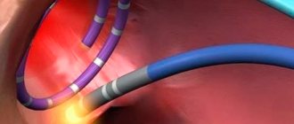
Fetal circulation and duct of Botalla
Ultrasound in early pregnancy: benefit or harm
An ultrasound of pregnancy in the 1st trimester is first of all necessary to confirm the fact of its presence. Indisputable evidence of successful conception is the presence of a fertilized egg in the uterine cavity. Then its location is determined, as well as the state of the reproductive system. Thanks to the obtained picture, doctors assess the risks of developing possible complications. If they are present, a treatment plan is developed that is aimed at preventing the threat of miscarriage.
But, despite the undeniable advantages of early diagnosis, women are afraid to do an ultrasound and think that it may negatively affect the health of the unborn baby. To reassure them, we’ll tell you about how ultrasonic waves affect the body.
Cavitation
The cavitation effect of ultrasonic waves has long been considered dangerous to the body, but recent research has shown that this ability of ultrasound can be used in other areas of medicine with even greater success.
The effect is the effect of ultrasound on fatty tissue. Thanks to its low frequencies, it passes through tissue and converts the fluid contained in fat cells into a gas-like substance. Gas bubbles collapse, fat cells die, and their breakdown products are excreted along with bile.
Cavitation cannot affect the development of the fetus, since its effect is superficial.
Thermal impact
The thermal effect is due to the absorption of energy by tissues during the procedure. By transmitting ultrasonic waves, our body converts them into heat. Ultrasound of pregnancy can lead to a slight increase in temperature up to 1-2 C, which in most cases is not felt and does not pose a danger. It is worth noting that this condition helps improve local blood circulation, which is certainly beneficial for the baby, because at this time he receives more nutrients.
Influence on biochemical processes
Ultrasound can slightly speed up biochemical processes in the body. Under its influence, enzyme activity changes, it normalizes metabolic reactions, improves tissue respiration, and affects the dynamics of nerve impulses.
Effect on immunity
Ultrasound does not affect the immune system in any way, because the effect of ultrasound during examination of pregnant women is very short-lived. All antibodies are produced as usual, and leukocyte mobility is not affected.
Fetal heart defects - how and when they are detected
Fetal valve defects may go undetected and may be acquired late in pregnancy due to gestational infection.
Some heart defects in utero may go undetected due to the small size of the fetal heart (about 10 mm at the second prenatal examination). Small holes in the septum, abnormal venous drainage, or coarctation of the aorta often go undetected.
In addition, heart health is affected by changes in pressure in the organ after birth, as well as the degree of closure of certain fetal circulatory structures - the foramen ovale, the ductus arteriosus, or the ductus venosus.
The fetus's lungs are filled with amniotic fluid, the fetus inside the uterus does not breathe, and oxygen comes through the umbilical cord from the mother. Therefore, fetal pulmonary circulation can only be assessed after birth, when the baby takes his first breath.
Limitations of the examination are associated with the small size of the fetal heart when examined during the period of 19-24 weeks of pregnancy, as well as with the hardware capabilities of ultrasound machines - their resolution and enlargement of structures. Therefore, all patients are recommended to undergo an additional third prenatal examination of the child at 28-33 weeks of pregnancy. The fetus is then much larger and certain structures, especially the fetal heart, are better visible.
Norm and interpretation of results
Fetal echocardiography is a non-invasive diagnostic method that is highly informative. An echocardiogram can only be interpreted by a cardiologist, since independent study of the indicators cannot clarify the full picture of the study.
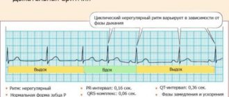
Any echocardiogram conclusion contains a number of mandatory indicators, the values of which reflect the structure and functions of the heart chambers. In the conclusion, the parameters of the right and left ventricles, atria, interventricular septum, and the condition of the heart valves and pericardium must be indicated. With the help of special medical aids, certain standards have been established, deviations from which may indicate the appearance of various heart pathologies.
The following indicators must be recorded in the protocol after this examination:
- value of left ventricular myocardial mass;
- value of left ventricular myocardium;
- end systolic size;
- short axis;
- long axis;
- end-diastolic volume of the left ventricle;
- aorta;
- right atrium;
- left atrium;
- myocardial thickness of the interventricular septum systological;
- myocardial movement;
- myocardial thickness of the interventricular septum diastological;
- miltral valve;
- aortic valve;
- ejection fraction;
- wall thickness in diastole;
- pulmonary artery;
- stroke volume;
- diastolic wall thickness;
- diastolic size.
The main indicators that determine the normal development and functioning of the heart muscle are data indicating the functioning of the ventricles, as well as the development of the septum between them. Decoding the data after the study may indicate a manifestation of heart failure or stenosis. Failure develops when the heart valve leaflets, which prevent the reverse flow of blood, fail to cope with their functions, because of this, blood is directed into an adjacent chamber, and the heart becomes less efficient. With stenosis, there is a decrease in the diameter of the valve opening, as a result of which blood pumping worsens.
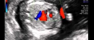
Interpretation of cardiac echocardiography necessarily includes data on myocardial contractility, as well as the pumping function of the left ventricle in dynamics, the presence of scars, aneurysms, tumors, blood clots, as well as their size and impact on the contractility of the walls. In addition, based on the study, the doctor has the opportunity to reliably assess the condition and functional characteristics of the heart valves, as well as the degree of hypertrophy of their walls. This technique allows you to examine blood flow through valves and large vessels, as well as detect the presence and existing degree of reverse blood flow through the valves.
When is it necessary to do an ECHO of the heart?
Although cardiac echo is extremely useful in detecting many cardiovascular diseases, the gynecologist orders fetal echocardiography first when she detects an arrhythmia or suspects that the baby may have a heart defect.
The risk of fetal heart defects among healthy pregnant women is approximately 4%. For this reason, every pregnant woman should undergo a complete fetal heart examination. The risk of karyologic problems is higher, including in children with increased neck transparency, abnormal karyotype, or generalized edema. This examination should also be carried out if:
- diabetes or gestational hypertension;
- phenylketonuria or other chronic diseases;
- autoimmune diseases such as Hashimoto's thyroid disease;
- connective tissue diseases, systemic lupus;
- taking antiepileptic drugs or other drugs used for chronic diseases;
- a family history of heart defects or the presence of a child with such a defect;
- rubella, coxsackie virus, toxoplasmosis, parvovirus B19 or other infections;
- aged from 35 years;
- burdened with family genetic defects;
- If the mother has Rh-positive blood, and the child’s father has negative blood, this is the Rh factor (serological conflict).
Diabetes mellitus in a pregnant woman
Indications for echocardiography also include a suspected genetic defect of the fetus, other previously identified fetal defects and abnormal ultrasound results, that is, increased arterial hypertension and abnormal biochemical results.
Main characteristics of the method
Fetal echocardiography is a non-invasive research method that can visualize the heart and blood vessels during the fetal development of a child using ultrasound waves. Modern technologies make it possible to use high-precision and safe devices in prenatal diagnostics, which guarantee safety during this study.
It is difficult for overweight women to perform high-quality screening, since a thick layer of fatty tissue transmits ultrasound less well
The basis for high-quality diagnostics is the presence of a program that allows you to determine the functional state of the cardiovascular system on a device equipped with a special sensor with a scanning frequency of at least 5 MHz, as well as color mapping of the circulatory system and Doppler.
Echo kg of the fetus allows you to determine the following parameters:
- heart rate;
- patency of blood vessels;
- speed and direction of blood movement through the vessels;
- the ratio of the volume of blood entering and exiting the aorta.
Fetal diagnosis should be carried out by perinatal experts who have a certified specialization to perform the technique. As a rule, these are cardiologists and obstetricians-gynecologists.
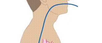
How is ECHO performed on the fetal heart?
A fetal echo is a completely safe, painless and non-invasive test. For a woman, it is practically no different from a standard ultrasound examination performed several times during pregnancy.
First, the doctor applies a special gel to the expectant mother’s belly, which improves the ultrasound. He then places the head of the ultrasound machine on the skin. The generated ultrasound reflects the structures of the heart and the fetal blood flowing in the heart, and is then returned to the device where the result is processed.
This will show the baby's chest and heart on the monitor. The doctor can evaluate the structure and function of the organ and check the heart rate. The equipment also provides images of blood flow through the heart (Doppler ultrasound). Thanks to this, the examination is even more accurate than standard ultrasound.
Echocardiography is usually performed between 19 and 24 weeks of pregnancy, when all the heart structures are developed and clearly visible. This takes from 15 minutes to 1 hour, depending on the specifications of the test.
It is also possible to do an early fetal heart echo at 14-18 weeks of pregnancy, especially in patients with abnormal double test results, serological conflict, after amniocentesis, infection during pregnancy. A complete fetal cardiac assessment should be repeated after 18 weeks of gestation.
Reviews
Ekaterina, 25 years old During the second screening at week 20, the doctor identified an additional chord. The gynecologist sent me for an echo kg, 7 days after the ultrasound. The procedure took about 15 minutes, after which the conclusion was issued. Based on the screening results, it turned out that this is a structural feature of the heart, and all parameters are normal.
Valentina, 36 years old This screening took place at the 24th week of pregnancy, since I belonged to a risk group (I have diabetes). The procedure was done in a specialized paid clinic, where a highly qualified cardiologist works. I had a Doppler echocardiogram done, it showed not only the structure of the heart, but also the flow of blood through the arteries leading to the heart. The results revealed no defects.
Anna, 27 years old During an ultrasound scan at week 22, the doctor discovered a defect in the interventricular septum. I was given a referral for an echocardiogram, which I underwent 12 days later. Based on the screening results, the diagnosis was confirmed; the child was diagnosed with a small membranous defect of the VSD. I was registered with a cardiologist, with whom I was constantly examined. Immediately after birth, the defect closed spontaneously, as it was small in diameter.
Benefits of detecting heart defects prenatally
First of all, this gives time to properly prepare for childbirth, both in its course and in the place of birth of the baby. A baby diagnosed with a heart defect should be born in a specialized center, where he will be under professional care.
Of course, we must not forget that modern medicine offers the possibility of prenatal treatment of cardiovascular diseases in a child, for example, the aforementioned arrhythmia or circulatory failure. Thanks to the so-called prenatal cardiology, pregnancy does not need to be terminated long before the due date, and the child has a chance to survive safely to the end in the best conditions - in the mother’s womb.
Intrauterine treatment of cardiovascular diseases in a child
Thanks to prenatal checkups, the fetus can be properly treated during pregnancy and delivered on time, avoiding premature birth.
Ultrasound during pregnancy: timing, harm and benefit
The examination is carried out in each trimester of gestation. Each procedure is aimed at the timely detection of possible pathologies and their early treatment. It is thanks to this diagnostic method that many women manage to bear and give birth to healthy children.
Doctors do not recommend doing an ultrasound in the early stages of pregnancy, more precisely before 9 weeks. During this period, the formation of all systems and organs occurs, so even the slightest impact can disrupt the development of the fetus.
A woman can choose where to have an ultrasound done during pregnancy; prices depend on the qualifications of doctors and the availability of modern equipment.
conclusions
Thus, fetal heart echo is the main cardiac examination of pregnant women. The test is used to evaluate the anatomy of the fetal chest and heart and its functioning.
During the examination, abnormalities in the structure (heart defects) and function of the heart, as well as arrhythmia, can be diagnosed. The information provided by this test can determine the further course of pregnancy and childbirth. Children with heart defects should be born in specialized centers where they will receive professional care immediately after birth.
MAKE AN APPOINTMENT
[contact-form-7 id=”296" title=”Untitled”]
Abortion and contraception clinic in St. Petersburg - department of the medical gynecological association "Diana"
Make an appointment, tests or ultrasound via the contact form or by calling +8 (812) 62-962-77. We work seven days a week from 09:00 to 21:00.
We are located in the Krasnogvardeisky district, next to the Novocherkasskaya, Ploshchad Alexander Nevsky and Ladozhskaya metro stations.
The cost of a medical abortion in our clinic is 3,300 rubles. The price includes all pills, an examination by a gynecologist and an ultrasound to determine the timing of pregnancy.
Stages of work during ultrasound examination
Conversation with an ultrasound diagnostic doctor.
Comfortable position on the couch.
Applying media gel to the study area or to the sensor (a special sterile condom is placed on the vaginal sensor).
With smooth movements, the ultrasound sensor moves in the study area, receiving images of the organs being examined through a piezoceramic transducer.
At the end of the study, the remaining media gel is removed with a disposable napkin.
Drawing up and issuing an ultrasound examination protocol to the patient.