Cervical spinal stenosis is a process of reducing the lumen of the spinal canal due to the development of various pathological structures. As a rule, the tendency to form stenosis most often occurs in people over 55 years of age. In a quarter of cases of the disease, cervical vertebral stenosis is diagnosed. In the absence of timely measures taken, spinal stenosis causes disability and disability.
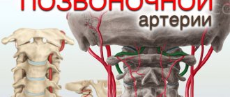
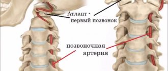
Vertebral artery syndrome with cervical osteochondrosis
Vertebral artery syndrome is a pathological condition that develops against the background of impaired blood supply in the vertebral artery basin; in practice, it manifests itself as symptoms of brain hypoxia in the places that it supplies.
Most often, vertebral artery syndrome occurs in combination with osteochondrosis of the cervical spine. Less commonly, the disease has other causes, sometimes not even of vertebrogenic origin. For a long time, vertebral artery syndrome was a disease of older people. But in recent years the disease has become much younger, and today such a diagnosis can be found even in children. The pathological condition is unpleasant due to its symptoms, which significantly worsen a person’s quality of life. In addition, as it progresses, it acquires consequences, which in most cases cause the development of transient ischemic attacks and even ischemic stroke.
With the right approach to the treatment of vertebral artery syndrome, specialists are able to alleviate the condition of a sick person and prevent many complications of the disease. Under no circumstances should the disease be ignored in the hope that its manifestations will subside over time. It is better to treat SPA immediately after the first signs appear, which will significantly increase the chances of recovery and provide a positive prognosis for the patient’s future life.
Why does cervical stenosis occur?
Cervical spinal stenosis develops for several reasons:
- Spinal fracture with compression of the vertebrae (compression);
- Congenital pathological changes in the structure of the vertebrae;
- Inflammatory diseases of the spine;
- Ankylosing spondylitis;
- Tumor processes;
- Intervertebral disc herniation;
- Chronic diseases of the articular surfaces of the spine;
- Adhesions after surgery;
- Overweight;
- Intervertebral disc displacement;
- Bone growths and osteophytes;
- Violation of the structure of the posterior yellow ligament;
- Osteochondrosis.
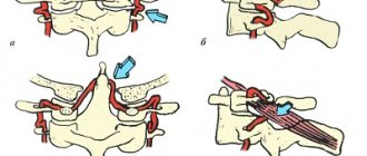
Stenosis of the neck vessels develops due to a decrease in the cavity where the spinal cord, nerves and vessels of the spine are located. First, complaints arise during certain turns and bends, then blood flow is disrupted and the situation worsens.
Over time, the pressure of the cerebrospinal fluid increases, provoking congestion and inflammatory processes.
Causes of development of vertebral artery syndrome
There are several main reasons for the development of cervical-vertebral syndrome in humans, among which are:
- osteochondrosis and other degenerative-dystrophic disorders in segments of the cervical spine;
- congenital anomalies of the structure of the bone elements of the neck;
- consequences of injuries in the area of the vertebral artery;
- pathological conditions that negatively affect the state of the sympathetic arterial plexus;
- atherosclerotic lesions of the vertebral arteries and other pathologies of the vascular wall;
- tonic muscle spasms in the neck;
- defects in the structure of the cervical vessels.
Qualified treatment for vertebral artery syndrome of the cervical spine should be carried out regardless of the causes of the development of the painful condition. Its scope and basic methods largely depend on the nature of the disease. Therefore, when symptoms of pathology appear, you should not self-medicate them, but immediately seek help from specialists who have experience in such clinical situations.
Causes
Over time, the walls of the carotid arteries can become thickened, their lumen narrows due to the formation of atherosclerotic plaques, which leads to a decrease in the volume of blood flowing through them. Atherosclerotic plaques consist of accumulations of cholesterol, calcium, fibrous tissue elements and cell debris that penetrate the artery wall through microscopic damage to the inner lining and form an atherosclerotic plaque in the area of which a blood clot (thrombus) can form.
Normal, healthy carotid arteries, like other normal arteries, are soft and flexible and allow blood to flow freely. If you place your finger on either side of the Adam's apple, you can feel the pulsation of the carotid arteries. The carotid arteries provide oxygen and nutrients to the cortex and other vital structures of the brain.
Symptoms
The following clinical symptoms are characteristic of vertebral artery syndrome:
- headaches, which in the case of vertebral artery syndrome are localized in the occipital zone with a transition to the front of the head and can appear after sharp turns of the neck or after sleep (Barré-Lieu syndrome);
- migraine pain of a basilar nature, which is often accompanied by loss of consciousness;
- sudden falls with throwing back of the head and loss of coordination;
- loss of normal stability and ability to maintain balance along with nausea, vomiting, dizziness, darkening of the eyes;
- development of transient ischemic attacks;
- short-term fainting states after sudden movements of the head;
- pain and feeling of “sand” in the eyes, dysfunction of the visual analyzer, loss of visual fields, etc.;
- hearing loss, tinnitus;
- hot flashes with excessive sweating;
- sometimes pain in the heart area.
Identifying the symptoms of vertebral artery syndrome is not a problem for an experienced specialist. Therefore, when the first signs of illness appear, you should not postpone a visit to the doctor, who will be able to conduct a high-quality diagnosis of the pathological condition and begin treatment on time.
Complications of cervical-vertebral syndrome
Cervical-vertebral syndrome is dangerous due to its complications, which often pose a threat to the patient’s life. This disease can cause the following consequences:
- ischemic stroke in the blood supply of the vertebral artery;
- myocardial infarction associated with dysfunction of the neurovascular bundle that supplies the heart muscle;
- suffocation as a result of impaired swallowing reflex.
All complications of SPA can be prevented with the help of timely and competent treatment, which can only be prescribed by a qualified specialist with extensive experience.
City Hospital reports!
Carotid artery stenosis is a condition in which there is narrowing (stenosis) or complete closure (occlusion) of the carotid artery. A person has two carotid arteries located in the neck (right and left). These blood vessels carry blood to the brain and face. Most often, the patency of arteries suffers as a result of the development of atherosclerotic plaques in their walls, narrowing the lumen.
Symptoms of carotid artery stenosis
Most people with carotid artery lesions have no symptoms. If symptoms are present, the risk of ischemic stroke increases several times. The most common symptoms are transient (i.e., temporary) ischemic attacks, which are sometimes called minor strokes.
During an ischemic attack, blood supply to certain areas of the brain is reduced. This may cause temporary dizziness, blurred vision, numbness and tingling of the skin of the extremities, and weakness in an arm or leg, which usually lasts no more than 30 minutes. The risk of stroke is very high in people who have had a transient ischemic attack.
A stroke occurs when there is a sharp decrease in blood supply to a vessel supplying the brain or when it is occluded. Depending on the area of the brain affected, a stroke manifests itself as paralysis of an arm and/or leg, visual and speech disturbances, and behavioral changes. The larger the area of the brain affected, the greater the risk to life. Cerebrovascular accidents are one of the main causes of death in Russia. Ischemic stroke confidently ranks first among diseases leading to permanent disability. Only 10 - 20% of patients who have suffered a stroke recover their ability to work. The rest become disabled and have persistent neurological defects. Along with this, the risk of developing a recurrent stroke remains, since the root cause (atherosclerotic narrowing of the carotid artery) has not been eliminated.
Risk factors for stroke:
- Atherosclerosis
- Diabetes
- High blood pressure
- Smoking
- Eating fatty foods
- Excess weight
- Increased thrombus formation
Diagnosis of carotid artery stenosis
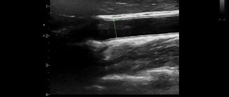
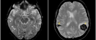
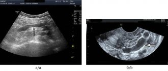
To determine whether you have stenotic lesions of the carotid arteries or not, your doctor will examine you. Even if you have no symptoms, your doctor may hear a murmur over your carotid arteries caused by blood flowing through the stenotic area. If necessary, Doppler ultrasound of the main arteries of the head (USDG MAG) will be prescribed first. It allows you to determine the localization of the narrowing, its degree and significance.
For a more detailed assessment of the condition of the carotid arteries, your doctor may recommend angiography (x-ray examination of blood vessels). This study is performed by catheterization, usually of the femoral or wrist artery, under local anesthesia in a special operating room equipped with an angiography unit.
Angiography of the carotid arteries
During this test, a very thin catheter is inserted into an artery in the leg or wrist and advanced toward the neck. A contrast agent is then injected through the catheter, making the carotid arteries and other arteries in the neck visible under X-rays.
On the eve of the examination, you should shave the skin in the groin area. You should also refrain from eating and drinking in the evening (except for medications). In the operating room, you will be covered with sterile sheets, which you cannot touch so as not to compromise sterility. During the test, your doctor will monitor your electrocardiogram (ECG) and blood pressure (BP). The catheter insertion site will be treated with an antiseptic and numbed. After this, the doctor will puncture your artery, through which he will pass a catheter to your neck. You won't feel it, but you will be able to see the catheter on the monitor.
You must follow all doctor's instructions. Sometimes you will need to hold your breath and not move. From time to time, from the administration of a contrast agent, you may feel warmth or a rush of heat in your head. The doctor will take pictures of the artery. If you have stenosis or occlusion of the carotid arteries, they will be detected.
Depending on the results obtained, you may be recommended a treatment procedure or scheduled for re-examination at a later date.
Treatment of carotid artery stenosis
There are two methods of surgical treatment. The first is an open endarterectomy performed by vascular surgeons. The second is a modern, minimally invasive, x-ray surgery - stenting. Both methods have their indications and contraindications. Therefore, the question of choosing one of them is always decided individually.
Carotid endarterectomy is a surgical operation aimed at removing the inner wall of the carotid artery affected by atherosclerotic plaque. The surgical technique is as follows: under anesthesia, an incision is made in the neck in the projection of the carotid artery. The artery is isolated and opened at the site of narrowing. The inner part of the artery wall is removed, along with the atherosclerotic plaque. Then plastic surgery of the artery is performed and the wound is sutured layer by layer.
Stenting is the installation of a stent, which is a metal tube consisting of cells, into a narrowed part of an artery. Expanding, the stent pushes the narrowed walls of the artery apart from the inside and constantly maintains them in a straightened state. Thanks to this, the internal lumen of the artery is restored and thereby the blood supply to the brain is improved. The following goals are pursued with stenting: elimination of chronic cerebral ischemia and prevention of ischemic stroke (or prevention of its recurrence).
The first stages of carotid artery stenting are carried out in the same way as an angiographic study: preparation, local anesthesia, puncture of the artery, insertion of a catheter and administration of a contrast agent. Before the operation, the patient is connected to special monitoring equipment that monitors parameters such as blood pressure and heart rate. The entire operation takes no more than an hour. From time to time you may feel hot flashes in your head.
Stenting of the carotid arteries is performed with protection against microembolism of cerebral vessels during surgery. Today, many of the world's leading experts prefer so-called filters. The filter is a metal frame on which a membrane is located (vaguely reminiscent of an umbrella). The filter retains microemboli without interfering with blood flow through the vessel: blood flows through micropores in the membrane that do not allow emboli to pass through. In some situations, according to indications, other microembolic protection devices are used.
After puncture of the artery, having installed a guide catheter into the carotid artery affected by atherosclerosis, the doctor passes the guide with a filter above the narrowing of the artery. Then a stent is installed along the guidewire into the area of stenosis. On the monitor, the doctor can see and evaluate the result. In some cases, it may be necessary to inflate the installed stent with a special balloon catheter. At this point, you may feel some discomfort in the neck and a change in heart rate. This is normal and should not be a concern. All manipulations are monitored using X-rays using a special high-tech angiographic device. The radiation dose is minimal and absolutely safe. Many such operations are performed around the world every year.
At the end of the intervention, the filter, balloon (if used) and guiding catheter are removed. The stent remains in the artery permanently, keeping it open. The doctor will apply pressure to the puncture site of the femoral artery for several minutes until the bleeding stops completely. The patient may be transferred to the intensive care unit for several hours to monitor vital signs. Strict bed rest should be observed for 24 hours after stenting. After returning to the room, you can eat and drink as usual.
The length of hospital stay depends mainly on the patient's well-being. After returning home, it is important to strictly follow all doctor’s instructions and regularly take prescribed medications. The further success of the operation depends on this. You should undergo regular examinations by a neurologist. If new complaints appear, you should immediately consult a doctor. For postoperative dynamic monitoring, Doppler ultrasound of the carotid arteries is very informative.
Following the doctor's recommendations after surgery improves treatment results and prognosis of the disease.
Prevention
The earlier stenosis of the artery carrying blood to the brain is detected, the easier it is to prevent the development of ischemic stroke and prevent disability of the patient. Therefore, at the first symptoms you should immediately consult a doctor.
Indications for intervention:
As has been described in detail above, candidates for carotid artery stenting are patients with hemodynamically significant stenoses of the internal carotid arteries. If the patient has already suffered an ischemic stroke or transient ischemic attack (TIA) in the territory of a stenotic artery, then the latter is considered symptomatic (that is, with a high degree of probability, it is the cause of the cerebral catastrophe that occurred). In such cases, stenosis of the internal carotid arteries (ICA) of more than 50% in diameter is subject to surgical treatment. If the patient has not suffered an ischemic stroke or TIA, but an ultrasound examination reveals ICA stenoses of more than 70% in diameter, the patient is also subject to surgical treatment. In cases where patients meet the indications described above, surgical treatment of targeted ICA stenoses has been shown to reduce the risk of ischemic stroke.
Clinical examples:
To illustrate clinical examples of performed stenting of the carotid arteries, there is no need to describe in detail the condition of the patients before and after the intervention, because the patient's well-being may not change significantly. The main goal of the intervention is stroke prevention.
Below are some illustrative examples of stenting:
Now patients have the opportunity to undergo examination and treatment at Medservice LLC by specialists with extensive experience in this area using the most modern angiographic complex Philips Allura Xper FD20 (Netherlands). This device with a new digital image processing system has a unique technology for suppressing noise and artifacts, which makes it possible to significantly increase image clarity without increasing radiation exposure and to see the thinnest vascular structures and stents. The latest generation X-ray tube minimizes radiation exposure to the patient.
You can ask questions related to angiographic and X-ray endovascular interventions at Medservice LLC:
Tel., email mail
Ivanov Andrey Gennadievich (head of the department of x-ray surgical methods of diagnosis and treatment, doctor of the highest category in the specialty “endovascular diagnostics and treatment”)
Diagnostics
Before treating vertebral artery syndrome, the doctor refers the patient to undergo a full diagnosis of the condition of his body. For this disease, diagnostic measures include a number of important methods:
- X-rays in the neck area, which helps determine areas of vascular compression;
- angiographic examination of the vessels of the neck to determine their patency, the presence of areas of narrowing of the lumen, deformations, etc.;
- ultrasound examination of the neck and diagnosis of the condition of blood vessels, muscles, determination of the nature of blood flow, the presence of areas of atherosclerosis;
- MRI in the neck with a more in-depth study of all the nuances and disorders;
- biochemical blood test to determine cholesterol levels and the degree of development of atherosclerosis.
Types of cervical stenosis
This pathology is represented by the following groups.
- Congenital (primary) stenosis, when the disease develops as a result of congenital abnormalities in the structure of the spine.
- Degenerative , acquired or secondary stenosis of neck vessels is a consequence of destructive-degenerative acquired changes.
- Combined or mixed stenosis is determined in the presence of various causative development factors.
Based on the area of the lesion, a distinction is made between relative and absolute stenosis (area less than 75 mm² or more than 75 mm², respectively).
Lateral stenosis is defined as a narrowing of the intervertebral foramen to 0.4 cm or less.
The term sagittal stenosis refers to a narrowing of the canal in the same plane.
Anatomically, the following types of cervical spinal canal stenosis are distinguished:
- lateral or lateral , when the exit site of the roots (radicular canal) of the spinal cord decreases in volume;
- central , when on the arch of the cervical vertebra (at the point where the roots exit) the distance from the posterior surface to the lateral surface decreases.
Drug treatment
Drug treatment of vertebral artery syndrome with spinal osteochondrosis and other disorders of the cervical region includes a number of medications, the action of which is aimed at restoring lost functions, eliminating pathological symptoms and preventing the development of complications. For vertebral artery syndrome the following are prescribed:
- decongestants that help eliminate compression of blood vessels by neighboring tissues;
- muscle relaxants to relieve compression and tension of muscle tissue;
- drugs to improve brain nutrition;
- vascular dosage forms;
- neuroprotectors that protect nervous tissue from ischemia;
- drugs for the treatment of atherosclerosis;
- B vitamins to strengthen nerve fibers;
- antispasmodics that relieve headaches.
Medicines for vertebral artery syndrome can be taken only with the permission of the attending physician, without changing the dosages prescribed by the specialist and without exceeding the prescribed course. It is important to understand that any self-medication and ignoring the doctor’s recommendations has dangerous consequences, which often pose a threat to the patient’s life.
Physiotherapy
Physiotherapeutic treatment helps eliminate many problems with impaired blood flow through the vessels that supply the brain. In such pathological conditions, a specialist can decide on the advisability of prescribing UHF therapy, electrophoresis, mud applications, magnetic therapy, etc. to the patient. These modern techniques can improve the nutrition of the central nervous system, eliminate the manifestations of the disease, normalize blood flow, reduce tissue swelling and relieve local inflammation. Before physiotherapeutic treatment, you should consult a doctor. For more detailed information, you can follow the link.
Exercise therapy
Exercise therapy for vertebral artery syndrome is an effective method of eliminating the main pathological symptoms, the effectiveness of which is based on strengthening the muscular structures of the neck, which create a framework for the neurovascular bundle. With the help of special therapeutic exercises, it is possible to achieve a positive effect of therapy and prevent complications of the process. The doctor will tell the patient in more detail about the duration and scope of such treatment after studying the clinic’s problem, the extent of the disorders and the presence of complications.
Treatment of carotid artery stenosis
Conservative treatment includes:
- taking medications (satins, antispasmodics, venotonics, antiplatelet agents, neuroprotectors);
- physiotherapeutic procedures;
- lifestyle changes (quitting smoking, dieting, physical activity, losing excess weight).
If there is a high risk of stroke, surgery is indicated:
- endarterectomy – plaques are removed from the artery;
- angioplasty - the lumen of the vessel is expanded using a canister filled with compressed air.
After restoring the diameter of the vascular lumen, a stent is placed inside the artery to maintain the walls in the desired position.
Primary appointment (examination, consultation) with a cardiovascular surgeon
1850 rub.
Sign up
Why is it better to treat carotid artery stenosis at Major Clinic?
Our clinic’s specialists have extensive knowledge and many years of experience in the treatment of stenosis and other vascular pathologies. They have a powerful diagnostic base at their disposal, which facilitates making an accurate diagnosis and obtaining detailed information about the condition of the blood vessels. Based on the diagnostic results, doctors at the Major Clinic Medical Center (Moscow) individually select a comprehensive treatment that gives a positive effect even in advanced cases.