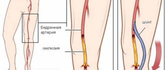
May-Turner syndrome is a pathological condition that is characterized by compression of the left iliac vein of the small pelvis by the iliac artery. Experts consider it difficult to diagnose, provoking a number of serious diseases in men and women in the form of varicocele and varicose veins of the small pelvis.
Where is May-Turner syndrome treated? If you are faced with this dangerous disease or suspect its development, contact the phlebology department of CELT. Our clinic is multidisciplinary and has a powerful diagnostic and treatment base, which allows us to accurately diagnose and treat May-Turner syndrome in Moscow in accordance with international standards.
At CELT you can get advice from a phlebologist.
- Initial consultation – 3,000
- Repeated consultation – 2,000
Make an appointment
Causes of May-Turner syndrome
The iliac veins are common paired blood veins located at the level of the sacroiliac joint and provide blood flow from the legs to the pelvic region. They are divided into right and left from the abdominal vein and aorta in the sacral region and diverge into external and internal. The external ones pass into the femoral veins, the internal ones are designed to ensure the outflow of blood from the pelvic organs.
May-Turner syndrome in men and women develops due to compression of the common iliac vein, located on the left side, by the right artery to the spine. It provokes disturbances in the outflow of blood from the left leg and pelvis and can occur against the background of atherosclerosis of the walls of blood vessels. As a result, blood stagnates in the leg and genitals and varicose veins develop.
Case description
Patient G.
, 30 years old, was admitted to the clinic on the referral of a gynecologist with complaints of severe chronic pelvic pain, more in the left iliac region, limiting activity, worsening in a sitting position, as well as with slight straining; feeling of heaviness and discomfort in the abdomen after sitting; frequent urination with a feeling of incomplete emptying of the bladder; coital and post-coital pain; heavy and painful menstruation; periodic swelling in combination with increased painful sensitivity of the perineum and external genitalia. The above complaints bothered me for about six months with gradual progression of symptoms. According to the results of ultrasound angioscanning (USAS) of the pelvic veins, performed at the prehospital stage, dilatation of the left ovarian vein (LOV), pampiniform, parametric and uterine veins on both sides was detected. She was observed for a long time by a gynecologist for endometriosis, but did not receive any specific treatment at the time of admission. There is a history of two independent births (the last in 2021). On the eve of hospitalization, she was examined by a gastroenterologist, surgeon, and gynecologist. No significant concomitant pathology, with the exception of endometriosis, was identified. No specific treatment was prescribed.
On admission: general condition was satisfactory. Consciousness is clear. Height - 165 cm, body weight - 56 kg, body mass index - 20.57. Skin and visible mucous membranes are of physiological color. In the lungs, breathing is vesicular, there are no wheezes, the respiratory rate is 16 per minute. Heart sounds are clear and rhythmic. Blood pressure - 115/80 mm Hg, heart rate (HR) - 68 beats/min. The abdomen is soft, not swollen, participates in the act of breathing, is painful on palpation in the iliac regions, more on the left. There are no symptoms of peritoneal irritation. The lower extremities are physiological in color, warm, not enlarged in volume, the pulsation of the arteries is clear. There are no varicose veins on the lower extremities and perineum.
Preliminary diagnosis: “Pelvic varicose veins. PTS, painful form. Dilatation of the PLV, grape-shaped, parametric and uterine veins on both sides. Ad, Po, r10. Endometriosis."
Laboratory data. Complete blood count: hemoglobin - 127 g/l, erythrocytes - 4.12·1012/l, hematocrit - 37%, leukocytes - 7.2·1012/l, platelets - 317·109/l. General urine analysis: specific gravity - 1.02, protein - negative, leukocytes - negative, bacteria - negative. Biochemical study: total protein - 79.6 g/l, total bilirubin - 11.8 µmol/l, ALT - 21 U/l, AST - 26 U/l, urea - 6.4 µmol/l, creatinine - 71 µmol /l, total cholesterol - 5.32 mmol/l, glucose - 5.9 mmol/l. Coagulogram: fibrinogen - 2.3 g/l, APTT - 28.6 s, prothrombin time - 11 s.
ECG: sinus rhythm, heart rate - 78 beats/min, normal direction of the electrical axis of the heart, alpha angle value - +64°.
Ultrasound of the kidneys and adrenal glands: signs of 1st degree nephroptosis on the right.
Ultrasound of the pelvic organs: signs of pelvic endometriosis (adenomyosis, endometriosis of the uterosacral ligaments, endometriosis of the bladder). Dilatation of intrapelvic veins and adhesions in the pelvis.
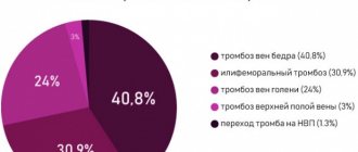
UZAS of the pelvic veins: dilation of the pelvic veins. Dilation of the ovarian veins (up to 9.1 mm on the left, up to 4.3 mm on the right), uterine veins (up to 8.6 mm on the left, up to 6.1 mm on the right), parametric veins (up to 7.7 mm on the left, up to 7.3 mm on the right), grape-shaped veins (up to 10.0 mm on the left, up to 10.6 mm on the right). Pathological reflux in the indicated veins - more than 1 s (Fig. 1a,
Rice. 1. Ultrasound picture of varicose veins of the small pelvis in B-mode of patient G. a - left ovarian vein (indicated by the arrow); b — veins of the pampiniform plexus (indicated by the arrow). b).
Ultrasound examination of the veins of the lower extremities: the IVC is patent and located up to the renal veins, diameter 16.2-18 mm, distally (along the aorta) - the IVC and common iliac veins on both sides are not visualized (Fig. 2a,
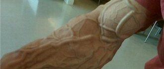
Rice. 2. Ultrasound scan of the veins of the lower extremities of patient G. Aplasia of the proximal segment of the inferior vena cava (its distal segment is indicated by the arrow). a — B-mode; b — color flow mode. b). The external iliac veins are patent on both sides. In the iliac regions, many collaterals are visualized (the IVC communicates with the above-described collaterals through the left renal and ovarian veins). Incompetence of the saphenofemoral and saphenopopliteal anastomosis of both lower extremities. Valve insufficiency of the right femoral, both popliteal and posterior tibial veins. Dilatation and short-term reflux of blood through the saphenofemoral and saphenopopliteal anastomosis on both sides. There were no signs of thrombosis in the examined vessels at the time of examination.
Multislice computed tomography of the IVC and pelvic veins: signs of occlusion of the IVC and both common iliac veins. Expansion of the PLN up to 15.4 mm, uterine and grape-shaped on both sides - up to 11 mm (Fig. 3a,
Rice. 3. Multispiral computer venogram of patient G. - expansion of the left ovarian vein to 15.4 mm (indicated by the arrow). a - direct projection; b - transverse projection. b).
X-ray contrast venography: occlusion of the IVC and common iliac veins on both sides, dilatation of the veins of the pelvic plexuses and PLJ (Fig. 4a,
Rice. 4. X-ray contrast venogram of patient G. - occlusion of the IVC and common iliac veins with drainage of the external iliac veins into the pelvic veins (1), lumbar veins (2) (indicated by arrows). a - right; b - left. b).
When assessing the quality of life, 24 points were obtained using the CIVIQ questionnaire, and 93 points using the PVVQ questionnaire. The severity of the disease was determined according to clinical scales: VCSS scale - 0 points, Villaltа scale - 2 points, PVCSS scale - 21 points, visual analogue scale - 64.3 points.
Based on the diagnostic studies performed, a clinical diagnosis was established: “Aplasia of the infrarenal portion of the inferior vena cava and common iliac veins. Varicose veins of the pelvis. PTS, painful form. Enlargement of the PLV, grape-shaped and uterine veins on both sides. C0A, EC, AS2,3,4,D6,7,10,13,14,15, PR, O. 01/11/2019. LIII. Pelvic endometriosis. Adhesive disease of the pelvis. Nephroptosis 1st degree.”
Analysis of diagnostic findings made it possible to establish that the patient’s compensatory collateral outflow occurs along two pathways—deep and median [7]. Considering the secondary compensatory expansion of the PLJ, which, along with the paired and semi-gyzygos veins, served as one of the main collaterals of the venous outflow in the absence of a functioning IVC, surgical treatment of the SVT was not considered indicated. Elimination of the primary cause of the disease using endovascular stenting of the IVC was technically impossible. Carrying out reconstructive bypass surgery is inappropriate due to significant surgical trauma in a patient without signs of chronic venous disease (CVD), in the absence of edema syndrome, venous claudication and trophic changes in the soft tissues of the lower extremities. Thus, the patient was prescribed conservative treatment: wearing class 2 compression tights, taking a micronized purified flavonoid fraction at a dose of 1000 mg/day. A repeat examination by a gynecologist is recommended. Considering that, according to the literature, the patient belongs to an increased risk group for the development of DVT, she underwent PCR diagnosis of congenital thrombophilia, followed by an examination by a gynecologist.
The patient was re-examined after 1.5 months.
Blood tests for thrombophilic mutations: GG-homozygous normal according to indicators F2: 20210 G>A, F5: 1691 G>A, F7: 10976 G>A, FGB: 455 G>A; TT-homozygous norm according to the ITGB3 indicator: 1565 T>C; 4G-4G homozygous pathology according to SERPINE1 (PAI-1): 675 5G>4G; GT-heterozygous pathology according to F13 indicator: 20210 G>T; CT heterozygote pathology according to the ITGA2 indicator: 807 C>T.
Consultation with a hematologist. Carriage of genetic prothrombotic mutations: plasminogen activator inhibitor - homozygote, integrin alpha-2 and F13 - heterozygote. Recommended: drink plenty of fluids, acetylsalicylic acid 50 mg/day.
Consultation with a gynecologist: endometriosis. Hormone therapy (diferelin intramuscularly) was prescribed.
From the point of view of the authors of this article, the interpretation of the results of the study of thrombophilic conditions was not entirely correct. The only indicator that in this patient could increase the risk of venous thromboembolic complications (VTEC) with the risk category “uncertain” is PAI-1. ITGA2 is practically a variant of the population norm, does not increase the risk of either venous or arterial thrombosis, and does not require constant use of acetylsalicylic acid. Carriage of the F13 mutation is also a weakly protective factor against the development of VTEC.
In addition, it is possible to challenge the gynecologist’s prescription of hormonal therapy against the background of severe pelvic varicose veins.
At the same time, not being specialists in the field of gynecology and hematology, the authors of this article do not believe that they have the right to insist on canceling the treatment prescribed to the patient.
Against the background of complex treatment, including phlebotropic and compression therapy, as well as taking diferelin and acetylsalicylic acid, the patient noted an improvement in her condition, a significant decrease in the intensity of pelvic pain and dyspareunia. The global quality of life index according to the CIVIQ and PVVQ questionnaires improved by 3 and 49 points, respectively. The results of reducing the median total score on disease severity scales are as follows: PVCSS scale - 15 points, visual analogue scale - 55.6 points. There was no decrease in the median on the VCSS and Villaltа scales.
Considering the positive dynamics during the treatment, the patient was recommended to continue conservative treatment and follow-up with a vascular surgeon or gynecologist.
Clinical manifestations of May-Turner syndrome
Unfortunately, the syndrome is not well studied because it is rare. Experts distinguish three stages of its development:
- First, there are no clinical manifestations;
- Second, the lumen of the iliac vein narrows, symptoms of venous insufficiency appear;
- Third, thrombosis develops.
The symptoms of the syndrome are as follows:
- Swelling of the left leg, often with a change in the color of the skin to bluish or purple;
- Intense pain symptoms in the affected area that cannot be eliminated with painkillers;
- Varicose veins visible to the naked eye on the thigh, testicle (in men) or labia (in women) on the affected side;
- Increased discomfort after physical activity, including minor ones;
- The appearance of hemorrhoids.
Both men and women suffering from the syndrome have problems with sexual intercourse. So, in the first case, pulling pains appear in the scrotum, which radiate to the urethra and head of the penis and become more intense after sexual intercourse. In the second case, sexual contacts become completely impossible due to the pain of the node.
How does the condition manifest?
The inferior vena cava is compressed by the enlarged uterus when the woman is lying on her back. At long gestation periods or with polyhydramnios, this can also occur in an upright position of the body.
The first symptoms appear at about 25 weeks. It becomes difficult for a woman to lie on her back, and she may experience dizziness, shortness of breath, and weakness. Blood pressure decreases. In some cases, even collapse with fainting occurs.
In severe cases, a woman quickly turns pale 2 to 3 minutes after turning on her back, complains of dizziness and darkening of the eyes, nausea and cold sweat. More rare signs are ringing in the ears, heaviness behind the sternum, a feeling of strong fetal movement.
Suddenly developing pallor and hypotension are very similar to signs of internal bleeding, so the doctor may mistakenly suspect placental abruption, uterine rupture, or myocardial infarction in such a pregnant woman.
The appearance of a vascular pattern and varicose veins in the legs is also associated with the described syndrome. One of the common manifestations of this condition is hemorrhoids.
The described pathological condition leads to fetal hypoxia and disturbance of its heartbeat. The development of organs and systems of the unborn child suffers. If it occurs during childbirth, it can cause fetal asphyxia. The connection of this disease with premature detachment of a normally located placenta has been proven.
Our doctors
Malakhov Yuri Stanislavovich
Doctor - cardiovascular surgeon, phlebologist, Honored Doctor of the Russian Federation, Doctor of Medical Sciences, doctor of the highest category
Experience 36 years
Make an appointment
Drozdov Sergey Alexandrovich
Cardiovascular surgeon, phlebologist, Doctor of Medical Sciences
47 years of experience
Make an appointment
CT scan of the inferior vena cava with contrast
Contrast CT of the inferior vena cava takes place in two stages. First, the patient is placed on the table and a series of native images are taken using a tomograph. Then an iodine-containing solution, characterized by high radiopacity, is injected intravenously. The photographs clearly show the system of the inferior vena cava with ducts. This diagnostic method is highly informative; it can be used to determine the condition of blood vessels and surrounding tissues.
CT angiography with contrast is used in the diagnosis of inflammatory diseases of the inferior vena cava, to identify neoplasms and blood clots in the lumen of the vessel. The method allows you to assess the nature and site of surgical intervention in the case of surgical treatment, as well as monitor regeneration processes in the recovery period.
Nephrectomy with removal of thrombus from the inferior vena cava on computed tomograms
There is an emergency CT scan of the inferior vena cava, which is performed in case of conditions that threaten the patient's life.
Treatment of May-Turner syndrome
The syndrome requires surgical intervention because it is caused by a mechanical cause (i.e., compression). The goal of the operation is to eliminate it through stenting. To do this, a self-expanding stent is delivered to the area affected by the syndrome, with the help of which the narrowed area is opened.
The stent itself will serve as a framework for the affected area and will be able to provide the required level of vein patency. In order to achieve the desired result, special reinforced stents are used, since conventional ones are not able to cope with high pressure. If, in addition to the syndrome, the patient suffers from severe varicose veins, he undergoes microphlebectomy - an operation aimed at removing damaged areas of blood vessels.
You can make an appointment with specialists from the phlebology department of CELT for the treatment of May-Turner syndrome online or by contacting our operators: +7 (495) 788-33-88
Make an appointment through the application or by calling +7 +7 We work every day:
- Monday—Friday: 8.00—20.00
- Saturday: 8.00–18.00
- Sunday is a day off
The nearest metro and MCC stations to the clinic:
- Highway of Enthusiasts or Perovo
- Partisan
- Enthusiast Highway
Driving directions
Preparing for the study
The CT angiography procedure of the inferior vena cava is performed using a contrast agent, and therefore requires preliminary preparation. After making an appointment for a CT or MRI, the patient can receive a free consultation, where he will be told how the examination is carried out and what needs to be done before the tomography.
Before performing MSCT, it is necessary to check the level of creatinine in the blood. This test is performed to assess the status of kidney function. If necessary, you can do an express test before the procedure - the Magnit clinic in St. Petersburg provides this opportunity.
Individual intolerance to iodine-containing drugs and a tendency to allergic reactions, hyperfunction of the thyroid gland, pregnancy and lactation, monitoring blood sugar levels using Metformin and its analogues require prior consultation with a doctor.
Diagnosis of pathology
Activities for diagnosing pathology include:
- X-ray examination;
- Venocavagraphy;
- CT;
- MRI;
- Endoscopy with biopsy.
The doctor needs to determine what caused the pathology. This is important for the selection of treatment methods. So chemotherapy is prescribed only if cancer is confirmed.
Lung cancer: symptoms