During a consultation with a cardiologist or surgeon, the patient may be diagnosed with an enlarged jugular vein in the neck; the causes of this phenomenon vary. Depending on the predisposing factors, a treatment regimen is prescribed.
The function of the jugular veins is to be responsible for the process of blood flow from the brain to the neck. Thanks to these blood veins, unpurified blood flows to the heart muscle so that the filtration process can take place.
Jugular veins are divided into several types:
- Internal. It is located at the base of the skull, and its end is in the region of the subclavian fossa. At this site, the vein pours unpurified blood into the brachiocephalic vessel.
- The external one begins under the auricle, goes down to the sternum and collarbone, enters the internal jugular vein, as well as the subclavian vein. This vessel has valves and processes.
- The anterior one originates from the outer part of the mylohyoid muscle and flows near the midline of the neck. This vein enters the subclavian and external, thereby forming an anastomosis.
A little anatomy
The main volume of blood is drained from the head and neck through the largest of the jugular ones - the internal one. Its trunks reach a diameter of 11-21 mm. It begins at the cranial jugular foramen, then expands to form the sigmoid sinus, and descends down to the place where the clavicle connects to the sternum. At the lower end, before connecting with the subclavian vein, it forms another thickening, above which, in the neck area, there are valves (one or two).
The internal jugular vein has intracranial and extracranial tributaries. Intracranial - these are the sinuses of the dura mater with the veins of the brain, orbits, hearing organs, and skull bones flowing into them. Extracranial veins are vessels of the face and the outer surface of the skull that flow into the internal jugular along its course. The extracranial and intracranial veins are connected to each other by ligaments that pass through special cranial foramina.
The internal jugular vein is the main line that drains blood saturated with carbon dioxide from the head. This vein, due to its convenient location, is used in medical practice to place catheters to administer medications.
The second most important is external. It runs under the subcutaneous tissue along the front of the neck and collects blood from the outer parts of the neck and head. It is located close to the surface and is easily palpable, especially noticeable when singing, coughing, screaming.
The smallest of the jugular veins is the anterior jugular, formed by the superficial vessels of the chin. It runs down the neck, merging with the external vein under the muscle connecting the mastoid process, sternum and clavicle.
Clinical significance[edit]
| Actual Accuracy This clinical sign |
The parrot sign is a sensation of pain when pressing on the retromandibular area. [ citation needed
]
Catheterization
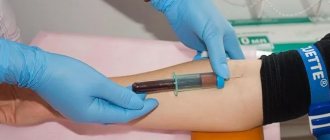
For venous access in medical practice, the right internal jugular vein or the right subclavian vein is usually used. When performing the procedure on the left side, there is a risk of damage to the thoracic lymphatic duct, so it is more convenient to perform manipulations on the right. In addition, the left jugular trunk carries blood outflow from the dominant part of the brain.
According to doctors, puncture and catheterization of the internal jugular vein is preferable to that of the subclavian veins, due to fewer complications, such as bleeding, thrombosis, and pneumothorax.
Scheme of jugular vein catheterization
Main indications of the procedure:
- Impossibility or ineffectiveness of drug administration into peripheral vessels.
- Upcoming long-term and intensive infusion therapy.
- The need for diagnostics and control studies.
- Carrying out detoxification using plasmapheresis, hemodialysis, hemoabsorption.
Catheterization of the internal jugular vein is contraindicated if:
- history of surgery in the neck area;
- blood clotting is impaired;
- there are ulcers, wounds, infected burns.
There are several access points to the internal jugular vein: central, posterior and anterior. The most common and convenient of them is the central one.
The technique for vein puncture using central access is as follows:
- The patient is placed on his back, his head is turned to the left, his arms are along his body, the table on the side of his head is lowered by 15°.
- The position of the right carotid artery is determined. The internal jugular vein is located closer to the surface parallel to the carotid vein.
- The puncture site is treated with an antiseptic and limited with sterile napkins, lidocaine (1%) is injected into the skin and subcutaneous tissue and the search for the location of the vein begins with an intramuscular search needle.
- The course of the carotid artery is determined with the left hand and the needle is inserted 1 cm lateral to the carotid artery at an angle of 45°. Slowly advance the needle until blood appears. Inject no deeper than 3-4 cm.
- If it is possible to find a vein, the search needle is removed and a needle from the set is inserted, remembering the path, or the needle from the set is first inserted in the direction found by the search needle, then the last one is removed.
Catheter placement is usually done using the Seldinger technique. The injection technique is as follows:
- You need to make sure that blood flows freely into the syringe and disconnect it, leaving the needle.
- A guidewire is inserted into the needle approximately half its length and the needle is removed.
- The skin is incised with a scalpel and a dilator is inserted through the guidewire. The dilator is taken with the hand closer to the body so that it does not bend and injure the tissue. The dilator is not completely inserted; it only creates a tunnel in the subcutaneous tissue without penetrating the vein.
- The dilator is removed, the catheter is inserted and the guidewire is removed. They do a test for an allergic reaction to the drug.
- By the free flow of blood, you can understand that the catheter is in the lumen of the vessel.
Links[edit]
This article incorporates public domain text from page 646 of the 20th edition
"Grey's Anatomy"
(1918).
- Thompson, Stephen H.; Jung, Alison Y. (2016-01-01), Hupp, James R.; Ferneini, Eli M. (ed.), "4 - Anatomy Relevant to Head, Neck, and Orofacial
Head, Neck, and Orofacial
Infections , St. Louis: Elsevier, pp. 60–93, doi: 10.1016 /b978-0-323-28945-0.00004-1, ISBN 978-0-323-28945-0, received 11/11/2020 - Cunningham, Larry L.; Card, Aaron Sterling (01/01/2012), Bagheri, Shahrokh K.; Bell, R. Brian; Khan, Hussain Ali (ed.), "Chapter 38 - Subcondylar fractures of the mandible", Current Therapy in Oral and Maxillofacial Surgery
, St. Louis: W. B. Saunders, pp. 298–304, doi: 10.1016/b978-1- 4160-2527-6.00038-4, ISBN 978-1-4160-2527-6, received 11/11/2020 - Lukota, Richard A.; Abdel-Galil, Khalid (01/01/2017), Brennan, Peter A.; Schlifak, Henning; Gali, G. E.; Cascarini, Luke (ed.), "6 Condylar Fractures", Oral and Maxillofacial Surgery (Third Edition)
, Churchill Livingstone, pp. 74-92. DOI: 10.1016/b978-0-7020-6056-4.00006-х, ISBN 978-0-7020-6056-4, received 11/11/2020 - ^ ab Kramer, Gregory D. (01/01/2014), Kramer, Gregory D.; Darby, Susan A. (ed.), "Chapter 5 - Cervical Region," Clinical Anatomy of the Spine, Spinal Cord, and ANS (Third Edition)
, St. Louis: Mosby, pp. 135-209. Doi: 10.1016/b978-0-323-07954-9.00005-0, ISBN 978-0-323-07954-9, received 11/11/2020 - Drake, Richard L. (Richard Lee), 1950- (2005). Gray's Anatomy for Students. Fogle, Wayne., Mitchell, Adam WM, Gray, Henry, 1825-1861. Philadelphia: Elsevier/Churchill Livingston. ISBN 0-443-06612-4. OCLC 55139039 .CS1 maint: multiple names: authors list (link)
- Poznick, Jeffrey K. (2014-01-01), Poznick, Jeffrey S. (ed.), "39 - Aesthetic Modification of Prominent
Ears: Evaluation and Surgery",
Orthognathic Surgery
, St. Louis: W.B. Saunders, pp. 1703-1745 , DOI: 10.1016/b978-1-4557-2698-1.00039-3, ISBN 978-1-4557-2698-1, received 11/11/2020
Pathologies of the jugular veins
The main diseases of these veins include pathologies characteristic of all large vessels:
- phlebitis (inflammation);
- thrombosis (formation of blood clots inside vessels that obstruct blood flow;
- ectasia (expansion).
Phlebitis
This is an inflammatory disease of the vein walls. In the case of the jugular veins, three types of phlebitis are distinguished:
- Periphlebitis is inflammation of the subcutaneous tissue surrounding the vessel. The main symptom is swelling in the area of the jugular gutter without impairing blood circulation.
- Phlebitis is an inflammation of the venous wall, accompanied by dense edema, while the patency of the vessel is maintained.
- Thrombophlebitis is inflammation of the vein wall with the formation of a blood clot inside the vessel. Accompanied by painful dense swelling, hot skin around it, blood circulation is impaired.
There may be several causes of phlebitis of the jugular vein:
- wounds, bruises and other injuries;
- violation of sterility when placing catheters and during injections;
- penetration of drugs into the tissue around the vessel (often occurs when calcium chloride is administered in addition to a vein);
- infection from neighboring tissues that are affected by harmful microorganisms.
For uncomplicated phlebitis (without suppuration), local treatment is prescribed in the form of compresses and ointments (heparin, camphor, ichthyol).
Heparin ointment is used for phlebitis to prevent the formation of blood clots
Purulent phlebitis requires a different approach. In this case the following are shown:
- anti-inflammatory drugs (Diclofenac, Ibuprofen);
- medications that strengthen the walls of blood vessels (Phlebodia, Detralex);
- drugs that prevent thrombosis (Curantil, Trental).
If therapeutic methods do not bring results, the affected area of the vein is surgically excised.
Phlebectasia
This is what is called in medicine the expansion of the jugular vein. As a rule, at the beginning of the disease there are no symptoms. The disease can last for years without showing itself in any way. The clinical picture unfolds as follows:
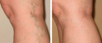
- The first manifestations are a painless enlargement of the vein in the neck. A swelling forms below, shaped like a spindle; at the top, a bluish bulge appears in the form of a sac.
- At the next stage, a feeling of pressure appears when screaming, sudden head movements, or bending.
- Then there is pain in the neck, breathing is difficult, and the voice becomes hoarse.
Ectasia can develop at any age, and the main causes are:
- Head and neck contusions, concussions, traumatic brain injuries.
- Sedentary work without breaks for a long time.
- Fractured ribs, spinal and back injuries.
- Disruption of the valve apparatus, which cannot regulate the movement of blood, as a result of which it accumulates and stretches the vascular walls.
- Hypertension, coronary disease, myocardial diseases, heart defects, heart failure.
- Prolonged immobility due to pathologies of the spine or muscle tissue.
- Leukemia.
- Tumors (benign or malignant) of internal organs.
- Endocrine disorders.
Most often, the jugular veins are dilated for several reasons.
Treatment of ectasia depends on the general condition of the patient, the severity of the disease and how dilated the vessel is and how this affects the surrounding tissue. If the normal functioning of the body is not threatened, the patient will be under observation and no special treatment will be required.
If an enlarged jugular vein negatively affects your health, surgical treatment will be required. An operation is performed to remove the pathologically enlarged area, and healthy areas are connected into one vessel.
As for complications, there is a possibility of vessel rupture and bleeding, which most often ends in death. Although ectasia ruptures are rare, the disease should not be left to chance. It is necessary to constantly monitor the doctor so that if the disease progresses, he can prescribe surgery in a timely manner.
Jugular vein thrombosis
With thrombosis, a blood clot forms inside the vessel, which obstructs blood flow. Jugular vein thrombosis can be congenital, acquired or mixed.
Hereditary risk factors include:
- special structure of veins;
- antithrombin-3 deficiency;
- blood clotting disorder;
- lack of proteins C, S.
For purchased:
- surgical intervention and condition after surgery;
- tumor;
- elderly age;
- postpartum period;
- prolonged immobility during a long trip or flight;
- chemotherapy;
- antiphospholipid syndrome;
- injuries resulting in compression of the vein;
- intravenous administration of narcotic drugs;
- gypsum bandage;
- catheterization of veins;
- acute heart attack, stroke;
- menopause;
- lupus erythematosus;
- smoking;
- stomach ulcer, sepsis;
- hormone therapy;
- thrombocytosis;
- severe dehydration;
- endocrine diseases;
- taking hormonal contraceptives.
Causes
There are many reasons that provoke pathological changes in the jugular vein bulb.
Most often, the condition of blood vessels worsens under the influence of the following factors:
- recent chemotherapy;
- taking hormonal medications;
- injuries and mechanical damage to the neck;
- tumors;
- infectious diseases;
- osteochondrosis of the cervical spine;
- cardiovascular diseases;
- high blood pressure;
- blood clotting disorder;
- intoxication, etc.


