Associate Professor V. K. Milkamanovich
Belarusian State University
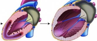
Myocardial infarction (MI) is a severe form of coronary heart disease (CHD), in fact, its dangerous complication. The danger of MI to life is emphasized by the unofficial synonym for the diagnosis - “cardiovascular accident”. This is necrosis (death) of one or more areas of the heart muscle as a result of an acute deficiency of its blood supply, which persists for more than 20 minutes (Fig. 1). Necrosis finally forms within 2–4 hours or more, depending on the collateral (bypass) blood flow and the sensitivity of cardiac muscle cells to decreased blood supply.
IHD is an independent heart disease that can manifest as MI, angina pectoris, post-infarction cardiosclerosis, cardiac arrhythmia, heart failure, and sudden death. The term “ischemia” (from the Latin ischemia - I delay, stop the blood) means a local deterioration in blood supply, most often caused by a narrowing or complete blockage of the lumen of the artery, leading to temporary dysfunction or permanent damage to the tissue or organ.
The main cause of IHD is atherosclerosis of the coronary arteries. Atherosclerosis (from the Greek athera - gruel and sclerosis - hardening) is a systemic chronic disease of large and medium-sized arteries, resulting from persistent chronic inflammation and dysfunction of the endothelium of the vascular wall, local accumulation of lipids in the area of inflammation, as well as blood components and compaction of the connective tissue tissue in its inner and middle layers.
Externally, an artery affected by atherosclerosis resembles a metal pipe corroded by rust. Throughout its entire internal surface, numerous atherosclerotic plaques of various shapes and sizes, clots of blood clots on the surface of atherosclerotic plaques, fatty spots and stripes, ulcerations, deposits of calcium salts (calcinosis), etc. are visible.
There are two phases in the development of the atherosclerotic process.
The first phase is atherogenic. The lipid plaque grows (up to about 40 years of age). Atherosclerotic plaques are accumulations of cholesterol endothelial cells - the inner lining of blood vessels, responsible for their elasticity. As it is deposited, cholesterol mixes with calcium to form dense plaques that increase in size and rise above the surface of the artery wall (Figure 2). Over time, this leads to blocking of the lumen of the vessel.
The second phase is thrombogenic. It is characterized by the presence of an atherosclerotic plaque, prone to the formation of thrombotic masses on its surface.
Under certain conditions (for example, a sudden increase in blood pressure), the morphological structure of the surface layers of the atherosclerotic plaque may be disrupted, and then the preconditions are created for a rapid disruption of blood flow in the coronary artery, which leads to a complete or partial cessation of blood supply to the area of the myocardium that this line supplies. Rupture of an atherosclerotic plaque and/or thrombus formation in the lumen of a coronary artery are the most common causes of MI.
Atherosclerosis is characterized by a slowly progressive cyclic course: periods of activity of the process are replaced by periods of calm.
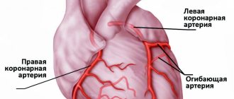
The left coronary artery, having a diameter of 3.4–4.9 mm (usually 3.9), supplies blood to the bulk of the left ventricle, the left atrium, part of the anterior wall of the right ventricle, and the anterior part of the interventricular septum. Therefore, interruptions in the supply of cardiac muscle cells with oxygen during circulatory disorders in the left coronary artery lead to dystrophic, necrotic and sclerotic changes in large areas of the myocardium, clinically manifested by a decrease in myocardial contractility, chronic heart failure, detraining of the patient and psychological disorders.
Most often with MI, the left ventricle of the heart suffers. The right ventricle is very rarely affected in isolation.
The diagnosis of “myocardial infarction” is established on the basis of characteristic complaints, objective clinical signs of heart damage, as well as electrocardiography, echocardiography and markers of myocardial necrosis.
The diagnostic problem of acute MI lies in the fact that in 50% of cases it develops suddenly, without any previous clinical symptoms, in a person in the prime of life, outwardly completely healthy and without bad habits.
Subjective clinical signs of myocardial infarction
Pain in the chest for more than 15–20 minutes, which spreads along the inner surface of the left arm with a tingling sensation in the wrist and fingers. Other possible areas of pain distribution (irradiation) are the shoulder girdle, neck, jaw, interscapular space (also mainly on the left).
The pain during a heart attack is very strong, perceived as dagger-like, tearing, burning, “a stake in the chest.” This feeling can be so unbearable that the person screams.
Sometimes the sensations may resemble an attack of angina pectoris, that is, instead of severe pain - discomfort in the chest, a feeling of strong compression, squeezing, a feeling of heaviness (“pulled with a hoop,” “squeezed in a vice,” “crushed with a heavy slab,” etc.).
Some people experience only a dull ache or numbness in the wrists combined with severe and prolonged chest pain or chest discomfort.
Pain in acute MI occurs suddenly, often at night or in the early morning hours. Painful sensations develop in waves, periodically decreasing, but not stopping completely. With each new wave, the pain (discomfort) in the chest intensifies, quickly reaches a maximum, then weakens.
Unlike an attack of angina, pain during MI does not stop and does not decrease at rest, or when taking nitroglycerin (even repeatedly).
Objective clinical signs of myocardial infarction
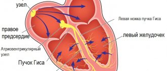
ECG changes that indicate acute myocardial ischemia (ST-T changes or complete block of the left bundle branch, the appearance of pathological Q waves).
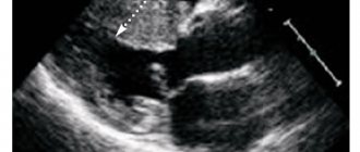
Changes in the echocardiogram, which record the loss of viability of a section of the myocardium (thinned and not contracting), or disturbances in its local contractility.
Increase and/or decrease in the level of cardiac-specific cardiac biomarkers - cardiac troponins I and T, cardiac form of creatine phosphokinase (CF-CPK). Cardiac troponin reflects even minimal myocardial necrosis weighing less than 1 g, which is not detected by other diagnostic methods.
According to the size of the damage to the heart muscle, MI can be microscopic (focal or point damage), small (damage involves less than 10% of the muscle mass of the left ventricle), medium-sized (10–30% of the left ventricular myocardium) and large (damage occupies more than 30% of the left ventricular myocardium). ventricle).
Echocardiography is usually used to determine the size of the infarction.
MI is classified according to the nature of its course:
- acute (primary) – in the absence of signs of a previous MI;
- repeated – when a patient who has already suffered an MI in the past develops new foci of necrosis.
Depending on the clinical consequences, MI can be uncomplicated or complicated by acute left ventricular failure (pulmonary edema), cardiogenic shock, rhythm and conduction disturbances, acute left ventricular aneurysm, etc.
The entire healing process of an MI usually takes about 5–6 weeks.
In modern conditions, control over the process of restoration of damaged myocardium is carried out using the following imaging methods:
- echocardiography;
- radionuclide ventriculography;
- myocardial perfusion scintigraphy;
- single photon emission computed tomography;
- magnetic resonance imaging;
- positron emission tomography;
- computed tomography.
The basis of treatment for acute MI is the destruction of a thrombus in the coronary artery by any available methods. Typically, a thrombolytic drug is administered as early as possible (eg, 1.5 million IU streptokinase intravenously over 30 minutes).
If there are contraindications to thrombolytic therapy, highly effective methods of percutaneous coronary interventions are used - balloon angioplasty and coronary artery stenting, which account for 95% in the treatment of acute MI in a number of advanced countries. Angioplasty and stenting of the coronary arteries is the removal of obstructions to blood flow by expanding the artery from the inside with an air balloon or a stent, a device that prevents the artery from collapsing.
The optimal result can be obtained by combining these treatment methods.
To “save” the myocardium, the time window is approximately 6 hours from the onset of a pain attack. In this case, the first hour of the disease is considered the golden hour for successful recanalization of the infarct-related coronary artery, which can prevent further development of MI.
The drug of choice for pain relief is narcotic analgesics, in particular morphine hydrochloride.
In the absence of narcotic analgesics, you can resort to neuroleptanalgesia. For this purpose, the combined administration of the narcotic analgesic fentanyl and the neuroleptic droperidol is used.
In order to reduce the heart rate to 50 per minute, β-blockers are prescribed.
IHD plays a leading role among the causes of morbidity and mortality due to diseases of the circulatory system. The problem of the incidence of myocardial infarction, which is one of the main causes of mortality and disability and reduces the quality of life of patients with coronary artery disease, deserves special attention.
In recent years, the main age of onset of MI is 40–70 years, but 30-year-olds are also often found among patients. At a younger age, men suffer from MI much more often than women, but people over 70 years of age get sick equally often.
According to foreign and domestic data, after suffering an MI during the first year of illness, every tenth sick person dies.
Recurrent MI develops in 32–40% of patients. The prognosis for repeated MI is more severe than for acute MI, due to the more pronounced development of heart failure and cardiac arrhythmias. Repeated MI more often than acute MI leads to disability, temporary and permanent loss of ability to work, and higher mortality. This makes the issue of rehabilitation of patients after acute MI particularly relevant. Preventing recurrent myocardial infarction is a vitally important goal of care for the patient and his relatives.
It is important for all participants in the adaptation and rehabilitation process to realize that recurrent MI can be prevented in most cases. The fact is that a patient who has suffered an acute MI is already very well aware of his disease and the high risk of its recurrence if the basic principles of prevention are not followed.
Basic principles of prevention of complications and recurrent myocardial infarction at the outpatient stage
The outpatient-polyclinic stage of rehabilitation of persons who have suffered an MI begins after completion of treatment in a hospital or department of a cardiological sanatorium and continues for almost the rest of their lives. It includes a number of activities whose task is to prevent complications of acute MI, prevent recurrent MI, maintain physical condition at a satisfactory level and return to normal life.
The basic directions of the restoration process are:
- formation of adherence to treatment of coronary artery disease and self-monitoring of the cardiovascular system;
- restoration and maintenance of necessary physical activity;
- assistance in psychological adaptation to new living conditions (teaching the ward to help himself in stressful situations, emotional states such as fear and/or depression, developing the ability to psychologically adapt to the consequences of the disease);
- healthy and healthy lifestyle (fight against smoking, balanced nutrition, etc.).
Formation of adherence to treatment of coronary heart disease and self-monitoring of the cardiovascular system
Adherence to treatment is a disciplined and responsible approach to fulfilling the prescriptions of a medical professional and the person’s attitude towards regular self-assessment of the clinical condition. If necessary, family members should be involved in this work.
The presence of chronic ischemic heart disease, against the background of which MI developed, requires constant or periodic medication to prevent possible complications and recurrent MI.
The dosage of drugs should be selected individually for each patient, especially for antihypertensive drugs. An excessive decrease in blood pressure in a patient who has had an MI may be the cause of an ischemic stroke.
With great caution, a patient after an MI should consume nitrates, which will only help ease and temporarily muffle the pain. An overdose of nitrates can cause recurrent MI. They are mainly used as adjuvant drugs in suspected stressful situations or unexpected angina.
When women reach menopause, replacement therapy, including estrogen, can be prescribed. In this case, the specific treatment regimen must be agreed upon with an endocrinologist and gynecologist.
A sick person after suffering an acute MI should be able to independently measure the respiratory rate, pulse and blood pressure level, which will allow timely detection of signs of a pathological reaction to physical activity, psychogenic factor, etc.
Heart rate is a very significant indicator. Both a frequent pulse and a rare one are dangerous, since the heart muscle cannot fully fill with blood with such a pulse.
All deviations in physiological parameters must be recorded in a personal diary. For example, before performing physical activity, a sick person counts the pulse on the radial artery for 15 seconds at rest. Then he counts the pulse every 5 minutes during the exercise, before the exercise on the simulator, immediately after it and 5 minutes after the end of the exercise; All data and unpleasant sensations that appeared during the load are recorded in a diary.
At the moment of a heart attack, the ward is obliged to immediately provide himself with help, without waiting for it from the outside.
So, if pain occurs during physical activity, you must immediately stop it, sit more comfortably, and place your feet on the floor. If pain occurs at rest, in a lying position, you should immediately sit comfortably. Provide access to fresh air (open a window, unbutton clothing that makes breathing difficult). Take a nitroglycerin tablet under the tongue or spray one dose of nitroglycerin aerosol under the tongue (without inhaling). If there is no effect, repeat taking the tablet after 3 minutes, and the aerosol after 1 minute. Call an ambulance and chew 0.25 g of aspirin before it arrives.
Restoration and maintenance of necessary physical activity
It has been established that people who maintain the necessary physical activity after an acute MI suffer repeated MIs 5–7 times less often and die 5–6 times less often than sick people leading an inactive lifestyle.
The famous Belarusian physiologist Academician Nikolai Ivanovich Arinchin proved that skeletal muscles are, first of all, suction-discharge micropumps that self-supply with blood. These are peculiar peripheral hearts, effective helpers of the heart.
When the muscles perform this or that physical work, the micropumps contained in them are activated, which suck in arterial blood and then return venous blood to the heart, increasing its filling.
If a person moves little, his skeletal muscles work poorly as pumps, so the heart does not receive proper help from them.
A sick person after an acute MI should adhere to such physical activity in which the relationship between the heart and its many assistants will be most favorable.
That is why patients who have suffered an MI or simply suffer from angina pectoris should, at a minimum, perform various household activities that are not contraindicated for them.
Patients on an outpatient basis are usually prescribed exercise therapy, exercise on an exercise bike, walking and skiing, climbing stairs, swimming in a pool, cycling, etc. Ideally, sick people should undergo exercise therapy courses under a physical rehabilitation program in special centers. The purpose of this or that load is determined by the physical therapy doctor. The higher the load, the more control is required over the conduct of classes.
In the process of conducting individual physical training and home exercises, at least once every 3–4 months, it is necessary to evaluate resistance (tolerance) to physical activity in order to determine the further level of activity and its optimal pace. This is done in special conditions under the supervision of a cardiologist during a test with a dosed load. Bicycle ergometry is most widely used.
The simplest and most accessible type of systematic training is measured walking at a moderate pace for 30 minutes 3-5 times a week. This is not just a walk, but a physical training session that must be performed as a medical procedure.
For walking, choose calm weather, without precipitation. If you can determine the number of steps per minute, after which there will be no unpleasant sensations, then it is better to adhere to the appropriate framework.
It is better to start with 80 steps/min, if this amount does not cause pain in the chest, severe shortness of breath, suffocation or difficulty breathing in the sick person; Only the slightest shortness of breath is acceptable. Then, gradually adding steps, you need to bring their number to 130 per minute. This usually takes about 2–3 months. Frequency
Observation, rehabilitation and care 13
3’2019
MEDICAL KNOWLEDGE
heart rate during physical activity should increase by no more than 10–15 beats/min.
The distance a sick person travels should be increased very carefully. After increasing the load, the patient must measure blood pressure and pulse. If the indicators differ from the norm, the load needs to be reduced.
In second place in terms of frequency of use is therapeutic exercises, in third place are sedentary or active games.
Ways to train your peripheral hearts daily
- Learn to wiggle your toes non-stop, make cycling movements with your legs without lifting them off the floor. Contracting muscles will lift venous blood to the heart, facilitating its work.
- Contract the gluteal muscles and anus to avoid stagnation of venous blood in the pelvis.
- Contract the abdominal muscles, thereby massaging the internal organs.
- Make various movements with your body.
- Periodically do deep breathing movements. In this case, the descending diaphragm presses on the internal organs of the abdominal cavity, squeezing venous blood from them into the chest cavity, and then to the heart, while simultaneously massaging the internal organs. The expansion of the chest straightens the lungs, blood flows to them, which also facilitates the work of the heart. In this way, the thoracic, abdominal and diaphragmatic pumps are trained - assistants to the heart, eliminating blood stagnation in the internal organs.
- Move your right and left shoulders alternately or together. Contract the muscles of the back and shoulder blades, bringing them closer and apart.
- Work with your fingers, clench your hands into a fist, contract the muscles of the shoulder and forearm.
- Contract the neck muscles with deviation and rotation of the head in different directions. Do self-massage of the muscles of the neck, base of the skull and head.
- Massage the ear canal and ears, which increases blood supply to the brain and hearing.
These movements, invisible to others, can be diversified, performed while sitting in the cinema and during work breaks, making them automatic, as necessary as breathing, blinking, etc.
It is advisable to perform these exercises at least 3 times a day (before breakfast, before lunch and before dinner), and be sure to monitor the sensations in the heart area.
Help in psychological adaptation to new living conditions after myocardial infarction
In most cases, the psychological state of sick people improves several months after the development of MI and is practically no different from their usual state before MI.
A variant of normal psychological adaptation is a general satisfactory psychological state with periodically recurring, not sharply expressed pathological phenomena (depressed mood, anxiety with increased frequency of angina attacks, etc.).
In a certain proportion of people who have suffered an MI, at the outpatient stage, irritability, mood instability, sleep disturbances, often pronounced fear for the heart (cardiophobia), and fear of sudden death are observed. This can change the behavior of a sick person: he is afraid to go out unaccompanied, to take a trip out of town where there may not be a doctor nearby, etc. In some cases, hypochondria develops (increased concern for one’s health). A sick person “goes into illness”, convinced that he has a serious and incurable disease that requires round-the-clock medical supervision.
Unfortunately, after an MI, the patient’s relatives often begin to consider him disabled, surround him with excessive care and try to limit his physical activity. This attitude has a bad effect on the psychological state of a sick person and makes it difficult for him to return to a full life.
The most accessible form of working with relatives is group or individual classes and conversations, during which they are given the necessary medical and hygienic information. During classes, it is advisable to explain the principles and methods of rehabilitation, the role of exercise therapy, psychological aspects, hygiene of sexual life after an MI, preparation for the return of a sick person to work, lifestyle after an MI, the need to quit smoking, diet, etc.
Autogenic training is considered an effective means of psychological rehabilitation, for which patients with MI are taught self-hypnosis techniques against the background of muscle and mental relaxation. It is important to note that when conducting classes with patients with coronary artery disease, significant attention should be paid to developing in them the correct attitudes towards disease prevention. As a result of mastering autogenic training, patients gain the opportunity to inspire themselves with everything that is reasonable, useful and necessary for health. Under the influence of such training, the patient’s mood improves, sleep normalizes, and various pain sensations decrease. Classes are conducted individually or in groups of up to 10 people using tape recording, preferably after lunch.
Anti-smoking
The primary goal at all stages of patient management after acute MI is smoking cessation. This is a long and complex process. The nurse should make every effort to motivate the patient to permanently stop smoking all types of tobacco. She must
Observation, rehabilitation and care 14
3’2019
MEDICAL KNOWLEDGE
give patients practical advice in a simple and personalized way: don’t smoke or even try. The risk from secondhand smoke should also be discussed.
Anti-smoking policies will be more effective if they are aimed at the entire family as a whole, rather than at an individual. In addressing the issue of effectively combating smoking, a nurse can use the European principle of “5 A”.
- Ask (survey): systematically identify all smokers at every opportunity.
The nurse should ask the patient about the patient's smoking habits (whether he smokes or whether anyone close to him smokes) and be sure to keep appropriate medical records.
- Assess (situation assessment): limit the extent of the habit and encourage the patient's tendency to quit smoking.
To ensure that a smoking cessation intervention is effective, the nurse should:
- strengthen the positive image of non-smoking;
- advise all smokers to quit smoking;
- explore the patient's attitude towards smoking and correct misperceptions (for example, that it is too late to start);
- support the patient's motivation and prepare him to take action, but do not force him if he is not yet ready for this.
- Advise (discussion, consultation): strongly encourage people to quit smoking.
To immediately combat a dangerous habit, the nurse must:
- at every consultation, meeting with a patient or home visit, look for an opportunity to raise and discuss issues related to smoking, the patient’s attitude towards smoking and the health status of all smokers; give a complete, clear, specific and personalized statement about the risks and harm caused by smoking to health, repeat and reinforce it;
- Help patients who are ready to quit smoking by drawing up an individual strategy plan: giving advice, distributing self-help booklets and brochures and providing the necessary support (set a specific date, involve a spouse and friends, avoid “smoking situations”, etc.);
- provide constant monitoring, in case of resumption of smoking, try to convince the patient to quit again.
- Assist: Arrange for smoking cessation to implement a strategy that includes behavioral counseling, nicotine replacement therapy, and/or pharmacological assistance.
First, it is necessary to determine the form of tobacco addiction in order to choose the most effective way to quit smoking. Conventionally, there are two main forms of smoking addiction – psychosocial (non-pharmacological) and nicotine (pharmacological).
It is almost impossible to get rid of the psychosocial form of tobacco smoking on your own. Therefore, such a patient should be provided with regular psychological assistance and support in quitting smoking.
With nicotine (pharmacological) dependence, the body gets used to it and requires maintaining a certain concentration of nicotine in the blood. Smokers regulate it by the depth and frequency of puffs. Therefore, they should be recommended replacement therapy with drugs containing nicotine. The main purpose of their use is to prevent withdrawal symptoms.
You can reduce your dependence on tobacco with the help of calming and mobilizing breathing exercises, manipulating various objects (fingering a rosary, squeezing a wrist expander, etc.).
- Arrange (coordination): agree on a program of subsequent visits.
Nursing care is provided as follows:
- a short one-on-one consultation regarding smoking cessation, during which the patient may be given explanatory brochures or leaflets;
- repeated consultations or invitations to a series of meetings at regular intervals (after 1, 3, 6 months), during which the anti-smoking attitude is strengthened.
Balanced diet
In the process of restoring a patient’s health after an acute MI, great importance is given to proper nutrition. Diets may be different, but they all have common principles:
- reducing the calorie content of food;
- limiting fatty, flour and sweet foods;
- avoidance of hot and spicy foods;
- minimum salt consumption – no more than 5 g per day;
- the amount of fluid consumed is about 1.5 liters daily;
- eating frequently, but in small portions.
The diet must include foods containing fiber, vitamins C and P, polyunsaturated fatty acids, and potassium.
The following foods are allowed: low-fat meat, fruits and vegetables (except spinach, sorrel, radish), vegetable oils, vegetable soups, compotes and juices without sugar, weakly brewed tea, bran and rye bread, cereals, low-fat fish, low-fat dairy products fat content, omelette.
It is advisable to refrain from fatty meat, sausages, smoked foods, fried foods, mushrooms, legumes, fresh bread, any baked goods, confectionery, spicy dishes (marinades, pickles, canned food, etc.), and alcoholic beverages.
It is necessary to strictly control the caloric content of food. The daily norm should not exceed 2500–3000 kcal. If you are overweight, caloric intake should be reduced to 1800–2000 kcal. However, you need to change your diet very carefully, since rapid weight loss is a new stress for the body. It is very important to avoid fasting. First, you need to try to reduce the intake of fast carbohydrates into the body, that is, give up sweets (cakes, chocolate, pastries, etc.). Fasting days are recommended (for example, eat only apples or cucumbers, or a lean piece of meat with a vegetable side dish).
An example of a light diet for a sick person after an MI:
- two light breakfasts (cocoa, milk, cabbage salad);
- lunch (cabbage soup, lean meat, peas, apples);
- afternoon snack (cottage cheese and rosehip decoction);
- first dinner (boiled fish and vegetable stew);
- second dinner (yogurt and rye bread).
Nursing functions
The nursing process for myocardial infarction begins with the first detection of disturbances in the functioning of the cardiovascular system. Because a history of cardiac pathologies, under certain circumstances, creates a risk of developing a heart attack. The main functions of a nurse at this stage include:
- explaining to the patient the specifics of his disease with the possible outcome if medical recommendations are not followed;
- familiarizing all family members with the clinical manifestations of an attack for its timely identification and immediate call for an ambulance;
- the correct use of nitroglycerin and medications prescribed by a doctor is stipulated.
If the attack cannot be avoided, it is not possible to bring the patient out of a heart attack at home. The main tactic in this case is early hospitalization. During intensive care, the medical worker is assigned the following functions:
- The nurse must be able to quickly assess the clinical picture and independently respond to it (resuscitate the patient). To do this, she must not only be able to carry out all medical orders, but also know the basics of providing care for a heart attack.
- After medical intervention to eliminate the blood clot and restore blood supply to the organ, the nurse provides continuous monitoring of the patient’s vital signs for all possible warnings and identification of possible complications. Full monitoring is carried out: measuring heart rate and respiratory movements, monitoring blood pressure, body temperature and heart rate.
- Treatment measures are being carried out.
- Careful and attentive attitude towards the patient will improve the process of treatment and recovery.
The main causes of the development of pathology of the heart muscle
In 90% of cases, the trigger for the formation of areas of necrosis in the myocardium is atherosclerosis of the coronary vessels of the heart. Due to various external and internal factors, the lumen of the arteries supplying the heart with blood is blocked by atherosclerotic plaques and salt compounds.
In some cases, the cause of myocardial infarction is embolism in the vessels of the heart. Various inflammatory diseases of the cardiovascular system lead to this process: from obliterating endocarditis to nodular periarthritis.
The cause of myocardial infarction can also be a congenital pathology. This disease develops when the patient has abnormal origins of the coronary vessels from the aorta and other heart defects. This happens extremely rarely; patients are identified in adolescence and do not affect the medical statistics of this heart disease.
Risk factors for myocardial infarction include:
- various diseases of the body (staphylococcal and streptococcal infections, rheumatic carditis, diabetes mellitus, obesity and hypertension);
- social aspects of the occurrence of myocardial infarction (gender, age, alcoholism, smoking, lifestyle and environment);
- the presence in the blood test of high levels of LDL cholesterol and low levels of HDL cholesterol;
- An unconditional risk factor for the occurrence of cardiac muscle necrosis is the patient’s history of treated myocardial infarction and any other symptoms of atherosclerosis.
During your hospital stay
After resuscitation measures and normalization of cardiac activity, the patient is admitted to the hospital. After experiencing a myocardial infarction of varying degrees, the patient needs careful care and rehabilitation assistance, which is mainly provided by a nurse. It is as follows:
- Maintaining psychological and physical peace. With this disease, during the recovery period the patient must observe strict bed rest. The position of the body is changed with the help of a nurse and only after the permission of the attending physician. Even independent trips to the toilet are excluded. The patient is also explained the importance of limiting activity and avoiding sudden movements.
- Maintaining hygiene for the patient - carrying out hygienic procedures: cleansing the skin, keeping the mouth, nose, and ears clean.
- Maintaining cleanliness in the ward - regular ventilation, wet cleaning.
- Monitoring the patient's specialized diet, which ensures regular bowel movements, prevents flatulence. The volume of liquid drunk is also controlled.
- Control over the implementation of the treatment prescribed by the doctor, timely administration of medications, measurement of physiological parameters and preparation of the patient for the necessary studies.
- The nurse also conducts regular conversations to help the patient understand the importance of following a diet at home, about possible physical activity in this case and the ability to independently count the pulse and measure blood pressure.
Solving possible problems during the rehabilitation period
During the rehabilitation period, the patient is in the so-called risk zone for complications. This occurs because the affected muscle needs time to return to normal function. In this regard, in the post-infarction period, the manifestation of some symptoms is possible, for which the patient must be prepared. Typically, these complications should be expected within six months after a heart attack (early complications), or after 6 months (late complications). These include:
- disturbances of heart rhythms and conduction of nerve impulses;
- acute heart failure (cardiac shock, pulmonary edema, etc.);
- pericarditis;
- parietal thromboendocarditis;
- post-infarction angina.
Timely recognition of complications contributes to their permanent elimination. During patronage, the nurse carefully listens to all changes in the patient’s health status and helps to overcome existing problems.
Life after a heart attack is possible if you follow the clear recommendations of a medical professional, which includes:
Myocardial infarction is a rather dangerous condition in which a minute of delay can result in death. Therefore, it is very important to consult a doctor if there are any changes in the condition, any pain in the heart area. This will save life and avoid serious complications.
Basic actions of a nurse in the early stages of myocardial infarction
An attack of myocardial infarction usually occurs suddenly and requires an emergency response to the situation. If at this moment there is a nurse next to the patient, then her actions should be aimed at limiting the area of necrosis of the heart muscle and preventing possible complications with the further development of the disease.
First of all, medical personnel or household members must ensure complete rest for the patient. To do this, he should be given a semi-sitting position, access to oxygen or fresh air should be provided, and the patient should be reassured. Before starting drug therapy, it is necessary to carry out the simplest studies: measure blood pressure, evaluate the frequency and filling of the pulse.
Among the medications in these first minutes of illness, you can use nitroglycerin 5-10 mg or validol. The effect of nitroglycerin usually begins within 2 - 3 minutes; if there is no effect, the medication can be repeated. If relief does not occur within 10 minutes, then angina can be ruled out.
Some experts recommend using cold compresses or mustard plasters on the heart area to relieve pain and reduce the area of necrosis. Resuscitators do not recommend conducting such experiments on your own, since the risk in this case exceeds the expected result.
Nursing care: patient problems during myocardial infarction
Mortality from cardiovascular pathology ranks first in the world among all diseases. To successfully combat this formidable disease, it is necessary to clearly understand the patient’s problems with myocardial infarction and know the protocol for providing first aid to the patient. This pathological process is a violation of the patency of the coronary vessels and the formation of areas of necrosis in the heart muscle.
Myocardial infarction often occurs in men aged 55–65 years, but recently this disease is rapidly becoming younger. This is due to increased psycho-emotional stress, abuse of bad habits and the constantly deteriorating state of the environment.
Actions before the doctor arrives, emergency care
Providing first aid during an attack is a very important set of sequential actions that help save a person’s life and protect him from related complications. Sister action sequence:
- Call an ambulance and describe the symptoms.
- Open the front door (ask relatives to do this), so there will be no loss of time when the doctors arrive.
- It is convenient to place the patient in a horizontal position, put a pillow under the head - it should be on an elevation.
- If possible, remove outer clothing, unfasten buttons - nothing should interfere with breathing.
- Provides a flow of fresh air.
- If a sick person experiences a panic attack, he needs to be calmed down.
- The nurse measures blood pressure and counts pulse on average every 5 minutes.
- Heat pads are placed on the patient’s hands and feet, and mustard plaster is applied to the heart area.
- Nitroglycerin - taken under the tongue. Provides blood flow to the heart muscle, slightly dampens the pain syndrome;
- Aspirin – used to thin the blood. For rapid absorption, the tablet must be chewed;
- Corvalol, Validol - as a sedative.
In case of loss of consciousness or complete cardiac arrest, when there are no respiratory movements or pulsation, the nurse performs cardiopulmonary resuscitation:
- indirect cardiac massage;
- artificial respiration.
Manipulations should be carried out until independent breathing movements and pulsation appear, or until an ambulance arrives. Next, the patient is urgently hospitalized.
Classification and clinical manifestations of myocardial infarction
In the medical literature, over more than 110 years of studying this pathology, a large number of classifications of cardiac muscle necrosis have accumulated. Experts systematize the clinical and laboratory manifestations of a heart attack by stages of development, anatomy of the affected areas, volume and depth of necrosis, course of the disease and localization of tissue breakdown processes in the heart muscle.
However, for providing first aid and stabilizing the patient’s condition in the early stages of the disease, all these subtleties are not of interest. To successfully combat the manifestations of myocardial infarction, the stages of development of this process should be distinguished:
Potential problems with myocardial infarction often depend on the course of the disease. More than 100 years ago, these three forms of myocardial infarction were described by the famous Russian clinician V.P. Samples.
The relevance of this classification for stopping the acute onset of MI is recognized by doctors today:
- The anginal form of myocardial infarction manifests itself as severe pain. The pain is acute, begins in the heart area and radiates to the left shoulder and arm. The intensity of the attack can lead to a serious complication of this disease - cardiogenic shock.
- If, with the development of necrosis of the heart muscle, the main symptoms are signs of respiratory failure and pulmonary edema, we can talk about the asthmatic form of the disease. In this case, there is no pain, and the severity of the patient’s condition is caused by a lack of oxygen in the body’s tissues.
- In the abdominal form of heart disease, gastrointestinal disorders, epigastric pain, nausea and vomiting come to the fore. When carrying out differential diagnosis, this condition is often confused with intestinal infections, which leads to late initiation of drug therapy for myocardial infarction and various complications in the future.
Understanding that acute myocardial necrosis can occur with various symptoms should help in choosing the right tactics when providing first aid in stopping the initial stage of the disease.


