- How does decompression sickness occur?
- Consequences of decompression sickness
- Severity of decompression sickness
- Chronic decompression sickness
- Symptoms of divers' disease
- Complications of DCS
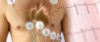
- Diagnostics
- Treatment
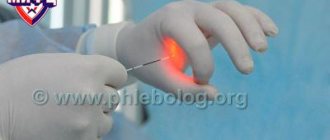
Caisson disease is a pathological condition in which gas bubbles form in the vessels and tissues of the body.
This occurs due to the rapid decrease in atmospheric pressure. Otherwise, the disease is called decompression sickness (DCS). The name "caisson" comes from the word "caisson". This device was invented in the 19th century for underwater work. The design was a chamber in which a person descended under water. At first, decompression sickness was diagnosed by underwater specialists. Over time, its distribution became wider. Sometimes this condition occurs in pilots who, when changing their flight altitude, are exposed to changes in atmospheric pressure. However, divers are most susceptible to this disease. Fans of scuba diving cannot always cope with the transition from high pressure to normal, which is why they develop “diver’s disease.” According to statistics, up to 4 cases of decompression sickness are recorded per 10 thousand dives. It can be not only acute, but also chronic.
To prevent the disease, you should use high-quality breathing mixtures when diving, avoid a sharp rise from the depths to the surface, observe intervals between dives or flights, and undergo preventive examinations if a person is working underwater.
How does decompression sickness occur?
The main reason for the formation of air bubbles in organs and tissues is a sharp decrease in atmospheric pressure when rising to a height or the surface of the water after a dive. However, there are factors that increase the risk of developing “diver’s disease”:
- Age-related changes. With age, it becomes more difficult for the heart and lungs to cope with stress, so decompression sickness is more common in middle-aged and mature people than in young people.
- Hypothermia. Cold impairs blood supply to organs and tissues. This is especially true for peripheral vessels. Because of this, the pulmonary vessels receive less blood, which leads to gas retention and the formation of bubbles.
- Increased blood viscosity. This condition occurs when dehydration occurs. Blood flow slows down, and blood stagnation occurs in peripheral vessels.
- Intoxication. Drinking alcohol before diving is life-threatening. Alcohol provokes dehydration, and when there is alcohol in the blood, air bubbles become larger and can clog the lumen of the vessel.
- Overweight. If the body contains a high percentage of adipose tissue, bubbles form faster due to the hydrophobicity of the fat. In addition, fats tend to dissolve inert gases from the breathing mixtures used by divers.
- Increased carbon dioxide concentration. This condition is called hypercapnia. It occurs when using poor quality mixtures or when breathing improperly under water. As CO2 concentration increases, more inert gases dissolve in the blood.
- Exercise stress. During exercise, blood flow becomes uneven. Gases in the blood dissolve more intensely and air bubbles appear. As a rule, they are very small in size and localized in the joint area. On subsequent dives, decompression sickness may become more severe.
1.General information
Caisson, or decompression sickness, is the notorious “caisson” that cost the health (and sometimes the lives) of many, many divers, scuba divers, submariners, high-altitude pilots and representatives of other professions associated with changes in environmental pressure and inhaled air.
The essence of this dangerous condition is the intense release of gas with the formation of many bubbles in the blood vessels and tissues. When such bubbles merge or group together, an embolism (blockage) may occur; in addition, increased mechanical pressure from the “gas clusters” often leads to damage to conductive nerves, muscle fibers and, in general, internal organs (the ligamentous-articular apparatus and the central nervous system are primarily affected).
The incidence of decompression sickness is currently 2-4 cases for every 10,000 dives to depth, rapid ascents to altitude, stays in caisson chambers, etc.
A must read! Help with treatment and hospitalization!
Consequences of decompression sickness
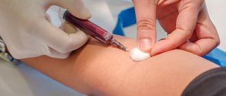
When immersed in water, atmospheric pressure increases. Because of this, the gases of the respiratory mixtures dissolve in the blood of the capillaries of the lung tissues. After ascent, when the pressure returns to normal limits, the opposite phenomenon occurs. Gases dissolved in the blood form bubbles. If a diver ascends quickly, that is, the body does not have time to adapt. If the rate of ascent is not observed, the blood seems to “boil.” At this moment, not only small but also large bubbles are formed. They attract platelets to themselves, increasing in size. These compounds can cause thromboembolism - blockage of the lumen of the vessel.
When a large number of such bubbles with platelets appear in the blood, a gas embolism develops. Circulating through the bloodstream, these compounds can damage the walls of blood vessels, causing hemorrhage.
In addition to blood vessels, bubbles can be found in joint cavities and soft tissues. Gas compounds compress nerve endings, causing pain throughout the body. Foci of necrosis may also occur in the muscles and internal organs, which is also caused by compression.
Text of the book “On the Edge of Possibility: The Science of Survival”
Bubbles in the blood
The cause of decompression sickness was discovered in 1878 by the French scientist Paul Ber. He proved that "writhing" occurs when a diver or caisson worker breathing compressed air rises too quickly to the surface, and then gases dissolved in the blood and tissues are released in the form of bubbles, blocking the blood vessels. The gas inhaled under pressure dissolves in body fluids in a larger volume: for example, for every 10 m of descent, an additional liter of nitrogen is absorbed (as we will see below, this process is not fast). As long as the gas is present in liquids and tissues in a dissolved state, excess does not create problems. The difficulty arises from the insufficient rate of removal of dissolved gas during decompression. If a diver rises to the surface slowly, the excess gas dissolved in the blood is expelled by the lungs when exhaling and does not pose a danger, but if the ascent occurs quickly, the lungs simply do not have time to remove the gas out, so the tissues and blood become oversaturated and at some point the gas breaks out of the solution in the form of bubbles{13} 13
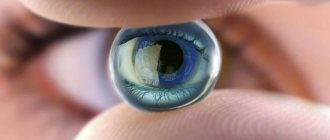
One of the first to describe this phenomenon was Robert Boyle, who in 1670 observed the formation of an air bubble in the eye of a viper during decompression.
[Close]. This phenomenon is familiar to anyone who has opened a bottle of sparkling water (or champagne): as soon as the pressure disappears, chains of bubbles rush out. If the cap is pulled off abruptly (fast decompression), the effect will be more impressive than if the cap is gently unscrewed and the gas is released slowly. However, if carbon dioxide is dissolved in carbonated water and champagne, then in divers breathing compressed air, bubbles in the blood are formed primarily by nitrogen, since the content of carbon dioxide is extremely low, and oxygen is quickly consumed by the tissues.
Why do sperm whales not suffer from decompression sickness?
Many marine mammals are capable of diving to depths inaccessible to humans. A dead sperm whale was once found at a depth of 1134 m, where it had caught its lower jaw on a transatlantic cable. Elephant seals are even more skilled divers, the record level they have reached is 1570 m, at this depth the pressure is 150 times higher than the pressure on the surface. This is far beyond human capabilities. In addition, elephant seals can dive repeatedly without experiencing any ill effects. In fact, the elephant seal would be more accurately called a “floater” rather than a diver, since it spends 90% of its time underwater. One of the elephants spent no more than six minutes on the surface during 40 days of observation. How do sperm whales and elephant seals avoid decompression sickness?
The thing is that marine mammals have developed a way to reduce the amount of nitrogen dissolved in the tissues of the body. Unlike humans, elephant seals and sperm whales exhale before diving. In this way, they limit the supply of air that they take with them, so somewhere at a depth of 50 m the alveoli are already completely compressed and no additional gases penetrate into the bloodstream. Pressure at depth forces the sperm whale's lungs themselves to completely compress, displacing all the air into the upper respiratory tract, which is reinforced by round cartilage discs and is less compressible. Blood flow to the lungs is also significantly reduced. Thus, during a dive, the gas practically does not enter the blood from the lungs, and the residual amount of nitrogen dissolves in body fluids, so when ascending, the formation of bubbles in the blood of the mammal does not threaten.
The formation of bubbles in the blood is fraught with serious consequences. Once formed, they continue to grow due to new portions of gas. As a result, they grow to such a size that they clog the thinnest blood vessels and prevent blood flow to the tissues, causing a lack of oxygen and nutrients. As a result, the cells die. In addition, air bubbles can activate blood cells that respond to air flow, such as platelets, which are involved in the formation of blood clots. Finally, the formation of bubbles within tissues can lead to deformation or rupture of tissue cells and impair their function.
Divers have developed a rich vocabulary that describes the symptoms of bubbles in various tissues. “Choke” – interruptions in breathing when large bubbles get stuck in the capillaries of the lungs, reducing the surface area necessary for gas exchange and causing sensations similar to asphyxia. “Wobble” occurs due to bubbles in the vestibular apparatus, which is responsible for balance. Bubbles in the knee and shoulder joints (the places most vulnerable to decompression sickness) lead to “writhing.” Being in the spinal cord, they lead either to numbness of the limbs or to paralysis, and in the most serious cases they can provoke degeneration of nerve fibers. Their appearance in the brain leads to speech and vision disorders, sometimes irreparable.
There is one curious story (possibly fictitious) about how, when digging one of the first tunnels under the Thames, the management decided to mark the passage to the middle mark with a dinner party directly in the tunnel. Since construction had not yet been completed, compressed air was supplied to the tunnel, and the guests had to dine “under pressure.” Much to their disappointment, the champagne did not shoot or “play” when opened, since the pressure in the bottle turned out to be the same as in the tunnel. And yet they drank champagne. The champagne that was drunk began to sparkle only later, when the leaders and guests came to the surface...
You need to climb slowly
Soon the caisson workers themselves discovered that increased atmospheric pressure compared to their working conditions relieved unpleasant symptoms. This gave Sir Ernest Moir the idea of a recompression chamber to treat decompression sickness. A similar camera was first used around 1890 in the construction of the Blackwall Tunnel under the Thames and the East River Tunnel in New York, where it proved its worth. However, the injured worker had to spend more than one hour in the cell to recover. It was clear that efforts should be directed, first of all, to the prevention and prevention of the disease. Thanks to the work of Paul Beer, the solution became obvious: the diver or caisson worker must ascend (or decompress) slowly enough to allow the lungs time to expel the gas dissolved in the blood. The most difficult thing remained - determining a safe ascent speed. By 1906 the problem had become so acute that the British Navy turned to Professor John Scott Haldane of Oxford University, a physiologist already known for his research on respiration (see Chapter 1), for help.
Together with Lieutenant G. Damant and Professor A. Boycott, Haldane conducted a series of experiments at the Lister Institute in London with a large steel chamber in which the pressure could be easily adjusted. During experiments on goats, it turned out that with a sharp decompression from 6 to about 2.6 atmospheres, nothing bad happens to the animal. However, if the pressure was reduced by the same amount, but from 4.4 to 1 atmosphere (i.e., to sea level), things took a different turn. Only 20% of the animals managed in this case to avoid decompression sickness, which sometimes took the most severe forms, including death. After some trial and error, the researchers found that they could quickly reduce the absolute difference in pressure by up to half, but then they had to reduce the difference as slowly as possible. Thus, the maximum diving depth that did not require decompression was identified - 10 m (pressure of 2 atmospheres). As has long been customary among physiologists, the researchers then conducted the test on themselves, fortunately, without consequences. The final stages of the experiment were carried out at sea off the Isle of Bute, off the western coast of Scotland, from the HMS Spanker. Haldane took the whole family to the sea and allowed his 13-year-old son Jack, who later also became interested in studying respiratory processes, to dive to a depth of 12 m {14} 14
As J. B. S. Haldane later wrote, it was still entertainment. The sleeves of the wetsuit ended with tight rubber cuffs that did not allow water to pass through. But the suit turned out to be too big for the boy, so water seeped inside and filled the suit up to the neck. Fortunately, the air pumped into the helmet did not allow the water to rise higher, but John was thoroughly chilled.
[Close].
Haldane was aware that the rate of dissolution of nitrogen in different tissues varied. Fat cells, for example, have a greater storage capacity, while brain cells store less nitrogen (this, in turn, means that women and obese people take longer to decompress than the average man). In addition, the rate of nitrogen accumulation depends on the rate of blood flow to tissues, so tissues with lower blood supply accumulate nitrogen more slowly. Thus, it takes more than five hours to completely saturate the body with nitrogen. During decompression, nitrogen dissolved in fluids and tissues must be eliminated through the bloodstream. The safe rate of its removal depends on the storage capacity and the rate of blood supply to various tissues, that is, simply put, the longer the gas accumulates, the longer it takes to be eliminated. It follows that the optimal situation for a diver is a quick dive, a limited time at depth, then a slow, gradual ascent to the surface.
The rapid dive recommended by Haldane and his colleagues was contrary to accepted practice, but it was quite justified from a physiological point of view: the less time a person spends at depth, the less gas will have time to dissolve in the tissues. During the first, fast stage of the ascent, the diver must overcome half the depth - this, as experiments have shown, is completely safe. The ascent should then proceed smoothly, with stops for a certain time at a certain depth to ensure gradual decompression. The meaning of this phasing is that the gas always increases in volume the same way, regardless of whether the pressure drops from eight atmospheres to four or from two to one (recall that the product of pressure and volume is a constant value, therefore, when the pressure is halved the volume will double). The research gave divers the benefit of a quick and unimpeded ascent to half depth, thereby allowing more time for decompression during further ascents. As Haldane himself noted, “the traditional method of recovery is ‹…› unduly slow at the beginning and dangerously accelerated towards the end.”
By 1908, Haldane and his colleagues had provided the Navy with detailed decompression tables outlining how long a diver should stay at a certain depth during a gradual ascent, depending on the depth and duration of the dive. Thanks to these tables, the number of cases of decompression sickness decreased sharply; they were observed only when the diver, for some reason, neglected the recommendations and ascended faster than prescribed. Not everyone immediately realized the importance of Haldane's research. As he himself said ten years later: “It is a great pity that in some countries it is not possible to introduce staged decompression due to the rigid rules of old-fashioned ascent either gradually or slowly at the beginning and accelerating as one approaches the surface and atmospheric pressure.” Fortunately, the results of his research spoke for themselves, and now the Haldane method is widely used. Nevertheless, tragedies still occur - as a rule, in case of neglect of recommendations. Among the most notorious accidents is the death of Chris and Chrissy Rous, quite experienced divers who died from decompression sickness in 1992 while examining a sunken German submarine.
It is interesting to compare how much time it took for caisson and tunnel workers to decompress in the past and how much time Haldane and his colleagues spend on decompression. The caisson workers, exposed to three times the atmospheric pressure (i.e. 3 bar), rose to the surface in ten minutes or less. Haldane recommended that after three hours of work, decompression should be at least an hour and a half. It is not surprising that so many caisson workers suffered from cramps.
In addition, divers are not recommended to remain in the air for some time after diving, since the pressure in an airplane is less than at sea level (see Chapter 1), and a further decrease in pressure can also cause the formation of bubbles in the blood. After a single dive, the diver must refrain from flying for 12 hours, and even longer after multiple dives or dives requiring gradual decompression. Marine recreation enthusiasts who are not familiar with the problems of decompression can develop decompression sickness if, after swimming with scuba gear in the morning, they fly home in the afternoon. Even military pilots flying unpressurized fighter jets risk succumbing to decompression sickness if they climb too quickly from sea level.
Scuba diving and decompression sickness
Divers without special equipment who immediately dive to great depths do not suffer from decompression sickness, since they do not stay at depth for long and the amount of nitrogen that is dangerous for ascent does not have time to dissolve in body fluids. Repeated deep dives are a completely different matter, as military doctor P. Pauleu, who served in the Danish Navy, found out from his own experience. In the early 1960s, he completed about 60 two-minute dives at intervals of one or two minutes in a submarine evacuation training tank (depth - 20 m). About half an hour after the final dive, he felt a sharp pain in his left thigh. At first he decided not to pay attention to her, but two hours later he began to experience severe chest pain, fog in his eyes, shortness of breath, and his right arm became paralyzed. In a state of painful shock, he was discovered by a colleague, who immediately placed him in a compression chamber, lowering the pressure in it to six atmospheres. The symptoms quickly passed. The subsequent decompression took over 19 hours, but, fortunately, Pauleu made a full recovery and subsequently described everything that happened to him.
Pearl divers on the Tuamotu Islands in the Pacific Ocean also often fall into a state similar to what Dr. Pauleu suffered. In their language it is called "tarawana" and translates as "crazy fall", and symptoms range from visual disturbances to loss of consciousness. Sometimes divers experience paralysis or even death (after all, unlike Dr. Pauleu, they do not have a decompression chamber). As one of the guests of the archipelago noted: “On the shore of any island, the largest group of buildings will most likely turn out to be a cemetery for dead divers.” Taravana is a common disease and is greatly feared. In one day alone, 47 of the 235 divers developed symptoms, some very severely, as six were paralyzed and two died. Fortunately, such extreme manifestations do not occur every day, but the incidence rate is still very high.
Although the etiology of taravana remained a mystery for many years, the work of Pauleu and his followers suggests that it is a type of decompression sickness. Tuamotu divers do not spare themselves, making two-minute dives to depths of up to 40 m (pressure - 5 bar). They make from 6 to 14 dives per hour with a scanty interval of 4–8 minutes. During this time, nitrogen, dissolved in tissues during a dive, does not have time to be eliminated from the body and accumulates with each new dive, therefore causing decompression sickness during ascent (taravana has never been observed at depth, only on the surface). It should be feared primarily by those who make repeated dives at short intervals. It should be noted that on the neighboring island of Mangareva, where the taravan has not even been heard of, tradition tells the diver to spend at least ten minutes on the surface between dives.
At the entrance to the water
Decompression sickness is not the only difficulty a diver faces. Even simply immersing the body in water up to the neck already causes physiological changes. When you stand on the seashore, the blood tends to your feet under the influence of gravity. If you are submerged in water up to your neck, the external pressure of the water will cause about half a liter of blood to rush up to the chest, filling the large veins and the right atrium, and increasing the volume of blood flow. The stretching of the atrium wall signals two hormones that affect the kidneys' water absorption and urine production. This is why we often want to go to the toilet after diving into water.
Ama - Japanese divers
The most famous divers in the world are the Japanese ama, who collect seafood (clams, sea slugs, octopus, starfish and algae) from the seabed. In Japan, unlike Western cuisine, all this goes into food. In addition, the Ama collect pearl shells called akoya-gai, which are used to grow artificial pearls. The profession of ama divers has existed for more than 2,000 years. This traditionally female activity is immortalized in prints by artists of the ukiyo-e school, depicting beautiful topless girls diving for the most valuable shells of the abalone shellfish. The engravings, however, somewhat embellish the reality, since ama work until they are 50 years old. And their work is not all sugar. This is how Sei-Syonagon, a court lady of the Japanese Empress Sadako, describes it: “The sea frightens even the prosperous. What horror must the unfortunate divers feel who have to plunge into the abyss for the sake of a piece of bread. It’s best not to even think about what will happen if the cord tying a diver’s waist breaks. While the women are underwater, the men sit in their boats and sing songs so as not to get bored, watching the crimson cord floating on the surface. An amazing sight is the complete indifference of men to the danger threatening women. Before rising to the surface, the diver pulls the cord, and the men, with haste that I understand, pull her out of the water. And now the diver is already clinging to the side of the boat, convulsively gasping for air. Even an outside observer cannot help but cry at the sight of this picture, and there is hardly a person who dreams of such work.”
Girls watch divers on Enoshima. From a triptych painted by the great ukiyo-e artist Utamaro around 1789.
The description of Sei-Syonagon is still relevant today, although a lot of water has passed under the bridge since then.
There were once thousands of ama in Japan (the 1921 census, for example, recorded 13,000), but their numbers have declined sharply in recent years. By 1963 it had dropped to 6,000, and now there are barely more than a thousand. Most modern ama are already aged, since few young people are attracted to such exhausting work. In addition, many types of shellfish are now grown artificially. Apparently, the ama profession will soon die out, surviving as a sad echo only in the names of villages like Amamati.
It so happens that among the Ama there are two varieties - katido and funado. Katido are young girls, students who dive without assistants to a depth of 5–7 m and spend about 15 seconds at the bottom. Although a katido can make about 60 dives per hour, it is not at risk of decompression sickness due to the shallow diving depth. The most experienced and skillful divers are funados, who dive to much greater depths - on average about 20 m. As can be seen from the Sei-Syonagon description, each funado has an assistant in the boat. After taking a series of rapid breaths to fill its lungs with air, the funado dives vertically to the bottom with a heavy load in its hands, squeezing its legs tightly for better streamlining. At the bottom, she releases the weight and begins to collect her prey in a small wicker basket. Before surfacing, she pulls a cord attached to the weight, and an assistant pulls the diver out by a rope tied around her waist. Each dive lasts about a minute, and half of that time is spent at depth. Between dives, the funado also rests for about a minute in the water, at the side of the boat. An experienced diver makes about 50 dives in the morning, then the same number in the afternoon, however, like a katido, after a series of dives she needs rest.
Caisson disease is not common among the Ama, but they suffer from ear diseases much more often than representatives of “land” professions. According to a 1965 study, 60% of funados experience hearing loss after the age of fifty. Other common ailments include ringing in the ears and ruptured eardrums.
Physiologically, women are better suited for the role of divers - they can hold their breath longer and freeze less in the water, but it is unlikely that only for these reasons all amas are exclusively female.
Even by simply dipping our face into water, we thereby cause a physiological reaction in the body - the heartbeat slows down. This phenomenon is known as the diving reflex, and although not very developed in humans, it is extremely important for marine mammals such as seals, as we will see below. You can verify the existence of this reflex yourself by immersing your face in a bowl of cold water and asking one of your friends to count your pulse and compare it with normal. However, this experiment does not always work, since nervousness (or excitement) causes the release of adrenaline, which increases the heart rate.
When we surface, the body is deprived of water support, and the blood is again redistributed from the chest to the legs. This must be taken into account. History knows many cases when drowning people rescued by helicopter developed collapse after rising from the water: a person stays afloat quite actively, and after being lifted into the helicopter he suddenly experiences cardiac arrest. Physiology came to the rescue here too, proving that when immersed in water, blood flows out of the legs and they cool more than the upper part of the body. Until recently, people were rescued from the water in an upright position by threading a rescue belt under their arms. As a result, when pulled out of the water, the blood instantly rushed to the legs, where it immediately cooled and, returning to the heart, caused it to stop. Lifting in a horizontal position helps to avoid this, using a second belt that wraps around your legs. In this case, blood redistribution does not occur. It is also important to keep the person lying down until the limbs warm up evenly. Since the British Water Rescue Service adopted this method, the number of cases of cardiac arrest after water rescue has dropped sharply.
Severity of decompression sickness
Depending on the manifestation of symptoms, DCS is divided into four degrees of severity:
- Easy. With a mild degree of pathology, the patient experiences pain in the muscles and joints, which is associated with pressure on the nerve endings of air bubbles. Due to blockage of superficial vessels and sweat glands, the skin begins to itch and becomes oilier.
- Average. Moderate pathology causes deterioration in coordination of movements, blurred vision, and disruption of the gastrointestinal tract. This is due to the accumulation of gases in the vessels of the mesentery and intestines.
- Heavy. The main symptom of the pathology is damage to the spinal cord due to compression of the nervous tissue. In some cases, the brain is involved in the pathological process. This is manifested by disturbances in the functioning of the heart and respiratory system.
- Lethal. Decompression sickness can be fatal if large bubbles clog vital vessels. The patient's blood supply to the lung tissue stops and acute heart failure develops.
Even with moderate severity of the pathology, acute “diver’s disease” can lead to severe damage to organs and systems. If left untreated, these conditions are life-threatening.
Book "Eye Diseases"
Chapter 19
- Exposure to ultraviolet and infrared rays
- Exposure to chemical factors
OCCUPATIONAL DISEASES OF THE VISUAL ORGAN
Occupational diseases are considered to be those pathological processes that arise under the influence of certain factors in the external production environment.
Occupational diseases of the body, incl. eyes, are caused by physical factors (ultraviolet, infrared, ionizing, light radiation, etc.), chemical and physico-chemical (industrial poisons - lead, silver, arsenic, trinitrotoluene, naphthalene, etc.) and less often biological factors (infections, invasions) . Occupational injuries and burns were discussed in the corresponding chapter. Occupational hazards have a multifaceted effect, affecting the entire body with various changes in individual organs and systems.
<< CONTENTS
Eye damage may be the leading symptom in some cases. Eye damage from radiant energy is observed in welders, smelting shop workers, electrical engineers, people involved in industrial radio and radiography, and medical personnel working in X-ray and laser rooms.
Ultraviolet rays cause electroophthalmia, which is expressed in the appearance of acute pain, blepharospasm, lacrimation and hyperemia of the conjunctiva, pericorneal injection, corneal edema and minor erosions.
Treatment: cold lotions with water, instillation of 0.5% dicaine solution, 0.1% adrenaline solution, 20% sodium sulfacyl solution, Actovegin jelly, solcoseryl.
For prevention - shields, helmets with appropriate light filters, light-protective glasses, portable protective screens, worker training, sanitary education.
Infrared rays most often cause clouding of the lens. This lesion occurs in smelters, metallurgists, steelworkers, glassblowers, etc. First, dust-like deposits appear at the posterior pole, which merge and then spread anteriorly along the axis of the lens, gradually capturing the entire lens. With thermal cataract, the zonular plate of the anterior lens capsule peels off and upon examination it looks like a film floating in front of the lens. This is a characteristic sign of thermal cataracts. When there is significant clouding of the lens, it is difficult to distinguish thermal cataracts from senile cataracts.
Treatment: in the initial stage, medication, and then, if indicated, surgery. Infrared irradiation can lead to swelling and hemorrhages of the retina, hemorrhages into the vitreous body. Prevention – wearing protective glasses and filters, eliminating occupational hazards.
Ionizing radiation causes damage to eye tissue both through direct exposure and through general exposure to the body. The lens is especially sensitive to this radiation. Radiation cataract resembles thermal cataract. In addition, ionizing radiation can cause dermatitis, swelling of the eyelids and loss of eyelashes, infiltration, ulceration and necrosis of the conjunctiva, keratitis, edema, hemorrhages and degenerative foci in the retina, secondary glaucoma.
Treatment is the same as for the same lesions caused by other reasons. Prevention – possible elimination of occupational hazards, preventive medical examinations of persons working in conditions of these hazards.
Light of high brightness (when exposed to high-power artificial light sources and when observing a solar eclipse without eye protection) causes changes in the retina. Direct sunlight burns the macula area, resulting in retinal edema, then pinpoint lesions that reduce central vision, and a central scotoma appears.
Ultrasonic waves are used in various fields of science and technology, as well as in medicine. The intensity of the waves depends on the distance to the irradiation source, the degree of absorption of the medium and the frequency of ultrasound vibrations. The biological effect is manifested in changes in the mechanical, thermal and chemical state of the irradiated tissues and depends on the intensity of the energy used, the frequency of vibrations, the duration of exposure and the properties of the tissues. At moderate intensity of ultrasonic waves, hyperemia and swelling of the conjunctiva and swelling of the corneal epithelium appear, which can result in the formation of a cataract. At low ultrasound frequencies, cavernous (perenuclear) cataracts form within 2 minutes. These opacities represent an accumulation of air bubbles and, after the cessation of the ultrasound, quickly resolve. There may be irreversible liquefaction of the vitreous body and with ultrasound exposure of 2 MHz or more (i.e., high frequency), irreversible clouding of the lens also appears after 2-3 minutes. Cataracts are associated with an increase in the temperature of the lens. Treatment of cataracts is surgical. Prevention: strict adherence to safety precautions when working with high-power generators.
Ultrashort waves are electromagnetic waves. Electromagnetic waves of different ranges are used in radio engineering, radar, radio electronics, and medicine. They propagate at the speed of light waves. Part of the active energy is reflected from the surface of the human body, and part is absorbed. Waves in the centimeter range (and the wave range from 1 m to 1 mm - decimeter, centimeter, millimeter - corresponds to an ultra-high frequency from 300 to 3000 MHz) have a cataractogenic effect on the lens. The energy absorbed by the body causes thermal and specific biological effects. An increase in the power of the electromagnetic field and the duration of its exposure has a more intense biological effect. In addition to clouding of the lens, there may be angiopathy of the retinal vessels, combined with arterial hypotension. Treatment is medication and then surgery – cataract removal. Prevention - shielding of installations, safety glasses, periodic medical examinations, in the case of initial cataracts after 6 months, with progression - transfer to another job.
Currently, optical quantum generators—lasers—are widely used in medicine. When working with lasers, subcapsular opacities of the lens are formed. The eye is mainly affected by reflected light rays. In the retina, under the influence of direct laser beams, degenerative processes can also develop in the cornea.
CHANGES IN THE VISUAL ORGAN CAUSED BY EXPOSURE TO CHEMICAL FACTORS
Benzene and its compounds. In the rubber, chemical, pharmaceutical, printing, paint and varnish industries, as well as in the production of synthetic rubber, artificial leather, benzene is used as a raw material for the production of paints, explosives and medicinal substances. It enters the body in the form of vapor through the lungs and through the skin. When exposed to benzene, acute and chronic intoxication can occur. In case of poisoning with nitro derivatives of benzene, anemia develops due to the destruction of red blood cells and hemoglobin and the formation of methemoglobin. The liver is affected. Benzene is excreted unchanged through the lungs and oxidized to phenol and dioxybenzene in the urine. Violation of accommodation is an early symptom of intoxication; then retinal hemorrhages, retrobulbar neuritis and visual acuity may decrease. In case of nitrobenzene poisoning, there may be dilatation of the retinal veins, the phenomenon of congestive optic disc, photophobia, conjunctival hyperemia, conjunctival injection of the eyeball, keratoconjunctivitis, pupil dilation, changes in the fundus in the form of a dark red color change, blurred contours of the optic disc, narrowing retinal vessels can be caused by poisoning with aniline vapors. General symptoms of benzene poisoning are fatigue, drowsiness, headaches, dyspeptic disorders, loss of appetite, prolongation of bleeding time, acceleration of ESR. Differentiate with diseases of the blood system, in which leukopoiesis changes, phenomena of hemorrhagic diathesis and anemia. Treatment is restorative and symptomatic.
Intoxication with dimethyl sulfate is caused through the respiratory system, and the vapors of this substance also have a toxic effect on the eye. On the part of the eyes, subjective sensations arise 3-16 hours after poisoning. Photophobia, lacrimation, blepharospasm, pain, swelling and hyperemia of the conjunctiva appear, and sometimes the cornea becomes dull. There may be chemosis of the conjunctiva, swelling of the corneal epithelium, the appearance of bubbles and erosions, and precipitates. Liquid dimethyl sulfate causes severe burns if it comes into contact with the eyes. Treatment. The patient is immediately taken out into fresh air, then placed in a darkened room. Providing assistance and treatment if a substance gets into the eye, as with chemical burns. Prevention is the strict implementation of safety precautions.
Arsenic and its compounds. Arsenic compounds are very toxic (arsenic disulfide, sodium arsenous, calcium, calcium arsenate, lead arsenate, arsenous anhydride, arsenic acid salts), and trivalent compounds are more toxic than pentavalent ones. Arsenic and its compounds enter the body through the gastrointestinal tract and respiratory system. It is able to linger in the body and form depots in the bones, liver, stomach walls, kidneys, skin, hair, nails, and brain tissue. In nervous tissue it causes disruption of metabolic processes. The clinical picture is accompanied by redness and peeling of the skin of the eyelids, and their swelling. Necrotic areas of the conjunctiva, similar to Bitot's plaques, may form. With large doses, keratitis is possible, and with general intoxication, damage to the optic nerve is possible. In this case, a concentric narrowing of the visual field appears, and then a decrease in visual acuity. Sometimes, bilateral damage to the optic nerve is the only sign of poisoning and is expressed by a picture of neuritis with subsequent atrophy. There may be retinal edema, vitreous opacification, uveitis, arsenic polyneuritis, changes in the skin and nails, the digestive tract, sometimes ataxia and deep sensitivity disorder. In the treatment of acute and chronic poisoning, specific antidotes are used - British anti-lewisite (BAL) in the form of intramuscular injections of a 5-10% solution in nut oil, at a dosage of 2.5 mg/kg. The first 2 days are administered 4 times a day, in the next 8 days - 2 times. A 5% solution of unithiol is also prescribed, 5-10 ml intramuscularly or orally, 0.5 ml 2 times a day, 3-4 days. Intravenous 30% sodium thiosulfate solution, 10-15 ml. Vitamins B1, B6, B12 are used. The conjunctival cavity is washed with water and Vaseline or other indifferent oil is instilled. For conjunctivitis and keratitis, a 5% solution of British anti-lewisite in the form of drops is prescribed. Citral is instilled. You should not use lead lotion or cocaine on your skin as a painkiller.
Nicotine and its compounds. In agriculture and forestry, nicotine is used to control plant pests in the form of nicotine sulfate, nicotindust and nicotine base. Nicotine is found in the air of drying and other departments of tobacco factories in excess of permissible concentrations (acceptable - 0.1 mg/m3).
Absorbed through the lungs, mucous membranes and damaged skin, excreted by the kidneys, lungs, salivary and sweat glands. Has a toxic effect on cells of the central and autonomic nervous system. In acute intoxication, visual functions are temporarily reduced and miosis is observed. There is a burning sensation in the mouth, heartburn, nausea, vomiting, salivation and sweating, severe headache, weakness, difficulty breathing, pain in the heart, a drop in cardiac activity, convulsions, delirium, a decrease in body temperature, sometimes unconsciousness and coma.
Changes in the organ of vision during chronic intoxication develop slowly. The papillomacular bundle is selectively affected, retrobulbar neuritis develops, a central scotoma appears in red and green, and visual acuity decreases somewhat. In the fundus there may be slight hyperemia of the disc, in the later stages there may be temporal pallor, a bilateral process. There is a decrease in the sensitivity of the cornea.
Treatment boils down to gastric lavage with an aqueous suspension of coal in case of ingestion of nicotine, laxatives, or subcutaneous apomorphine. Inside, a 0.5% solution of tannin or 5-10 drops of iodine tincture to promote the loss of nicotine in the form of a difficult-to-absorb sediment. Heart remedies, strong sweet tea, coffee. For bradycardia - atropine subcutaneously, for pain in the heart - 1% aqueous solution of sodium nitrate orally. For chronic poisoning - elimination of occupational hazards, vitamins B1, B6, B12, ATP.
Lead and its compounds. Lead compounds used in industry are toxic and enter the body in the form of vapor or dust through the respiratory tract, and from the lungs into the blood. Tetraethyl lead can be absorbed through the skin. Along with other symptoms of lead poisoning (blue-black lead rim on the gums, chronic catarrhal gingivitis, lead deposition in the form of individual black-blue spots on the gums, cheeks, tongue, lips, palate, ulcerative stomatitis, intestinal colic, difficulty moving the finger joints and hands, sclerosis of the aorta and arteries, damage to the central nervous system, anemia, porphyrinuria) damage to the external muscles of the eye, ptosis, nystagmus, and sometimes disturbance of accommodation are observed. In severe cases, retinopathy appears with periarteritis, hemorrhages, exudate, more often with kidney damage and arterial hypertension. The typical lesion is retrobulbar neuritis with a central scotoma and slight narrowing of the visual field. As the process progresses, optic nerve atrophy develops. In some cases, in severe cases, lead amaurosis develops, apparently associated with vasospasm, which can last up to several days. Then vision is completely restored. As a result of lead encephalopathy, congestive discs may develop.
Tetraethyl lead intoxication leads to increased intraocular pressure. There is no glaucomatous excavation, visual functions are preserved, unlike primary glaucoma. Iphthalmotonus normalizes after the cessation of the toxic factor. Treatment consists of immediately eliminating contact with lead and its compounds. Substances that form low-toxic complexes with lead that are easily removed by the kidneys (for example, EDTA) are used. A diet rich in calcium is recommended - milk, fruits, vegetables. Inside - sodium thiosulfate, potassium iodide, phosphoric acid, bile salts. Sulfur baths are recommended. For anemia - ascorbic acid with iron supplements. For severe damage to the central nervous system and cerebral edema - lumbar puncture, for inflammation of the kidneys - sodium bicarbonate 20-30 g in 4-5 doses daily. Prevention is the reduction of occupational hazards and mandatory periodic medical examinations of workers.
Silver - causes argyrosis of the eyes, as well as general argyrosis. It develops in people who come into contact with silver for a long time while doing their work. Penetrates through the lungs, digestive tract and mucous membrane of the eyes. Silver albuminate is deposited in the elastic fibers of the skin and the mucous membranes of internal organs, giving them a dark color.
The conjunctiva, more in the lower half of the eyeball, the lower eyelid and the lacrimal caruncle darkens. With general argyria, argyrosis of the cornea appears. With prolonged exposure, silver is deposited under the anterior capsule of the lens, in the vitreous body, in the tissue of the retina and optic nerve. As a result, degeneration of nerve cells, the appearance of hemeropia and decreased visual acuity.
Because Treatment is ineffective, so the main thing is prevention.
Carbon disulfide - enters the body through the respiratory system and skin, it is easily soluble in lipids and easily penetrates the blood. Carbon disulfide causes degeneration of cells of the central nervous system, being a neurotropic poison. It is excreted unchanged with exhaled air, and inorganic sulfates formed during oxidation with sweat, feces and urine. Under the influence of carbon disulfide, retrobulbar-type neuritis develops, with a central scotoma, and sometimes simultaneous narrowing of the peripheral boundaries of the visual field. Color perception is impaired, especially for red. There is also paralysis of the external eye muscles, paralysis of accommodation, nystagmus, decreased dark adaptation, and there may be pinpoint superficial keratitis. In case of chronic intoxication with carbon disulfide, vegetative-vascular disorders, cerebral disorders, and disturbances in the main types of metabolism are observed. First aid for acute carbon disulfide poisoning is rest, clean air, camphor, caffeine, oxygen. For chronic intoxication, intravenous infusion of 40% glucose solution, intramuscular vitamins B1, B6, subcutaneous injections of proserin according to the scheme, internal neuroprotectors, sedatives, cardiac drugs, physiotherapeutic, sanatorium treatment.
Carbon disulfide is used in the viscose industry, in the cold vulcanization of rubber, to control agricultural pests, as a solvent for rubber products, etc.
Eye damage from mercury may be the first and even the only symptom of general intoxication. Mercury is used in industry in the form of metallic mercury and its compounds sublimate, calomel, mercury fulminate, and mercury nitrate. Mercury poisoning occurs in people involved in the production of barometers, aerometers, thermometers, X-ray tubes, electric lamps, in chemical and physical laboratories, in mines, in the pharmaceutical industry, etc. Mercury vapor is particularly toxic. They enter the body mainly through the respiratory tract, enter the blood and can circulate in the body for a long time. They form depots in the liver, kidneys, spleen, and brain tissue. Excreted through the kidneys, intestinal mucosa, salivary, sweat, mammary glands and bile. In case of poisoning, sleep and the emotional sphere are disturbed, and tremors appear, because mercury affects the autonomic parts of the central nervous system, general malaise, headache. With chronic intoxication, mercury is deposited in the lens and this may be the only symptom of chronic poisoning, retrobulbar neuritis develops, and mercury stomatitis develops in the oral cavity. First, damage to the gums is noted, then the interdental gingival papillae become covered with a grayish-white coating, gradually become necrotic, emitting a fetid odor, and hypersalivation. Regional lymph nodes are enlarged. Teeth marks appear on the mucous membrane of the cheeks and the lateral surface of the tongue due to swelling. There is a metallic taste in the mouth and throbbing pain. In case of mercuric chloride poisoning, the main symptoms of eye damage are hemorrhages and atrophic lesions in the retina.
Treatment is aimed at removing mercury from the body, a 30% sodium thiosulfate solution is injected intravenously along with a 40% glucose solution, four-chamber sulfur baths, diuretics, laxatives, unithiol, the introduction of table salt into the body, and general health measures. Prevention - measures aimed at creating normal production conditions (ventilation, automation and sealing of production processes, etc.).
Just as with mercury poisoning, eye damage from trinitrotoluene may be the first sign of general intoxication.
The drug penetrates the body through the skin, respiratory organs and partially through the gastrointestinal tract. Affects the central nervous system, eyes, liver, blood, forming methemoglobin. On the part of the eyes, cataracts may develop due to the deposition of trinitrotoluene in the lens. Rarely, there may be optic neuritis with decreased vision. Common signs of poisoning include hypochromic anemia and gastric achylia.
Treatment - iron supplements and vitamin C, intravenous glucose, lipotropic drugs - methionine, local thermal procedures. Sanatorium-resort treatment, surgical removal of cataracts.
Phosphorus poisoning affects the nervous and cardiovascular systems, as well as parenchymal organs.
With prolonged exposure to yellow phosphorus vapors, first of all, fat metabolism is disrupted, then carbohydrate and protein metabolism. In addition to dysfunction of the liver and gastrointestinal tract, phosphorus necrosis of the bones and, more often, the lower jaw is observed. In acute poisoning, fatty degeneration of internal organs and tissue necrosis are observed. Phosphorus enters the body through the respiratory system, gastrointestinal tract and skin, and is excreted through the lungs, gastrointestinal tract and sweat. On the part of the eyes, in chronic poisoning with phosphorus and its compounds, conjunctivitis, a yellowish discoloration of the conjunctiva due to toxic jaundice, is noted. There may be hemorrhages in the retina and areas of degeneration in it. The latter resemble changes in the retina in kidney diseases. Retrobulbar neuritis and trigeminal neuralgia are also noted. There are rhinitis, laryngitis, tracheitis, bronchitis, gingivitis, alveolar pyorrhea, pneumonia, anemia, hepatitis, necrosis of bones, especially the lower jaw, premature birth, and a tendency to miscarriage. In case of acute poisoning, frequent and repeated gastric lavages are recommended with a 1% solution of copper sulfate or 0.4% solution of potassium permanganate, orally with a 0.1% solution of potassium permanganate, saline laxatives, 1% solution of copper sulfate, one tablespoon 2-3 times at intervals 10-15 minutes. Do not give milk or fats. Cardiac medications, inhalation of oxygen and carbogen are indicated. If yellow phosphorus gets on your skin, wash these areas generously with a 5% solution of copper sulfate or a 3% solution of hydrogen peroxide, bandage with a solution of potassium permanganate (1:1000). For chronic poisoning - restorative treatment, good nutrition, vitamin C, calcium supplements, sanatorium treatment.
Prevention. If possible, replace yellow phosphorus with other non-toxic substances. Effective ventilation of premises, sealing of the production process. Replacement of workers at certain intervals. Compliance with the rules of personal hygiene: after work, take a shower, carefully care for your mouth and teeth, wash your hands and face, and conduct periodic medical examinations. Women and persons under 18 years of age should not be allowed to work with phosphorus.
Akrikhin enters the body in the form of dust through the respiratory system, digestive tract and skin. Excreted in sweat, urine and feces. Dyes fabrics. High doses cause stimulation of the central nervous system. On the part of the eyes, keratoconjunctivitis develops. The conjunctiva is hyperemic, bright yellow, the cornea is dull, the epithelium is edematous, yellow-green, eroded in places. The sensitivity of the cornea is reduced, and visual acuity is also reduced. The cause of “yellow vision” can also be the coloring of the deep media of the eye by endogenous means, i.e. through blood. Prevention is mechanization and sealing of the process, showering and changing clothes after work, wearing glasses, etc.
CHANGES IN THE VISUAL ORGAN CAUSED BY INFLUENCE OF PHYSICAL FACTORS
Nystagmus, a rapidly repeating movement of the eyeballs, has been noted as an occupational disease among miners. Most often it is rotatory, less often horizontal, vertical or mixed. Professional nystagmus may be accompanied by trembling of the head and eyelids, blepharospasm, increased convergence in bright light, and decreased adaptation. Visual acuity may decrease. General health worsens, headaches, dizziness, and tachycardia appear. Physical methods of treatment and transfer to another job are recommended. Prevention includes sanitary and hygienic measures.
Impact of atmospheric pressure. Increased atmospheric pressure may affect the eye. Eye lesions in decompression sickness are varied: there may be hemorrhages in the conjunctiva, retina, vitreous body, paralysis of the oculomotor nerves, and hemianopsia. The latter are associated with embolism. Low atmospheric pressure, as an occupational factor, causes a decrease in visual functions and dilated pupils as a result of changes in the central nervous system. In the fundus there is dilation of the veins. Common symptoms include muscle weakness, loss of coordination, dizziness, nausea, decreased attention and memory. Everything is restored after inhaling oxygen. There may be occupational infectious and parasitic eye diseases, the causative agents of which are microbes, parasites and fungi. The infection is transmitted through contact with a sick person, sick animals, infected material and bacterial cultures.
Questions
- What eye changes are most often caused by infrared rays?
- Which eye tissue is most sensitive to the action of ionizing radiation?
- What changes in the eye are possible when observing a solar eclipse and high-power artificial light sources?
- What eye changes are early symptoms of benzene intoxication?
- In what form do arsenic compounds enter the human body?
- Name the symptoms of lead poisoning.
- How does silver and its compounds penetrate into the body, and what changes do it cause in people working in the same industry?
- What kind of poison is carbon disulfide, how does it enter the body and what changes does it cause?
- How do mercury vapors enter the body and what changes in the eyes and oral cavity occur during chronic mercury intoxication?
- What is observed in the eyes during trinitrotoluene poisoning, how does it enter the body and what changes does it cause besides eye damage?
- What specific change occurs in the jaw bones during phosphorus poisoning?
Share on social networks!
0
Symptoms of divers' disease
Beginners are not always able to recognize the symptoms of decompression sickness, because they increase gradually. The exception is the most severe degrees of the disease, in which a person feels unwell from the first minutes after surfacing. For most people, the first signs of pathology appear within an hour and gradually increase over five to six hours. Delayed decompression sickness occurs most rarely. It appears 1-2 days after the dive.
Symptoms depend on the degree of the disease. Patients with a mild form of the pathology experience pain in the back and joints. Usually the shoulders and elbows hurt the most, and the pain intensifies with movement. A rash or a “marbled” pattern may appear on the skin. The changes are accompanied by itching. Some people have enlarged lymph nodes.
If the degree of damage is more severe, the patient feels dizzy and has a headache, hearing deteriorates, sweating appears, and the skin turns pale. A person cannot engage in usual activities due to spots and fog before the eyes. Abdominal pain also appears, which is accompanied by nausea and vomiting, loose stools.
Patients with severe decompression sickness experience loss of sensation in the lower body, spasms, and problems with urination and defecation. If the brain is involved in the pathological process, headaches appear, a temporary speech disorder develops, and hearing deteriorates.
Patients with severe DCS require urgent treatment due to impaired respiratory function and cardiac function. The disease manifests itself as weakness and shortness of breath, chest pain, and decreased blood pressure. In the absence of medical care, acute oxygen deficiency develops, pulmonary edema may also develop, and the risk of myocardial infarction increases. Breathing becomes shallow, the skin turns pale and becomes blue.
In the fatal form of the disease, death occurs due to severe heart failure, which is caused by impaired circulation in the lungs or inhibition of the respiratory center located in the brain.
If the disease is chronic, the joints and bones are the first to suffer. This leads to the development of deforming arthrosis. Submariners may develop heart problems. Experts have different opinions regarding cardiac pathologies associated with decompression sickness. Many are sure that regular stay at depth contributes to the earlier development of atherosclerosis and myodegeneration of the heart.
14.2. First aid for diving diseases and their prevention. Part 1
Home / Publications / Literature / Bookshelf / Diver's HandbookDuring diving descents, specific diseases may arise, the cause of which in most cases is a violation of the Diving Service Rules. All diving personnel must be well aware of the causes and conditions for the occurrence of these diseases and be able to provide first aid. To provide first aid to divers, each diving station must be equipped with a diving first aid kit with instructions for its use, and each diver must be able to provide first aid and know how to perform artificial respiration.
Regular medical equipment for completing diving first aid kits is issued in addition to the standards for the supply of medical equipment.
Responsibility for the condition of the medical equipment included in the diving first aid kit report card and the timely replenishment of spent equipment rests with the diving station foreman. All divers must be able to use the medical equipment of a diving first aid kit. It is prohibited to use the diving first aid kit for other purposes. Control over the condition, replenishment and proper use of first aid kits and medical bags is the responsibility of a physiologist, paramedic or other person responsible for medical support of the descents.
Barotrauma of the ear
occurs in a diver when the ambient pressure changes, when a difference is formed between the external pressure and the pressure in the middle ear cavity.
The cause of the pressure drop is the deterioration or absence of patency of the Eustachian tubes, as a result of which air from the oral cavity does not flow (or does not flow in sufficient quantities) into the middle ear cavity.
Signs.
Feeling of “fullness” in the ears with decreased hearing acuity. If there is strong pressure on the eardrums, there may be hemorrhage into the cavity of the inner and middle ear. In some cases, two to three hours after suffering ear barotrauma, a diver may experience a sharp deterioration in health, accompanied by headache, dizziness, nausea and vomiting. Rupture of the eardrum usually occurs when the pressure drop exceeds 0.2 kgf/cm2 and is accompanied by severe pain and bleeding from the ear.
First aid.
In case of ear barotrauma, to prevent infection, it is recommended to rinse the throat with a disinfectant solution (furatsilin solution or two drops of iodine tincture in half a glass of water). In severe cases, the patient is given complete rest and symptomatic treatment is carried out.
If the eardrum ruptures with bleeding from the ear, you should wash the auricle with alcohol (cologne), and place a clean (preferably sterile) gauze or cotton wool in the ear canal. The victim should not blow his nose.
A diver who has suffered ear barotrauma is exempt from diving and is subject to outpatient or inpatient treatment. The question of when to start working underwater after treatment is decided by an otolaryngologist.
Prevention.
If there is a feeling of “pressure on the ears” during a dive, the diver should pause the descent and make several swallowing movements to open the mouths of the eustachian tubes. If the feeling of “stuffiness” does not disappear, you should reduce the depth of the dive by 1-2 m and repeat these steps again. If in this case air does not enter the middle ear cavity, the diver must go to the surface. Descents by divers with a runny nose or a feeling of “stuffiness” in the ears are prohibited.
Barotrauma of the paranasal cavities
(frontal and maxillary sinuses or ethmoid sinuses) occurs with obstruction or incomplete patency of the nasal passages associated with disease of the upper respiratory tract.
Signs.
Pain in the sinus area that appears during a diver’s dive or ascent to the surface due to the formation of a difference between external pressure and pressure in the accessory cavities.
First aid.
Pain in the sinus area usually goes away within a few hours after rising to the surface without treatment. If the pain does not stop, the diver is released from the descents and, if necessary, is referred to a specialist doctor.
Prevention.
If pain appears, the diver must pause the dive and, if necessary, rise 1-2 m. If the pain does not go away, the diver is raised to the surface. Descents of divers with a runny nose and pain in the area of the paranasal cavities are prohibited.
Barotrauma of the lungs
(damage to lung tissue). The cause of pulmonary barotrauma is a sudden change in pressure in the lungs, which leads to rupture of the lung tissue and the entry of air bubbles into the circulatory system. Rupture of lung tissue in divers can occur either when the air pressure inside the lungs increases or decreases compared to the ambient pressure by more than 80-100 mm Hg. Art. Gas bubbles that enter the blood vessels are carried by the bloodstream throughout the body and can cause blockage of blood vessels (in particular, the vessels of the brain and heart), and enter the pleural cavity and body tissues. In the event of rupture of the pleura and the formation of pneumothorax, significant disruption of the cardiovascular and respiratory systems occurs.
Lung barotrauma most often occurs during descents in regenerative equipment, when the breathing apparatus and the diver’s lungs form a single closed breathing system.
An increase in pressure in the breathing system can occur from an excessive sudden supply of a large amount of oxygen into the breathing bag of the apparatus, as well as from a blow or strong pressure on the bag; rapid ascent (ejection) to the surface when the capacity of the bleed valve is insufficient.
An increase in pressure only inside the lungs can occur when holding your breath during a rapid ascent (it is especially dangerous to hold your breath in the last 10 meters at the surface, where the expansion of air occurs relatively more sharply) and spasm of the glottis during rapid ascent from depth (spasm of the glottis can occur if cold water gets into the respiratory tract or under the diving suit).
A decrease in pressure in the respiratory system occurs in the following cases: rapid release of the gas mixture from the breathing bag and the formation of a vacuum in the lungs as a result; throwing the mouthpiece out of the mouth (when descending in wetsuits with a voluminous helmet) and breathing from the space under the helmet; etching of the respiratory mixture through the nose. Barotrauma of the lungs can also occur when diving to great depths without equipment, where the air of the lungs is not compressed to the value of the ambient pressure or the complete consumption of air from the apparatus cylinders, as well as when the breathing machine malfunctions (excessive air supply per inhalation or high resistance during inhalation) and rupture diving hose, resulting in the cessation of air supply for inhalation.
Barotrauma of the lungs can also occur when emerging from under a bell without equipment, which is usually associated with the occurrence of a reflex holding of breath while inhaling when entering cold water. This circumstance does not allow the diver to exhale during ascent, which leads to an increase in intrapulmonary pressure.
Signs.
Loss of consciousness 1-2 minutes after rising to the surface; bleeding from the mouth or production of frothy, blood-stained sputum; chest pain; severe bluishness of the face, frequent unstable pulse, shallow breathing with difficulty exhaling; in some cases, subcutaneous emphysema in the neck and chest; paralysis and paresis of limbs and other symptoms.
First aid.
A diver raised from the water with signs of pulmonary barotrauma is quickly freed from equipment. A diver with pulmonary barotrauma is seriously ill. Sometimes the victim has almost no complaints, only aching pain in the chest and minor hemoptysis are noted. This should not reassure first aid providers, since the disease can worsen at any moment - loss of consciousness will occur, and serious disorders of cardiac activity and breathing will begin.
The main method of treating pulmonary barotrauma is therapeutic recompression (repeated exposure of the victim to high pressure), which is carried out according to therapeutic recompression regimens (see Appendix 15.8). The presence of a physiologist in the chamber in all cases of pulmonary barotrauma is mandatory.
To eliminate spasm of the glottis, atropine sulfate or adrenaline is used (the latter cannot be used in case of heavy bleeding).
Pulmonary bleeding is stopped using anti-diphtheria serum, 10% calcium chloride solution, vitamin K, etc. Cititon is used to stimulate the respiratory center. To prevent coughing, codeine and dionine are given.
If breathing stops, artificial respiration is performed according to the Kallistov, “mouth to mouth”, “mouth to nose”, Laborde methods. Methods of artificial respiration with pressure on the chest are prohibited.
If there is no recompression chamber at the place of descent, urgent measures are taken to evacuate the victim to the nearest recompression chamber, regardless of the condition of the victim. In this case, before placing the victim in the chamber, it is recommended to start breathing oxygen.
Patients with pulmonary barotrauma are transported only on stretchers, not allowed to move independently even in seemingly good condition. No earlier than 6 hours after the end of therapeutic recompression and the disappearance of signs of the disease, the victim is evacuated on a stretcher to a medical facility for further treatment. During this period, the patient should remain near the recompression chamber and be completely at rest.
Prevention.
In order to prevent pulmonary barotrauma, the following rules must be observed:
- go to the surface along the descent end slowly, and when swimming, do not allow a sharp change in depth and do not hold your breath. In the event of a forced rapid ascent with or without an apparatus, it is necessary to exhale during the entire ascent and in no case hold your breath. The ascent speed with the apparatus should not exceed the speed of gas bubbles emerging from the valve;
- for use; Only serviceable and thoroughly checked equipment should be allowed for descents;
— when descending in a chest apparatus with an open release valve, you cannot lie on your back; if this position is necessary due to the nature of the work, the bleed valve should be closed;
- do not allow divers who have a cough to descend;
— when descending in regenerative equipment, avoid hitting the breathing bag both on the surface and under water;
— descents in breathing apparatus that have a breathing resistance exceeding permissible standards are prohibited;
— do not let the mouthpiece out of your mouth when descending in regenerative equipment with a helmet-mask on your face;
- with a continuous supply of oxygen, a gas mixture or air under pressure, in the event of a breakdown of the breathing machine, you must quickly throw the mouthpiece out of your mouth and, without holding your exhalation, go to the surface.
Decompression (caisson) sickness
occurs due to the formation of bubbles of indifferent gas (nitrogen, helium) in the blood and tissues of the body with a rapid decrease in ambient pressure. The main cause of decompression sickness in divers is non-compliance with the regime of reducing external pressure (incorrect decompression).
Signs.
In mild forms of the disease: skin itching, rash, changes in skin color (blue-purple spots or “marbling”), pain in muscles and joints that do not cause suffering to the patient.
For a moderately severe disease: severe pain in the bones, joints and muscles, a sharp increase in pulse and breathing, sometimes abdominal pain, nausea and vomiting.
In severe forms of the disease: damage to the central nervous system (paralysis of the limbs), dizziness, cyanosis, hearing and vision impairment, loss of consciousness, Meniere's syndrome.
First aid.
The main method of treating decompression sickness is therapeutic recompression, i.e. repeated exposure of the victim to high pressure in order to transfer the gas bubbles formed in the body to a dissolved state.
Carrying out therapeutic recompression (see Appendix 15.8) under the guidance of a physiologist is mandatory for all forms of decompression illness. The sooner recompression is started, the faster and more effective its results will be. If recompression is not possible immediately after the onset of the disease, it is carried out as soon as possible (including after 1-2 days or more). In cases where pain under pressure does not go away, massage and heat (especially paraffin baths and applications) are used directly in the pressure chamber.
Prevention.
To prevent decompression sickness you must:
- strictly observe the established time of the diver’s stay on the ground, as well as the speed of ascent to the surface and the holding time at stops;
— when choosing a decompression mode, take into account the degree of physical activity on the ground, the individual characteristics of the diver and the conditions of descent (water temperature, current and nature of the ground).
If the descent conditions are unfavorable (hard work, cold water, strong currents, sticky soil, etc.), you should select extended decompression modes.
Diver Crimping
may occur in cases where the ambient pressure is greater than the pressure of the breathing gas mixture. This happens in cases:
— rapid immersion when the supply of breathing gas to the diver is insufficient;
— stopping or reducing the supply of breathing gas to the diver at depth;
— quick release of air by the head valve of ventilated equipment;
— the diver’s failure and fall from the descent (under the keel) end;
- turning a diver upside down in ventilated equipment.
Areas of the body located under the elastic covers of the equipment (diving shirt) are subjected to crimping. When the chest is compressed, inhalation becomes difficult, the pressure in the helmet decreases and blood rushes to the head and upper chest.
Signs.
Difficulty breathing, rush of blood to the head, bleeding from the nose, ears, production of blood-stained sputum. After reaching the surface, bruises under the eyes and redness of the white membranes of the eye may be noticeable.
First aid.
Raise the victim to the surface (into the bell) observing the decompression regimen. Release from equipment and provide peace. Stop bleeding from the nose and mouth. Apply cold lotions to the swelling. In severe cases, call a physiologist to continue medical care.
Prevention.
When descending to small and medium depths, strictly control the supply of the breathing mixture using the pressure gauge. Be careful to avoid falling into deep water (falling off the descent or under the keel end). Do not bleed air from the helmet with the head valve if the supply of breathing mixture from the surface has stopped. Keep a normal amount of air in the spacesuit, releasing excess air. Do not exceed the set dive speed.
When descending in masks and helmet-masks, periodically exhale through the nose into the space under the mask, thereby equalizing the pressure under the mask with the surrounding one. During deep-sea descents, immersion of the bell, as well as increasing the pressure in the chamber, should be carried out at a speed not exceeding the maximum possible speed of filling the spacesuits with breathing mixtures. Maintain the normal height of the gas cushion, for which purpose periodically fill the spacesuit with the breathing mixture.
Oxygen starvation.
For a diver working underwater at different depths, oxygen starvation occurs at the same or lower partial pressure of oxygen than on the surface, and at a significantly lower percentage (see Table 14.3). When working in regenerative breathing apparatus, the onset of oxygen starvation is possible in cases of improper inclusion in the apparatus without flushing the “device-lungs” system with oxygen three times.
The cause of oxygen starvation is a decrease in the oxygen content in the inhaled air (blood flowing through the lungs is not completely saturated with oxygen). A decrease in the oxygen content in the inhaled air to 16% (partial pressure 120 mm Hg) does not yet cause any abnormal phenomena in a person.
In such conditions a person can perform light work. If the oxygen content in atmospheric air falls below 16%, a person experiences oxygen starvation, the severity of which depends on the magnitude of the partial pressure of oxygen and the severity of the physical work performed.
Table 14.3. Oxygen content as a function of depth at constant partial pressure, which can cause oxygen deprivation
The oxygen content in the inhaled gas mixture depends on the quality of the regenerative substance, on the amount of oxygen in the mixture supplied to the diver, and falls in the following cases:
- when using a low-quality regenerative substance;
- when using devices that have already been used for diving under water without first “breathing” them for five minutes on the surface;
— failure to comply with the established number of flushes while the diver is working under water and accumulation of indifferent gas in the breathing bag of the apparatus;
— disruption of the oxygen supply to the breathing bag of the device;
- inhaling air through the nose after being switched on to an oxygen apparatus on the surface or from a volumetric helmet under water.
When descending in injection-regenerative equipment, oxygen deprivation can be:
- when a diver switches to breathing an oxygen-poor breathing mixture on the surface or at a depth of less than 20 m;
— in case of an erroneous connection to the control panel instead of a helium-oxygen mixture of pure helium;
— if the process of mixing gases (helium, nitrogen, oxygen) during the descent is disrupted.
Signs
acute oxygen starvation. With a slow decrease in the oxygen content in the gas mixture, the diver’s breathing and pulse become more frequent, a disorder in the movements of the fingers, dizziness, pounding in the temples appear, and a decrease in intelligence and clarity of thought. After some time, a feeling of heat appears throughout the body, and then loss of consciousness may occur.
In the event of a sharp decrease in the partial pressure of oxygen in the inhaled gas mixture, loss of consciousness in a diver while working under water occurs suddenly, without the appearance of any precursors. Having regained consciousness, the victim, as a rule, does not remember how he lost consciousness.
First aid.
Lift the victim out of the water carefully but quickly, without sudden jerks, since in this case oxygen starvation may be complicated by barotrauma of the lungs.
After rising to the surface, the diver is immediately freed from equipment and, if there is no breathing, artificial respiration is performed.
If there is an oxygen inhaler with a mixture of oxygen and carbon dioxide (carbogen), which excites the respiratory center, then this inhaler is used simultaneously with artificial respiration of the victim. When breathing and consciousness are restored, the victim is given peace, his body is warmed and allowed to breathe pure oxygen or air. Further assistance is determined by the doctor depending on the general condition, cardiac activity and breathing.
Prevention.
To prevent oxygen starvation during diving descents in regenerative equipment, it is necessary:
— carefully prepare and check breathing apparatus before descent; do a qualitative analysis of the composition of the respiratory mixture and regenerative substance;
- correctly switch on to the breathing apparatus;
— regularly perform the established number of flushes of the “device-lungs” system (Table 14.4);
- correctly calculate the safely permissible time spent under water based on the supply of air, oxygen (Table 14.5) or artificial breathing mixture.
Table 14.4. Frequency of single flushes of the “device - lungs” system
Note: On training descents for persons who do not have prior training in light diving, in order to practice the technique of single washings under water, the interval between washings can be reduced to 5 minutes as directed by the leader of the descents.
Table 14.5. Oxygen consumption for breathing and flushing the “device-lungs” system in l/min
To prevent oxygen starvation during descents in injection-regenerative equipment, it is necessary:
- after putting on the equipment, supply air to the spacesuit at normal pressure; transfer to breathing with a gas mixture with a low percentage of oxygen is carried out only after reaching the depth at which switching is provided;
— if the hose ruptures and there is no supply of the gas mixture to the suit, breathe through the injector window;
- if a breathing mixture with a low percentage of oxygen is incorrectly supplied, immediately switch the diver to breathing with a mixture with an oxygen content corresponding to the depth of the dive.
Oxygen poisoning.
The toxic effect of oxygen on the human body depends on the magnitude of its partial pressure and the time spent in this gaseous environment.
When breathing pure oxygen in a chamber at an absolute pressure of 3 kgf/cm2, convulsions and loss of consciousness may occur after 90 minutes. As oxygen pressure increases, the onset of seizures shortens. The cause of oxygen poisoning when performing diving work in regenerative equipment may be exceeding the permissible depth of descent under water when breathing pure oxygen and the permissible time of the diver's stay at this depth. Conditions conducive to the onset of oxygen poisoning in this equipment are: the accumulation of carbon dioxide in the apparatus-lung system (1-2% due to the poor quality of the chemical absorber or malfunction of the breathing valves), which accelerates the onset of oxygen poisoning by 4-5 times (at the same time carbon dioxide poisoning may also occur); intense muscle work; hypothermia of the body.
When descending in ventilated equipment and breathing compressed air, oxygen poisoning can occur only in emergency cases, when the time a diver spends at a depth of 50-60 m significantly exceeds the permissible limit, which can only happen in case of accidents with a diver (entanglement, blockage with soil when washing tunnels). in injection-regenerative equipment, oxygen poisoning can occur when the composition of the gas mixture does not correspond to the depth of descent (erroneous connection of cylinders with mixtures unsuitable for the given depth, violation of the proportion of gases mixed during the descent, etc.).
Signs.
The first signs of oxygen poisoning are a feeling of numbness in the fingers and toes, twitching of the muscles of the face (especially the lips) and eyelids, convulsive twitching of the fingers, and a feeling of anxiety. Then general convulsions and loss of consciousness occur quite quickly. After 1-1.5 minutes, the convulsions stop, but consciousness does not return. Breathing during this period is frequent and deep. After another 1-2 minutes, a second attack of convulsions may occur. If the diver is not raised to the surface, the attacks of convulsions become more frequent and longer, and the intervals between them become smaller. With a rapid increase in the partial pressure of oxygen, attacks of general convulsions with rapid loss of consciousness can occur suddenly, without the appearance of initial symptoms of poisoning.
First aid.
At the first sign of impending oxygen poisoning, the diver must immediately signal to ascend to the surface or enter the bell. After rising, the diver must be immediately freed from equipment and given the opportunity to breathe clean atmospheric air.
First aid in the event of oxygen poisoning in a diver who is underwater or in a chamber is to reduce the depth of descent, switch to the most shallow, safe stop possible, and immediately, as soon as possible, switch the diver to breathing air or oxygen-depleted gas mixture. In the case of a convulsive form of poisoning, it is necessary, as far as the conditions of descent allow, to hold the victim, protecting him from hitting hard objects.
If oxygen convulsions occur in a diver while working on the ground in injection-regenerative equipment, the second diver must hold him in a vertical position, open the switch valve, update the gas mixture and deliver the victim to the bell platform, and when lifting, place him in the bell.
If a convulsive attack occurs in the injured diver in the bell or recompression chamber, the supporting diver holds and protects him from bruises on surrounding objects. At the same time, he must take measures to ensure that the victim breathes a gas mixture with a small percentage of oxygen (in the bell, increase the ventilation of the spacesuit or the ventilation of the bell itself with a fresh gas mixture or air; in the chamber, disconnect the victim from the regenerative type breathing apparatus and demand increased ventilation of the chamber).
Subsequently, during the first 24 hours, the victim should be under the supervision of a doctor who prescribes symptomatic treatment. In case of prolonged exposure of a diver under the influence of increased partial pressure of oxygen, it is recommended to prescribe penicillin or sulfonamides to prevent pneumonia.
Prevention.
To prevent oxygen poisoning during diving, it is necessary: not to exceed the permissible depth when breathing pure oxygen; know exactly the percentage of oxygen in artificial respiratory mixtures; do not exceed the permissible (safe) time of stay at depth depending on the partial pressure of oxygen in the breathing mixture (Table 14.6); do not exceed the permissible time spent under high pressure in the decompression chamber while breathing oxygen (Table 14.7); prevent the diver from switching to oxygen breathing at depths of more than 20 m and accurately maintain the oxygen decompression mode.
Table 14.6. Allowable time for a diver to work underwater while breathing pure oxygen
* When performing heavy work at depths of 15-20 m, the individual predisposition of divers to the toxic effects of oxygen and the possible escape of carbon dioxide should be taken into account.
Table 14.7. Allowable time spent in a decompression chamber while breathing oxygen
Note.
When carrying out continuous oxygen decompression, the total time of breathing with pure oxygen in the apparatus should not exceed 3-3.5 hours, since the descending divers had already been breathing oxygen under high pressure for a long time. If there are breaks to breathe air during oxygen decompression, this time can be increased to 5 hours. Carbon dioxide poisoning.
Maintaining normal levels of carbon dioxide in the body is regulated by the central nervous system and its highest department - the cerebral cortex. The central nervous system is very sensitive to changes in carbon dioxide in the body: when carbon dioxide decreases or increases, breathing, blood circulation and the activity of other systems change, as a result of which the release of carbon dioxide from the body decreases or increases. Such regulation is possible up to a certain limit. With a high content of carbon dioxide in the inhaled air, the body cannot cope with the removal of this gas through increased breathing and blood circulation; the activity of individual body systems becomes abnormal, and severe disorders and death may occur.
When descending in ventilated, injection-regenerative or oxygen equipment in violation of the rules for its operation, the carbon dioxide content in the inhaled gas mixture increases so much that it can lead to poisoning. The reasons for the accumulation of carbon dioxide can be: insufficient ventilation of the spacesuit or complete cessation of air supply; insufficient injection or regeneration of the gas mixture in the injection-regenerative equipment (low pressure, malfunction of the injection-regenerative device, poor quality of the regenerative substance, improper charging of the regenerative box with the substance, etc.); malfunction or improper operation of regenerative equipment (malfunction of the inhalation and exhalation valve, poor quality of the chemical absorbent or regenerative substance, improper charging of the regenerative box with the substance); insufficient ventilation of the decompression chamber.
Signs.
Shortness of breath, feeling hot, headache, weakness, cold sweat, tinnitus, nausea, vomiting. At higher concentrations of carbon dioxide, loss of consciousness occurs, convulsions appear, breathing and blood circulation stop.
First aid.
If signs of carbon dioxide poisoning appear, it is necessary to reduce its content in the inhaled air (increase ventilation, injection, flushing, etc.). If after this the signs of poisoning do not disappear, immediately begin to lift the diver to the surface in compliance with the decompression regime and free him from the equipment. Breathing fresh air usually leads to a restoration of normal well-being. The victim is kept at rest, and symptomatic medications are given if indicated.
In severe cases of poisoning, accompanied by loss of consciousness, the victim is raised to the surface (also in compliance with the decompression regime) with the help of a safety diver, freed from equipment and immediate assistance begins: artificial respiration, cardiac massage, administration of respiratory and cardiac stimulants, etc. etc. In the future, until complete recovery, the victim should be under the supervision of a doctor.
If, during deep-sea descents, a diver loses consciousness due to carbon dioxide poisoning, the second diver is obliged to deliver him to the bell platform, report to the surface that he is ready for ascent and begin intensive ventilation of the victim’s spacesuit through the switch valve and the poisoning valves of the shirt. After reaching a depth of 60-80 m, he leads the victim into the bell, takes him on a suspension and, after draining the bell, gives him the porthole. If necessary, the bell is ventilated.
After the bell is raised to the surface, the injured diver is transferred to a flow-decompression chamber, where, in the absence of signs of decompression illness, decompression is carried out according to the regime. When indicated, begin therapeutic recompression.
Forward Contents Back
Complications of DCS
Most often, patients suffer from chronic Meniere's syndrome, in which pathology affects the middle ear. The person experiences dizziness and hearing gradually deteriorates. Another possible disorder is aeropathic myelosis, which is a lesion of bone marrow cells.
With moderate and severe variants of the disease, all kinds of cardiac pathologies of an inflammatory and degenerative nature occur. Among the most common are endocarditis and myocarditis, cardiosclerosis. Pneumonia may develop from the respiratory system. The most common neurological manifestations of the disease are paresis, muscle paralysis, and loss of sensitivity.
Caution: symptoms of DCS
Vladimir was flying home and could not understand what was happening to his left hand. Strange sensations appeared immediately after his last dive, but it soon went away. Now on the plane the numbness recurred and became stronger.
Experience
Vladimir. 41 years old. Recently received Open Water Diver certification on a local lake. While studying, I booked a diving tour to Egypt.
Dive
Vladimir completed 10 dives over a 4-day safari, but never went deeper than 23 m. During each dive, he used his brand new computer and strictly observed all decompression stops.
The final dive was on a shallow reef at a depth of 14 m. Without any visible problems. Within 30 minutes of surfacing, my left arm and wrist became numb. A slight numbness was also felt in the area of the right index finger, but it passed within 5 minutes. An hour and a half later everything was back to normal. Vladimir went on an excursion and relaxed. The plane home didn't leave until noon the next day.
Incident
26 hours after his last dive, Vladimir was sitting on a plane on his way home. Immediately after takeoff, my hand became numb again. This time to the shoulder. His arm became heavy and he felt pain in his joints.
On the next connecting flight, the joint pain and numbness continued. Vladimir took painkillers from his first aid kit.
The next morning he woke up dizzy and had pain in his elbow. Without wasting time, Vladimir went to his doctor. The doctor ordered an ECG and sent for an x-ray. The results showed no abnormalities. As a result, the therapist diagnosed decompression illness. Approximately 55 hours after his last dive, he underwent a 6-hour decompression procedure according to Table 6. His symptoms resolved. Vladimir was advised to refrain from diving for a month.
Analysis
Decompression sickness (DCS), or caisson in diver parlance, begins when bubbles form in the tissues and blood after a dive. Usually these are bubbles of nitrogen and sometimes helium, if the dive is carried out with a special gas mixture. The body's reaction and possible consequences depend on the size of the bubbles, the number and location in the body. Symptoms of caisson may appear within 24 hours. The only way to completely eliminate the risk of DCS is to not dive at all. Most tables and computer algorithms calculate a 1 percent chance of developing decompression sickness. So if you stay within the parameters they set, you still have a small chance of getting a caisson. At the same time, tables and dive computers do not always take into account individual characteristics, such as heart rate, nervous and physical condition, alcohol consumption and other risk factors.
When a diver complains of symptoms related to diving injuries, the doctor performs a neurological examination to record various signs such as weakness, tingling, and numbness.
Vladimir showed some symptoms, after which he flew home. The standard recommendation for flying or going to altitude after several days of diving is to wait 18 hours after the last dive unless symptoms occur. Any altitude after diving can cause health problems for the diver. Reduced atmospheric pressure pulls nitrogen from tissues and blood. Even though airplane cabins are pressurized, it is still less than the pressure on the surface. In Vladimir's case, the low pressure on the plane worsened his condition. His symptoms came to light.
Despite the fact that decompression sickness does not occur often, more than one procedure is required to eliminate its effects, and sometimes they cannot be eliminated completely. Identifying common symptoms and seeing a doctor are all very important. Sometimes divers are embarrassed to admit the symptoms of caisson. They believe that they did something wrong and broke the rules. But it is not always the case.
Like a runner who needs a rest after spraining his ankle in order to return to the sport. In this case, DCS is no different.
Lessons
- Learn the main signs and symptoms of decompression sickness.
- Report symptoms immediately.
- Take a dive accident response course.
The most common symptoms of DCS according to DAN :
- pain in joints and muscles,
- numbness of limbs/paresthesia,
- fatigue/malaise, nausea,
- dizziness/dizziness,
- muscle weakness,
- rapidly changing mood
- problems with the intestines and bladder.
Diagnostics
If there are signs of DCS, you should contact a traumatologist. Depending on the form of the disease and its manifestations, treatment is also carried out by cardiologists and neurologists. The doctor examines the patient and collects anamnesis. Hardware procedures include ECG, X-ray, ultrasound of internal organs, CT and MRI.
Without fail, the doctor evaluates the functioning of the heart and lungs, the condition of the spinal cord and brain. When examined on an X-ray, gas bubbles can be seen in the tissues and joints, which confirms decompression sickness.
3. Symptoms and diagnosis
Most often, the clinical manifestations of decompression sickness develop gradually, during the first six hours after returning to the natural environment.
However, both rapid and delayed (up to two days) development of symptoms is possible - the second happens, for example, during air travel after deep-sea diving.
There are two main types of decompression sickness. Type I, as a rule, is less dangerous: the damage mainly affects the lymph nodes and ducts, skin, muscle tissue and joints. Accordingly, lymphadenopathy develops, movement-dependent joint and muscle pain of varying intensity (very significant in severe cases), and an itchy skin rash, often in patches, occur.
The much more serious danger (and high mortality) of type II decompression sickness is associated with damage to the respiratory, cardiovascular and central nervous systems. In severe and severe cases, symptoms develop immediately after ascent and include one or another neurological symptom complex (for example, dizziness and headache, decreased hearing and/or vision, paresthesia, speech impairment, excretory functions and coordination of movements, etc., up to to a convulsive seizure resulting in a coma, which can be fatal). Sometimes there is suffocation, cyanotic pallor, and chest pain.
One of the long-term manifestations of decompression sickness is osteobaric osteonecrosis, a tendency to non-inflammatory degeneration and death of bone tissue in various structures of the musculoskeletal system, which can lead to severe disability. This trend is usually considered an occupational disease (caused by systematic compression-decompression over many years), since osteobaric osteonecrosis is rare among amateurs.
The diagnosis is established mainly by anamnesis (it is usually not difficult to trace the connection between symptoms and diving to depth). X-ray, MRI, CT are prescribed, but all these methods in this case have insufficient information content and low differential diagnostic sensitivity.
About our clinic Chistye Prudy metro station Medintercom page!
Treatment
For minor manifestations of the disease, the patient remains at home or in the hospital under the supervision of a doctor. In moderate and severe cases, it is necessary to carry out recompression, which takes place in a pressure chamber and allows the patient’s condition to normalize.
Recompression consists of several stages. First, the pressure rises, as if the person is again at depth. A person stays in such conditions for half an hour or more. After the condition has normalized, the pressure is gradually reduced, simulating a rise to the surface of the water. Oxygen is also supplied, which will displace nitrogen from the blood. In some cases, the procedure must be repeated.
If the patient develops complications, then symptomatic treatment and physical therapy are prescribed. According to statistics, 80% of people with “diver’s disease” return to their previous lives without health consequences if treated in a timely manner.
4.Treatment
Great importance is attached to the time factor: even with the apparent mildness of the symptoms, a patient with signs of decompression sickness should be taken to a medical facility as soon as possible.
The first-line treatment is usually hyperbaric oxygen therapy. General restorative, analgesic, anti-inflammatory, neuroprotective and other drugs are used symptomatically. Up to 80% of cases can be stopped without any special consequences.
However, due to the high risk of severe complications (both in the immediate and long-term period of decompression sickness), detailed methods of prevention have been developed - in the form of instructions for stepwise descent and ascent, monitoring the condition, monitoring over time, etc. These requirements must be followed strictly .