CT angiography of various vessels in St. Petersburg is becoming an increasingly popular appointment for neurologists, vascular surgeons, cardiologists and doctors of other specialties. Often, in order to examine the vessels, ultrasound or X-ray methods are used before CT angiography, but it is the angiography study that is the most informative and indicative, allowing one to accurately identify the cause of vascular disease and begin treatment as soon as possible.
Angiography
|
CT angiography: what is it?
Multislice computed tomography is a modern method of radiological diagnosis of the pathology of organs and their systems. During the study, several processes take place simultaneously:
- Forward movement of the table with the patient on it;
- Movement of the X-ray tube in a spiral around the subject being examined;
- Registration of an X-ray image by a sensor located on the opposite side and transferring it to a computer screen.
As a result, the doctor receives a series of layer-by-layer images of the examined area of the body, after which the information is processed by a computer and a two- or three-dimensional image is built. It often happens that after a standard study, contrast enhancement of the vessels of the pathological focus is necessary to determine the nature of its blood circulation - then CT angiography of the vessels appears in the diagnostic plan. This study is aimed at obtaining reliable data on the patency of blood vessels, the direction of their course and disturbances in blood flow.
The medical diagnostic center “Medicine of the Northern Capital” uses a modern multislice tomograph Siemens Somatom installed in 2016.
Cost and effectiveness of angiogram
The cost of an angiographic study depends on the method of its implementation and depends on the specialized equipment necessary for its implementation. The average price for these services in Moscow and various regions of Russia is as follows:
- Coronary angiography: 13.5-14 thousand rubles;
- Angiography of cerebral vessels: 11-12 thousand rubles;
- Angiography of the arteries of the upper extremities: 11-12 thousand rubles;
- Angiography of the renal arteries: 11-12 thousand rubles;
- Angiography of the lower extremities: 11-12 thousand rubles;
Today, angiography methods are the “gold standard” in diagnosing diseases of the circulatory system.
In the vast majority of cases, research using angiography gives reliable and accurate results, and reviews from patients and doctors only confirm this. These methods allow not only to diagnose the disease and choose the necessary treatment tactics, but also make it possible to monitor the performed surgical interventions on the vessels.
Types of angiography and purposes of the study
Angiography is based on the examination of blood vessels using X-rays and a contrast agent injected into the patient’s bloodstream, which is capable of blocking X-rays and thus enhancing the signal from the blood circulating in the vascular bed.
Depending on the organ that supplies blood to the vessel examined by angiography, it can be called cerebral, vertebral, superior and inferior cavagraphy in the study of the vena cava, celiacography in the study of the celiac trunk, aortography, etc.
Depending on the vascular imaging technique used, angiography can be:
- Classic X-ray;
- Digital subtraction;
- CT angiography;
- Magnetic resonance scanning.
Classic angiography
Classic X-ray contrast angiography, depending on the vascular area to be examined, is divided into:
- General is a type of angiography when you need to examine the entire vascular system or most of it;
- Selective - if you need to inspect the branches of the basin of a specific medium-sized vessel. To conduct selective angiography, a contrast agent is injected into the blood vessel that is planned to be examined;
- Superselective - this angiography is prescribed to study pathology in small vessels.
However, this type of vascular angiography is used quite rarely in modern medical practice. This is due to the need for hospitalization, the introduction of contrast into the puncture directly in the diagnostic area, the overlay of soft tissue shadows on the images of blood vessels, and the frequent development of complications. All these consequences of angiography in its classic version force doctors to choose three more modern varieties for diagnosing the vascular bed.
Digital subtraction
It differs from the classic one in that there is no overlay of shadows from soft tissues and organs, because The digital angiography processing method allows you to “remove” them from images. However, such angiography is an invasive procedure that requires mandatory hospitalization and observation by doctors for at least one day. After the angiography procedure, the patient is prohibited from getting out of bed, since iodine-containing contrast is injected through a catheter into the femoral artery, a reliable stop of bleeding from which requires several hours of immobility.
CT scan
For scanning, instead of a conventional X-ray machine, a multislice computed tomograph is used. With this type of angiography, a radiopaque contrast agent is injected into the cubital vein, like a regular injection. This significantly reduces the risk of adverse reactions associated with bleeding. After a successful angiography, the patient can immediately go about his business - no bed rest is required.
CT angiography examines vessels of almost any location, and its information content is comparable to the results of digital angiography. Therefore, in recent years the method has become increasingly popular.
Magnetic resonance angiography (MRA)
Like CTA, magnetic resonance angiography is a non-invasive study, that is, it does not penetrate the body. But it allows you to obtain a highly accurate image of the vascular network of the organ under study. There is no need for an operating room or any preliminary preparation of the patient, everything is simple: come and do it. For greater accuracy of the study, and when studying the vascular bed of a tumor without fail, the manipulation is carried out with the introduction of a contrast agent, which highlights the vessels against the general background. The contrast agent for MRA does not contain iodine, but it can also damage the kidneys, although to a lesser extent.
The power of a magnetic resonance imaging scanner and, consequently, the accuracy of the study is determined by the magnetic field created by the device. The higher the magnetic field voltage, measured in Tesla units, the better the image. The simplest “low-field” MRI scanners correspond to 0.5 Tesla, the most accurate are “ultra-high-field” 3 Tesla, but in clinical practice “high-field” 1.5 Tesla are quite sufficient. MRA is more accurate than all other angiographic techniques when examining calcified vessels, but the degree of vessel narrowing may exaggerate.
There are limitations in the use of magnetic resonance angiography in the form of the presence of pacemakers and defibrillators in the patient’s body, which go astray from the program, which threatens the patient with irreparable consequences. It is impossible to study obese patients and those who are terrified of closed spaces - claustrophobia; a magnetic resonance imaging scanner is a tube of limited diameter where the patient is located. The main difference between MRA and CTA is the absence of ionizing radiation - radiation, so it is possible to study children and pregnant women.
Book a consultation 24 hours a day
+7+7+78
Indications for prescribing angiography
Despite the significant information content when examining vessels of a particular location, strict indications are needed in order to perform CT angiography. All indications for angiography can be divided into:
- General, which determine the choice of research method;
- Particular ones, characteristic for the study of a certain vascular bed, which we will discuss below.
General indications for angiography include the need to:
- determining the exact location of the pathology;
- clarification of its nature: narrowing, blockage, compression of the vessel, aneurysm;
- calculating the effective lumen of the vessel;
- selection of the best treatment in this particular case: surgical or medicinal;
- assessing the effectiveness of the blood flow bypass.
Contraindications for angiography
Contraindications to manipulation are divided into relative and absolute.
However, if manipulation is necessary for health reasons, almost any contraindication can be circumvented. Limitations to diagnosis are associated with the presence of chronic or acute pathology, which may be aggravated by the administration of a contrast agent. Before angiography, you should tell your doctors if you know you have the following diseases or conditions:
- Acute or chronic kidney pathology;
- Increased thyroid function;
- Pregnancy at any stage;
- Weight more than 150 kg;
- Compounded allergic history;
- Pathology of blood clotting;
- Neurological diseases that do not allow a person to remain motionless while an angiography procedure is performed.
Indications and contraindications for examination
There is a fairly large list of diseases and pathological conditions for which doctors recommend performing such a diagnostic procedure as vascular angiography.
Listed below are just a few and the most significant of them:
- Atherosclerosis of the vessels of the head and coronary arteries;
- Thrombosis of deep and superficial vessels of the upper and lower extremities;
- Pulmonary embolism;
- Diagnosis of retinal pathology;
- Detection of vascular tumors and cysts;
- Assessment of renal function;
- As a preoperative diagnosis, and also a method of monitoring performed surgical manipulation on the heart or brain;
- and much more…
However, despite the sufficient safety and low traumatic nature of this procedure, there are a number of conditions in which this intervention is contraindicated:
- Decompensated heart, liver and kidney failure;
- Certain mental illnesses;
- Allergic reactions to iodine and its derivatives, as well as to other substances used for contrasting blood vessels;
- Diseases associated with disruption of the blood coagulation system;
- Pregnancy and lactation period.
How is vascular angiography done?
CT examination of blood vessels (angiography) is performed as prescribed by a doctor if there are indications, so the very first thing you need to do is visit your doctor. During this visit, the specialist will explain the purpose of the study and prescribe the necessary laboratory tests, including the mandatory determination of kidney function. If renal function cannot be assessed in advance, patients can have a rapid test done at our center.
If there is a concomitant pathology that may complicate diagnosis, consultation with specialists may be required before prescribing CTA. You should also tell your doctor if you are taking medications on a regular basis, because some medications must be stopped before the test.
Before the manipulation, you need to rest; if you have increased anxiety, you can take mild sedatives. You should have a light snack, but not overeat - the contrast is best tolerated after eating a light meal about a couple of hours before the procedure.
The study is carried out in a specialized equipment room in compliance with all rules of asepsis and antisepsis. The procedure takes place in several stages:
- The doctor or laboratory assistant instructs the patient, tells how the procedure will take place and answers questions;
- The patient is placed on the retractable tomograph table in a comfortable position; special cushions are placed under the head and arm to make lying comfortable throughout the entire scan;
- The healthcare worker treats the skin with an antiseptic drug in the area of vascular access, usually the elbow or forearm;
- The X-ray contrast agent is administered using a special device - an injector, which is synchronized with the operation of the tomograph and ensures that the contrast enters the bloodstream at a certain speed;
- The contrast is distributed along the vascular bed and a series of images is simultaneously taken;
- Once completed, the laboratory assistant helps the patient stand up and clarifies how long it will take to prepare the results.
Due to the fact that contrast is injected into the cubital vein, the way carotid artery angiography is done is no different from CT angiography of cerebral or limb vessels. Angiography is usually not accompanied by any unpleasant sensations and no more than 30 minutes pass from the start of the introductory briefing to the end of the scan.
If the examination is planned for a nursing woman, then on the eve of the examination it is necessary to express a sufficient amount of milk, because it is impossible to feed the child after a computed tomography scan for 2 days. You will need to pump at least twice after the procedure.
Features of the study of vessels of various locations
When prescribing examinations of vessels of various locations, there are some nuances that you should be aware of.
Examination of the vessels of the abdominal cavity and small pelvis
Indications for CT angiography of abdominal and pelvic vessels
- Anomalies in the development of blood vessels and organs;
- Neoplasm of vascular origin;
- Arterial hypertension presumably of renal origin;
- Signs of vascular obstruction.
When examining the renal artery, excretory urography is simultaneously performed, through which the functional abilities of the kidneys are assessed.
Order of conduct
The actual scanning takes up to two minutes. A catheter is first installed into the patient’s vein to administer a contrast agent. The total volume of the substance and the rate of delivery depend on the age, weight, and body type of the patient. As the contrast passes through the veins, you may feel hot and slightly dizzy. If the symptoms are severe and you feel unwell, tell your doctor.
To obtain images of the area under study, the patient lies on the manipulation table on his back. During the scanning process, the table will move horizontally to pass through the aperture of the tomograph. The doctor is monitoring the patient from the next office, you can communicate with him via speakerphone, it is constantly working.
It is advisable not to move while the tomograph is operating. Rolling over is prohibited. At a certain point you will need to hold your breath for 8-10 seconds, the doctor will warn you. After the scan is completed, the catheter is removed and the patient leaves the office. The results will be presented after processing the received data.
Angiography of coronary vessels
This is a method for assessing blood flow in the coronary vessels, or coronary angiography. Angiography in this case allows you to assess the patency of the arteries that supply the heart. The positive aspect is the possibility of performing diagnostics without hospitalization of the patient and the low invasiveness of the procedure. Angiography of the coronary vessels is possible using computed tomographs that perform 64 slices or more. Indications for CT coronary angiography:
- IHD;
- Various types of arrhythmia;
- Preparation for AV ablation or interventional treatment for additional pathways of electrical impulses in the myocardium;
- Significant disturbance of the electrolyte composition of the blood;
- Malignant arterial hypertension;
- Suspicion of endothelial dysfunction.
What is it for?
Angiographic research is used to clarify the diagnosis and prescribe the most effective treatment. Also, such an examination is carried out in preparation for surgery, making it possible to determine the location and condition of the vessels. Angiography is combined with endovascular interventions: angioplasty and stenting. Sometimes the decision about the need to implant a stent into a vessel is made immediately during an angiographic examination - in this case, stenting is carried out simultaneously with angiography.
Angiography of the arteries of the neck and brain
Two related vascular studies are related by the fact that the carotid (localized in the neck) and vertebral arteries are the main ones for the blood supply to the brain, so the indications for their examination are similar:
- Headache that persists for a long time;
- Dizziness of unknown etiology;
- Severe causeless drowsiness;
- Frequent or prolonged fainting;
- Traumatic injuries to the neck and skull;
- Presence of vascular pathology in the past.
Features of angiography of various organs
Angiography of cerebral vessels
Today, angiography of cerebral vessels is the most common method for diagnosing cerebral circulatory disorders, especially in diagnostically complicated cases, as well as before neurosurgical operations.
The appearance of the vascular pattern will help diagnose many pathological processes, including tumors, microstrokes, cysts and others. There are a number of pathological conditions that may require cerebral angiography:
- Persistent, prolonged headache that cannot be relieved with conventional medications;
- Nausea and dizziness;
- Regular short-term loss of consciousness;
- Before performing neurosurgical operations on the brain.
Angiography of the heart
Indications for cardiac angiography (coronary angiography) are the following diseases:
- History of myocardial infarction;
- Progressive angina;
- Heart rhythm disturbances;
- Angina pectoris, in which taking several medications does not give the desired effect, and other conditions.
Angiography of the lower extremities
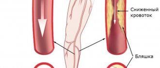
Almost every third person over the age of 65 has disease of the arteries or veins of the lower extremities. Smoking and a history of diabetes mellitus also aggravate this pathology. The main symptom of diseases of the arteries of the lower extremities is pain in the legs during long walking, occurring in different places, which depends on the level of vascular damage.
So, in what cases is angiography of the vessels of the lower extremities performed:
- Obliterating atherosclerosis and endarteritis of the vessels of the lower extremities;
- Deep vein thrombosis;
- Thrombophlebitis of the superficial veins of the lower extremities;
- a number of other pathological conditions.
Fundus angiography
Fundus angiography does not require special preparation, as with other types of angiography. The use of fundus angiography makes it possible to identify such unpleasant diseases as macular retinal degeneration, diabetic retinopathy and many others in the early stages.