The purpose of duplex scanning is to assess the health of the veins and their valves in the legs, track the speed of blood flow, the presence of lesions and narrowing of blood vessels.
With duplex scanning, veins are examined:
- Vein diameter
- Blood return point
- Presence of blood clots and compactions.
This type of research is one of the most modern, combining classical ultrasound and Doppler measurements. Most often it is prescribed in the following cases:
- The patient complains of pain, swelling, heaviness in the legs, as well as numbness or cramps
- The patient is bothered by itching or tingling in the legs, he quickly gets tired from even a short walk
- There are visual changes: the veins have darkened, began to protrude, the skin tone or its structure has changed, dryness or irritation has appeared that is not associated with external causes or diet, there are spider veins
- The patient is scheduled for surgery
Ultrasound duplex scanning of veins is prescribed, except for the specified cases, and when the patient already has a diagnosis - varicose veins, atherosclerosis of the lower extremities, angiopathy, thrombosis and others. This is done in order to assess the patient’s condition, understand how the disease develops, and find out whether the prescribed treatment is helping.
Color duplex scanning of the vessels of the lower extremities
Duplex scanning is a combined diagnostic method that includes two research modes, the first of which is a standard ultrasound examination of blood vessels, and the second is color mapping of blood flow in real time using the Doppler effect, that is, assessing the quality of blood flow in arteries and veins.
Duplex scanning of the lower extremities is an ultrasound examination in which the doctor analyzes not only the condition and position of the vessels, but also how blood flows through them. The specialist analyzes the speed and flow of blood flow inside the vessels, checks for the presence of obstacles (cholesterol plaques, blood clots), studies structural features (for example, excessive or pathological tortuosity), and checks the correct anatomical location of veins and arteries.
Who is the test prescribed for?
There are many indications for duplex scanning of leg vessels and they are very diverse. The patient may be concerned about the condition of his lower extremities, and then a duplex scan is prescribed to specifically search for the problem, or the general state of health suggests the likely development of vascular complications - in this case, the attending physician will also definitely recommend examining the vessels of the legs.
So, this type of diagnosis is prescribed for the following suspicious conditions of the lower extremities:
- a feeling of pain in them at rest or during physical activity
- swelling of the feet and/or legs
- thickening of the veins, sensation of “harnesses” under the skin of the legs
- pain in the muscles of the lower extremities
- occurrence of seizures
– change in skin color on the legs (also sudden hair loss on the legs)
- tingling sensation in the toes, feet or coldness
- impaired sensitivity, numbness in the legs constantly or during physical activity.
The doctor will also recommend a duplex scan of the lower extremities if the patient:
- diabetes
- trophic (vascular) ulcers on the body
- constantly increased cholesterol levels in the blood
- arterial and/or venous insufficiency
- Previous injuries or operations performed on the legs.
Who is indicated for vascular ultrasound?
A general practitioner or neurologist, as well as doctors of other specializations, can refer a duplex scan if the following symptoms and features are present:
- frequent headaches;
- dizziness;
- loss of consciousness;
- high cholesterol levels according to the results of laboratory blood diagnostics;
- hypertonic disease;
- atherosclerosis;
- osteochondrosis of the cervical spine;
- pain in the neck, shoulder girdle;
- suspected brain tumor;
- sleep disorders (drowsiness, insomnia, etc.);
- ringing in the ears, buzzing;
- “floaters” in the eyes, transient disturbances of visual perception.
In addition, the examination may be required as routine preparation for surgical interventions on blood vessels or the heart.
The procedure can be performed in the absence of symptoms and diseases, solely for preventive purposes. It is a safe and informative way to assess vascular function, so there are no restrictions on the number of times it can be performed.
It can and should be repeated after completing a course of treatment prescribed by a neurologist or therapist: this way the specialist can assess the dynamics of recovery and the response to medications and other therapeutic interventions.
Contraindications for the study
The method is based on obtaining the reflection of an ultrasound wave emitted by a sensor from the tissues of internal organs. Duplex scanning of the arteries and veins of the lower extremities is a procedure that does not injure the skin and mucous membranes of the body, and therefore is completely painless for the patient. Concomitant conditions or diseases are also not an obstacle to the examination, except for extensive skin damage (as, for example, with burns). It can be argued that there are no contraindications for this type of diagnosis. Duplex scanning is safe and can be used at any age.
Pregnant women are also often prescribed examinations of the blood vessels of the legs. Pregnancy involves the development of congestion in the veins and arteries of the legs, which can lead to varicose veins or thrombotic complications. There is no need to worry, even if repeated ultrasound examinations are necessary: ultrasound is harmless to the body of the expectant mother and her baby.
Is special training needed?
Since this type of study is not invasive, that is, it does not require intervention in the human body, the integrity of the skin and mucous membranes is not violated during diagnostics, there is no need to pre-take any tests specifically for duplex scanning of blood vessels.
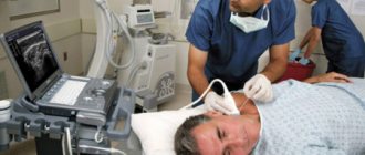
In the specialist’s office, before performing the manipulation, the doctor asks you to remove your outer clothing to expose your legs. A special gel is applied to the area being examined, which improves the conductivity of ultrasound, its access to tissues inside the body, and also facilitates the sliding of the sensor over the skin (especially important if there is a lot of hair growing on the skin). A specialist doctor places a sensor over the area under study, in this case, over the projection of the vessels of the lower extremities onto the skin. First of all, the general condition of the blood vessels is assessed using the standard mode. Then, using Doppler mode, the doctor evaluates the direction and intensity of blood flow.
After the examination and all necessary measurements, the patient is given a report on the condition of the arteries and veins of the lower extremities.
Indications and contraindications
Before being sent to the ultrasound room, the patient should understand what a duplex scan detects for veins located in the legs. Its main task is to identify possible vascular obstruction, taking into account the degree of neglect and localization features.
The technique also provides for the diagnosis of incompetent valves and perforating veins.
Typically, patients are sent for such an analysis if, at an appointment with a specialist doctor, they complained about:
- swelling in the ankle area;
- dilated saphenous veins;
- brown or purple spots on the skin;
- weeping wounds;
- suppuration;
- periodic loss of sensitivity;
- paroxysmal muscle contractions that occur involuntarily;
- pronounced itching that has no dermatological background.
Separately, doctors identify a risk group that includes people with a hereditary predisposition to varicose veins. They need to undergo preventive examinations and scans on a regular basis to check their results and generally accepted norms.
Also, all those who are scheduled for abdominal surgery can be sent for examination, and the patient is suspected of having blood clots.
If the victim complains of an accumulation of fluids in the tissues of the legs, then this is also a reason to do specific testing. Even with strange pain in the lower extremities, reminiscent of bloating from the inside, it is worth eliminating the risks of serious illnesses and undergoing a simple procedure.
It is also worth sounding the alarm if a person is worried about the bluishness of the skin or even their darkening from the foot to the knee.
The sooner the victim goes for examination, the higher the chances of a speedy recovery without the risk of worsening the quality of life in the future. With the help of a timely scan, it will be possible to detect and confirm a number of serious specialized pathologies such as:
- atherosclerosis;
- blood clots;
- varicose veins;
- endarteritis.
The doctor draws conclusions about a possible progressive disease from the list above based on the diagnostic report received. He prescribes in the certificate not only the current state of the vascular walls. The level of patency of deep and superficial veins and possible valvular insufficiency are also noted there.
If the patient has blood clots or related pathologies, then the overall picture will immediately show this.
There are no special contraindications for prescribing a duplex study. The only exceptions are a general severe condition of various origins, problems along the dermatological spectrum, or observations from a psychiatrist.
Advantages of the method
Duplex scanning of vessels allows you to obtain with high accuracy important information about the vessels and those processes that occur both in the lumen of the vessel and outside it. The method is a safe and painless way to identify disorders in the arteries and veins of the lower extremities even at the earliest stages, when there are no clinical signs of the disease.
The main advantage for the patient is the simplicity and ease with which this examination is carried out. Duplex scanning does not require special preparation, has no contraindications and does not lead to complications.
For a doctor, in turn, this method is an indispensable diagnostic method, as it allows one to detect possible problems in the early stages of their development, even in the smallest, so-called peripheral vessels of the legs (microcirculation difficulties).
The ability to assess blood flow, its dynamics and characteristics in real time makes it possible to analyze not only structural malformations, but also possible functional disorders.
Also, if the study is carried out several times over a certain period of time, a tendency towards the development of certain conditions or diseases can easily be identified, which allows timely treatment to be prescribed if necessary.
How it goes
No special preparation is required to assess the condition of extracranial vessels. On the day of the examination, it is advisable not to smoke or drink alcoholic or caffeinated drinks. You must warn your doctor if you are taking any medications. Diagnosis takes place in the supine position. There may be a small cushion under the neck. In some cases, the study is carried out with functional tests: turning the head, briefly pressing the arteries, holding the breath.
After the ultrasound, a conclusion is issued, which is deciphered by the attending physician. In some cases, other types of ultrasound or vascular examination using MRI may be prescribed.
What the results can show
In ultrasound diagnostic mode, duplex scanning of the arteries and veins of the lower extremities reveals any anatomical features or malformations: for example, increased tortuosity of blood vessels, their atypical location, deformation, expansion or narrowing.
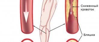
Assessing the state of blood flow in the lumen of a vessel in real time can reveal any difficulties in the bloodstream: stenoses, occlusions, blood clots, tumors, atherosclerotic plaques.
The doctor visually evaluates large vessels on the monitor screen and draws conclusions about their condition. By performing duplex scanning in Doppler mode, analyzing blood flow and making the necessary measurements, the specialist with very high reliability forms a conclusion about the condition of the smallest and deepest vessels.
Duplex scanning allows not only to detect existing pathological processes, but also to assess their nature and degree of development. Naturally, the final diagnosis, method of treatment or prevention depends on the results of the examination.
Advantages of diagnostics at the Innovative Vascular Center
Our medical center specializes in vascular and endovascular surgery, so it is important for us to obtain detailed information about the condition of the carotid arteries, because ischemic stroke is one of the most dangerous complications in vascular surgery. We perform ultrasound of the carotid arteries on all patients in our hospital and have developed an accurate ultrasound diagnostic algorithm. Prices for research in our center in Moscow are affordable to everyone. An appointment with a vascular surgeon is always accompanied by ultrasound diagnostics.
The advantage of ultrasound of the carotid arteries in our clinic is expert-level ultrasound scanners, evaluation of the results obtained by an ultrasound physician together with the operating vascular surgeon. The accuracy of the results of this study by our specialists is 96%, versus 80% in many other general practice diagnostic centers.