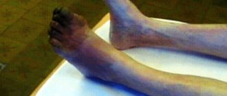
Occlusion of the femoral artery is a violation of its patency (blockage).
As a result of the occlusion, arterial, oxygenated blood stops flowing to the lower leg. Ischemia develops (local anemia).
Femoral artery occlusion occurs mainly in men. Persons over 50 years of age account for up to 75% of cases of the disease.
Femoral artery occlusion can be:
- segmental, affecting only a limited area of the artery;
- complete, when the entire artery is affected;
- combined with occlusions of other arteries of the lower extremities.
Causes of femoral artery occlusion
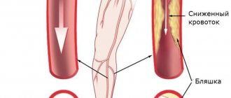
The cause of occlusion of the femoral artery in the majority (75-80%) of cases is obliterating atherosclerosis. Atherosclerosis is a disease in which cholesterol plaques are deposited on the walls of the artery, which over time block the lumen of the vessel. Also, occlusion of the femoral artery can be caused by injury, blood clot, and some other reasons.
Factors contributing to the development of occlusion
, are:
- smoking;
- high blood pressure;
- hereditary predisposition;
- improper diet (fatty foods);
- diabetes.
Diagnosis of acute vascular occlusion of the extremities
The diagnostic algorithm for suspected acute occlusion of the vessels of the extremities involves conducting a complex of physical, laboratory, and instrumental studies. Palpation of the pulse at typical points (on the dorsal artery of the foot, in the popliteal fossa, on the posterotibial and femoral arteries, etc.) reveals the absence of pulsation of the artery below the occlusion and its preservation above the affected area. Important information during the initial examination is provided by functional tests: marching (Delbe-Perthes test), knee phenomenon (Panchenko test), determination of the zone of reactive hyperemia (Moshkovich test).
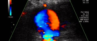
Laboratory blood tests (coagulogram) in acute occlusion of the vessels of the extremities reveal an increase in PTI, a decrease in bleeding time, and an increase in fibrinogen. The final diagnosis of acute occlusion of the vessels of the extremities and the choice of treatment tactics are determined by ultrasound data (duplex scanning) of the arteries of the upper or lower extremities, peripheral arteriography, CT arteriography, MR angiography.
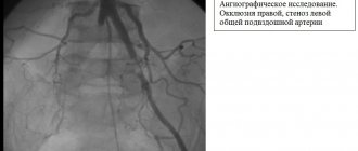
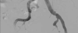
CT angiography of the abdominal aorta and its branches. Complete occlusion of the lumen of the left internal iliac artery
Differential diagnosis is carried out with a dissecting aneurysm of the abdominal aorta and acute thrombophlebitis of the deep veins.
Treatment methods for femoral artery occlusion
With limited occlusion, the body can compensate for the blood circulation of the limb using blood flow through the lateral branches of the arterial system (collateral circulation). In this case, conservative treatment is possible.
With increasing severity of ischemic symptoms, intermittent claudication occurring after less than 100 meters of walking, pain at rest, it is necessary to resort to surgical treatment. Such symptoms mean that circulatory compensation is insufficient, and this threatens the development of ulcerative-necrotic changes, gangrene and loss of limb.
Surgery
In the surgical treatment of occlusion, depending on the area of artery damage, the following are used:
- endarterectomy (removal of atherosclerotic deposits from the lumen of the artery);
- femoropopliteal bypass surgery;
- femoral-tibial bypass (if there is concomitant occlusion of the popliteal artery).
Make an appointment Do not self-medicate. Contact our specialists who will correctly diagnose and prescribe treatment.
Rate how useful the material was
thank you for rating
Symptoms of vascular occlusion
Acute occlusion of the vessels of the extremities is manifested by a symptom complex, designated in the English literature as the “complex of five Ps” (pain - pain, pulselessness - lack of pulse, pallor - pallor, paresthesia - paresthesia, paralysis - paralysis). The presence of at least one of these signs makes one think about possible acute occlusion of the vessels of the extremities.
Sudden pain distal to the site of occlusion occurs in 75-80% of cases and is usually the first sign of acute occlusion of the vessels of the extremities. If collateral circulation is preserved, pain may be minimal or absent. More often, the pain is diffuse in nature with a tendency to intensify, and does not subside when the position of the limb changes; In rare cases of spontaneous resolution of occlusion, the pain disappears on its own.
An important diagnostic sign of acute occlusion of the vessels of the extremities is the absence of pulsation of the arteries distal to the site of occlusion. In this case, the limb first turns pale, then acquires a cyanotic tint with a marbled pattern. Skin temperature is sharply reduced - the limb is cold to the touch. Sometimes, upon examination, signs of chronic ischemia are revealed - wrinkled and dry skin, lack of hair, brittle nails.
Disorders of sensitivity and motor sphere in acute occlusion of the vessels of the extremities are manifested by numbness, tingling and crawling sensations, decreased tactile sensitivity (paresthesia), decreased muscle strength (paresis) or lack of active movements (paralysis) first in the distal and then in the proximal joints . In the future, complete immobility of the affected limb may occur, which indicates deep ischemia and is a formidable prognostic sign. The end result of acute vascular occlusion can be gangrene of the limb.
Obliterating diseases of the aorta and arteries of the lower extremities
X
Chronic obliterating diseases of the aorta and arteries of the lower extremities (caused in most cases by atherosclerosis) account for more than 20% of all types of cardiovascular pathology, which corresponds to 2–3% of the total population [1]. Thus, in the Edinburgh study (1990) [2], patients with intermittent claudication accounted for 4.5% in the age group from 55 to 74 years, and asymptomatic lesions were noted in 8% of cases. It is significant that the attending physicians only 30–50% of patients knew about the presence of intermittent claudication in the latter [3].
The main feature of this pathology is its steadily progressive course.
, characterized by an increase in the severity of intermittent claudication and its transition to a constant pain syndrome or gangrene, which occurs in 15–20% of patients [4]. Perioperative mortality for amputations below the knee is 5–10%, above the knee – 15–20%. Mortality during the first two years after amputation ranges from 25–30%, and after 5 years – 50–75%. Moreover, after amputation of the lower leg, only 69.4% of patients walk on a prosthesis after 2 years, and only 30.3% of the hips.
Mortality after reconstructive surgery, which previously amounted to 2–13% [5], currently does not exceed 1.2% in leading Russian clinics [6]. When estimating the required number of operations for patients with obliterating diseases of the aorta and arteries of the lower extremities, we can cite the United States as an example, where in 1995 there were 400,000 hospitalizations for diseases of the peripheral arteries. 50,000 balloon angioplasties, 110,000 bypass operations, and 69,000 amputations were performed. Moreover, the costs of primary amputation in developed countries, such as the UK, were twice as high as the costs of successful revascularization [7].
According to L.A. Bokeria et al. [8], as of 1998, the need for reconstructive operations on the arterial system in Russia is 930 per 1 million population, no more than 22% of the required number is performed annually.
Pathomorphology and pathogenesis
The term “atherosclerosis” comes from the Greek words “athtre” - wheat gruel and “sclerosis” - hard. Despite the fact that the pathomorphology of atherosclerosis has been studied for more than 140 years, starting with the first works of R. Virchow (1856), the nature and features of the processes occurring in the vascular wall during this disease remain not completely clear. Even cellular and extracellular changes observed during microscopic examination in the vessel wall in the area of atherosclerotic plaque formation are interpreted differently. During the formation of atherosclerosis, the main changes occur in the endothelium and smooth muscle cells of the subendothelial layer of the intima.
There are 4 types of atherosclerotic changes in blood vessels:
1. Fatty spots or stripes, which are pale yellow areas containing lipids that do not rise above the surface of the intima. These are the earliest manifestations of atherosclerosis.
2. Fibrous plaques
- oval or round formations containing lipids, rising above the surface of the intima, often merging into continuous tuberous fields.
3. Fibrous plaques with various kinds of complications:
ulceration, hemorrhage, imposition of thrombotic masses.
4. Calcinosis
– deposition of calcium salts in fibrous plaques.
The most significant atherosclerotic changes are most often localized in places of greatest hemodynamic or mechanical impact on the vessel wall: bifurcation zones, places where the main arteries depart from the aorta and in tortuous sections of the artery.
According to JSA Fuchs [9], the leading risk factors for the development of atherosclerosis include arterial hypertension, hypercholesterolemia and smoking. To a lesser extent, obesity, diabetes mellitus, hypertriglyceridemia, sedentary lifestyle, stress and heredity influence.
Modern diagnostic methods
Modern methods for diagnosing peripheral arterial circulatory disorders are distinguished by their wide range - some are used to clarify the clinical diagnosis, the nature and extent of vascular damage, others are used to assess the effectiveness of treatment or dynamic monitoring of the patient. In order to study hemodynamics in the lower extremities and topical diagnosis of lesions of the arterial bed, the following instrumental research methods are used:
Doppler ultrasound sphygmomanometry, treadmill test, ultrasound angioscanning, including duplex, and radiopaque aortoarteriography. In addition, it is necessary to determine indicators of lipid metabolism, coagulation system and rheological properties of blood.
As the first stage, all patients with suspected occlusive-stenotic lesions of the aorta or arteries of the lower extremities undergo Doppler ultrasound with measurement of the ankle-brachial index
.
This index is the ratio of the maximum pressure on one of the tibial arteries to the pressure on the brachial artery. A decrease in this indicator to less than 0.9 requires closer attention to the patient. of Doppler ultrasound and standard treadmill test seems to be one of the most promising at present.
[10].
Non-invasive research methods also include ultrasound angioscanning
, thanks to which the degree of stenotic lesion can be determined with a high degree of certainty.
Recently, duplex ultrasound angioscanning
has taken one of the leading places in the diagnostic program algorithm due to its non-invasiveness and safety, as well as high sensitivity and specificity.
According to duplex scanning data, not only the structure of the atherosclerotic plaque is determined, but also the hemodynamic degree of stenosis is assessed, which is of fundamental importance. X-ray contrast angiographic examination
currently remains the main method for diagnosing obliterating diseases of the vascular bed. Using this method, it is possible to accurately determine the location, extent, degree and nature of stenosis, the multiplicity of occlusive lesions of the main arteries of the lower extremities, assess the condition of the collateral bed, predict the nature and volume of reconstructive surgery, and also monitor the effectiveness of treatment and surgery. In the arsenal of angiologists and vascular surgeons there are also such diagnostic methods as laser Doppler flowmetry, transcutaneous O2 monitoring, photoplethysmography, radioisotope study, computed tomography and nuclear magnetic resonance.
Terminology and clinical classification
“Leriche syndrome” is often used to refer to diseases of the abdominal aorta that lead to its narrowing or occlusion.
, which summarizes the picture of damage to the bifurcation of the abdominal aorta and iliac arteries.
The main features of the clinical course of this lesion are high intermittent claudication (pain in the limb when walking), bilateral absence of pulses in the arteries and impotence.
In approximately 30% of patients with chronic arterial insufficiency of the lower extremities, the atherosclerotic occlusive process is localized in the abdominal aorta, in 70% of patients - in the arteries of the femoral-popliteal segment.
Tactical issues in choosing one or another treatment method for atherosclerotic lesions of the aorta and arteries of the limb are based on the severity of chronic ischemic syndrome, which is classified into 4 stages of the disease. The predominant assessment system is the classification of R. Fontaine and A.V. Pokrovsky.
At stage 1 of the disease
pain in the lower extremities appears only with heavy physical activity; it is not related to the distance the patient walks.
For stage 2
Characteristic is the appearance of limiting pain when walking (limiting intermittent claudication). From a tactical point of view, this stage is divided into 2A (a distance walked without pain of more than 200 m) and 2B (the appearance of pain when walking at a distance of less than 200 m).
Pain in the limb at rest characterizes the 3rd stage
, the appearance of ulcerative-necrotic changes -
stage 4 of the disease.
Surgical tactics and determination of the degree of surgical risk
The fundamental generally accepted position in choosing a treatment method in accordance with this classification is the need to restore the main blood flow using reconstructive operations, starting from stage 2B.
When deciding on surgical treatment, it is necessary to take into account the multifocal nature of the atherosclerotic lesion and the presence of concomitant pathology that aggravates the condition of the patients. According to our data, about 70% of patients suffer from coronary heart disease, every 4th is diagnosed with post-infarction cardiosclerosis and chronic cerebral circulatory failure, half of the patients have hypertension in combination with chronic lung diseases. 35% have gastrointestinal tract diseases and every 7th person has diabetes mellitus.
Based on all of the above, treatment of patients with atherosclerosis should be comprehensive
, aimed both at restoring blood circulation in the aorta and main arteries of the limb, and at correcting concomitant pathology. The main goal - restoration of blood circulation - should be achieved with minimal trauma to the patient.
Principles of conservative treatment
One of the main directions of conservative treatment is to improve the rheological properties of blood.
And this is not accidental, since patients have pronounced deviations in rheological characteristics: an increase in the level of fibrinogen in plasma, an increase in platelet aggregation time, blood and plasma viscosity, a decrease in the fibrinolytic activity of the blood and a change in thromboelastogram parameters towards hypercoagulation.
Among the drugs used for conservative therapy, several groups are distinguished.
1. Antispasmodics:
peripheral myolytics (papaverine, drotaverine, bencyclane), drugs blocking a-adrenergic receptors or preganglionic impulse transmission (caffeine, prazosin), central cholinomyolytics (tolperisone, baclofen), substances with versatile effects (abana).
2. Disaggregants:
pentoxifylline, acetylsalicylic acid, xanthinol nicotinate, ticlopidine, rheopolyglucin.
3. Antiatherosclerotic agents:
drugs that block the absorption of cholesterol from the intestine (cholestyramine), inhibiting the biosynthesis and transfer of cholesterol and triglycerides (fibric acid derivatives - clofibrate, ciprofibrate) and statins (lovastatin, simvastatin), other drugs (nicotinic acid).
4. Metabolic drugs:
solcoseryl, actovegin, etc.
5. Angioprotectors:
pyricarbate, etc.
I would especially like to emphasize that dosed walking
– health path, which promotes the development of collateral circulation.
Purely conservative treatment is indicated for patients with chronic arterial insufficiency of stage 1 and 2A; in patients with stage 2B and critical ischemia with the development of ulcerative-necrotic lesions, the question arises about the need to restore the main circulation. Thanks to the capabilities of modern technologies, in recent years a lot of work has appeared on the use of balloon angioplasty.
in patients with different localization of occlusive-stenotic lesions of the arteries of the pelvis and lower extremities.
However, it is not possible to use balloon repair in all cases due to aortic occlusion or common arterial occlusions. Attempts at recanalization in these cases are dangerous by the development of thrombosis of the main arteries (often with thrombosis of the peripheral bloodstream), which inevitably leads to amputation of the limb in 60% of cases, and often to death.
Types of reconstructive surgical interventions
In case of high occlusion of the aorta, bilateral damage to the arteries of the extremities, depending on the severity of the patient’s condition, operations are performed from aortofemoral bifurcation or linear bypass to axillary or subclavian-femoral bifurcation bypass. If critical ischemia is present only on one side, then if the iliac and femoral arteries are affected on the contralateral limb, a unilateral cross-iliac-femoral, axillary or subclavian-femoral bypass
.
At the present stage, reconstructive operations occupy a leading place in the treatment of these patients. The number of such operations is constantly increasing, their scope is expanding significantly, which makes it possible to save a limb even in severe forms of chronic arterial insufficiency. Contractubex is effective for resolving keloid scars that occur after surgery.
, which has fibrinolytic, antithrombotic and keratolytic effects.
Meanwhile, performing a full-fledged reconstruction often conflicts with the patient’s ability to undergo surgery. Operations in these cases should be minimal in terms of trauma and duration, since the vast majority of this group of patients have severe concomitant diseases that sharply limit the functional reserve capabilities of the body [11]. Using the combined operations method
, including balloon angioplasty in combination with open surgery under epidural or local anesthesia, can significantly reduce the volume of intervention and eliminate complex surgical reconstruction in several segments.
Clinical case
Patient Z., 68 years old, was admitted with complaints
for aching pain in the right leg and foot at rest, intermittent claudication after 30 m.
Doppler ultrasound:
a significant decrease in the main blood flow in the right common femoral artery, collateral blood flow in the popliteal and tibial arteries.
Ankle-brachial index
left 0.59, right 0.35.
Aortoarteriography:
critical stenosis of the common iliac artery (CIA) on the right; stenosis of the deep femoral artery (DFA) on the right; occlusion of both superficial femoral arteries (SFA), segmental occlusion of the right popliteal artery for 5 cm (Fig. 1).
Rice.
1. Angiograms of patient Z.: a — stenosis of the abdominal cavity on the right; b — stenosis of the GBA on the right, occlusion of both SBAs; c — after balloon angioplasty (absence of an area of stenosis of the right PA). Atherosclerotic lesion of arteries (case history).
Concomitant diseases: coronary heart disease, atherosclerotic cardiosclerosis, angina pectoris, chronic bronchitis, pneumosclerosis, pulmonary emphysema.
coronary heart disease, atherosclerotic cardiosclerosis, angina pectoris, chronic bronchitis, pneumosclerosis, pulmonary emphysema.
The first stage was balloon angioplasty of the right abdominal organ, and the second stage, under epidural anesthesia, was lateral plasty of the gastrointestinal tract on the right (Fig. 2).
Rice.
Fig. 2. Scheme of stages of surgical treatment of patient Z.: a - before surgery; b — balloon angioplasty of the right PCA; c — condition after plasty of the GBA and balloon angioplasty of the abdominal organ on the right. As a result,
positive dynamics were noted: the ankle-brachial index on the right increased to 0.71 (initially 0.35). The patient was discharged in satisfactory condition for outpatient treatment.
You can find a list of references on the website https://www.rmj.ru
References:
1. Pokrovsky A.V., Koshkin V.M., Kirichenko A.A. et al. Vasaprostan (prostaglandin E1) in the treatment of severe stages of arterial insufficiency of the lower extremities. A manual for doctors. M., 1999; 16.
2. Fowkes FG, Housley E, Cawood EH et al. Edinburgh artery study: prevalence of asymptomatic and symptomatic peripheral arterial disease in the general population. Int J Epidimiol 1991; 20: 384–92.
3. Burakovsky A.I., Bockeria L.A. Cardiovascular surgery. M., 1989; 750.
4. Dormandy J., Mahir M., Ascady G. et al. Fate of the patient with chronic leg ischemia. J Cardiovasc Surg 1989; 30:50–7.
5. Stoffers HEJH. Kaiser V. and Knottnerus JA Prevalence in the general practice. In: Fowkes FGR, ed. Epidemiology of peripheral vascular disease. London: Springer Verlag. 1992; 109–13.
6. Spiridonov A.A., Fitileva E.B., Arakelyan V.S. Ways to reduce mortality in surgical treatment of chronic ischemia of the lower extremities. J. Annals of Surgery. 1996; 1:62–6.
7. Bead J.D. Amputation or reconstruction for critical ischemia. J. Angitology and Vascular Surgery 1998; 1 (4): 72–82.
8. Bockeria L.A., Gudkova R.G. Surgery of the heart and blood vessels in the Russian Federation. M., 1998; 43.
9. Fuchs JSA. Atherogenesis and the medical management of Atherosclerosis. In: Rutherford RB, ed. Vascular surgery. Philadelphia: W. B. Saunders Company. 1996; 1:222–35.
10. Zatevakhin I.I., Tsitsiashvili M.Sh., Yudin R.Yu. Treadmill in the diagnosis and treatment of chronic arterial insufficiency. M., 1999; 87.
11. Siskin G., Darling RC III, Stainken B. et al. Combined use of Iliac artery angioplasty and infrainguinal revascularization for treatment of multilevel atherosclerotic disease. Annals of Vascular Surgery. St. Louis. 1999; 13 (1): 45.
Prognosis and prevention
The most important prognostic criterion for acute occlusion of the vessels of the extremities is the time factor. Early surgery and intensive therapy can restore blood flow in 90% of cases. If treatment is started late or is absent, disability occurs due to the loss of a limb or death. With the development of reperfusion syndrome, death can occur from sepsis, renal failure, or multiple organ failure.
Prevention of acute vascular occlusion of the extremities involves timely elimination of potential sources of thromboembolism and prophylactic administration of antiplatelet agents.
Anesthesia for stenting
Most endovascular angioplasty procedures and lower extremity vascular stenting can be performed with mild intravenous sedation and local anesthesia at the access puncture site. Monitoring of blood pressure, electrocardiogram and blood oxygen saturation level (pulse oximetry) is mandatory. In case of unforeseen complications, the operating room is equipped with a breathing apparatus and a defibrillator. If the operation is performed for critical ischemia, then to make the patient comfortable, epidural anesthesia is performed (injection of an anesthetic drug through a catheter into the spinal area).
Observation after angioplasty and stenting
Our clinic has adopted a patient management scheme after surgery to avoid complications of angioplasty and stenting of peripheral arteries of the lower extremities:
- For better results, dual antiplatelet therapy is indicated, including taking Plavix and aspirin. Drug prevention plays an important role in the long-term results of the intervention and the life expectancy of patients.
- The first examination is carried out in the first 2 weeks of the postoperative period with mandatory ultrasound of peripheral vessels.
- Further examinations are carried out after a few months.
- One year after surgical treatment, MSCT angiography of the legs is mandatory.
Balloon angioplasty and stenting of the arteries of the lower extremities is an effective method of restoring blood flow in most occlusive lesions except the popliteal artery. In terms of its immediate results, this method is not inferior to open bypass surgery if performed according to strict indications. The advantage of endovascular surgery is low trauma, lack of pain, the possibility of repeated interventions, and a lower risk to life.
With the improvement of instruments for angioplasty and stenting, minimally invasive interventions are occupying an increasing place in the management of patients with critical ischemia and gangrene.
Endovascular technologies at the Innovative Vascular Center
The main mission of our clinic is the treatment of critical ischemia and gangrene of the extremities. We are committed to using the latest approaches to solve this problem. Since 2011, endovascular surgery methods for critical ischemia have been introduced into the practice of our vascular surgeons. Noting the advantages of the endovascular approach, every year we expand the capabilities of our clinic in the use of these methods.
The role of minimally invasive technologies is growing - now more than 50% of patients with critical ischemia and gangrene are operated on endovascularly, and even more than 40% use a hybrid approach. This is bypass surgery with angioplasty and stenting of the arteries of the lower extremities. The Innovative Vascular Center is a clinic where more than 500 angioplasty and stenting operations of lower extremity arteries are performed per year.
The capabilities of endovascular surgery are developing synchronously with the creation of innovative treatment tools, so in our practice, puncture operations are increasingly replacing open interventions.
Preparing for stenting
Before performing stenting surgery, the patient must be properly examined for vascular lesions and risks of complications. A set of laboratory tests and a coagulogram must be performed before surgery. Considering the patient's load on antithrombotic drugs, it is necessary to exclude possible sources of bleeding (stomach ulcer, bleeding hemorrhoids).
On the eve of the intervention, a light sedative is administered, allowing the patient to sleep well and not be nervous. Before the operation, the patient will shave the site of the intended access. In the preoperative room, the nurse will place a urinary catheter and an intravenous needle for infusions. In the operating room, a pressure cuff is placed on the shoulder and sensors for continuous ECG recording are fixed on the chest.
Types of angioplasty
- Subintimal balloon angioplasty of vessels - the conductor is passed under the modified inner wall (intima) of the vessel and then extends into the free lumen.
- Intraluminar angioplasty is a type of intervention where the conductor passes through the natural lumen of the artery, sliding through narrowed and blocked areas.
- With laser angioplasty, the atherosclerotic plaque is burned out with a special laser catheter.
Balloon angioplasty of lower extremity arteries without stenting is recommended for the treatment of lower leg lesions. It is imperative to stent the iliac arteries, since the frequency of repeated narrowings (restenosis) without a stent is very high.
After angioplasty and stenting, control angiography is required to evaluate the result.
Treatment of acute vascular occlusion of the extremities
If acute occlusion of the vessels of the extremities is suspected, the patient requires emergency hospitalization and consultation with a vascular surgeon. For tension ischemia and degree IA ischemia, intensive conservative therapy is carried out, including the administration of thrombolytics (intravenous heparin), fibrinolytic agents (fibrinolysin, streptokinase, streptodecase, tissue plasminogen activator), antiplatelet agents, and antispasmodics. Physiotherapeutic procedures (diadynamic therapy, magnetic therapy, barotherapy) and extracorporeal hemocorrection (plasmapheresis) are indicated.
In the absence of positive dynamics within 24 hours from the onset of acute occlusion of the vessels of the extremities, it is necessary to perform an organ-preserving surgical operation - thromboembolectomy from a peripheral artery using a Fogarty balloon catheter or endarterectomy.
In case of ischemia of degrees IB–IIB, emergency intervention is necessary to restore blood flow: embolism or thrombectomy, bypass surgery. Prosthetic replacement of a segment of a peripheral artery is performed for short-term acute occlusions of the vessels of the extremities.
Ischemia of degrees IIIA–IIIB is an indication for emergency thrombus or embolectomy, bypass surgery, which must be supplemented with fasciotomy. Restoring blood circulation with limited contractures allows for delayed necrectomy or subsequent amputation at a lower level.
In case of ischemia and IIIB degree, vascular surgery is contraindicated, since restoration of blood flow can lead to the development of post-ischemic syndrome (similar to traumatic toxemia in the long-term crush syndrome) and the death of the patient. At this stage, amputation of the affected limb is performed. In the postoperative period, anticoagulant therapy is continued to prevent rethrombosis and re-embolism.