The essence of the study
Special vessels, the brachiocephalic arteries (BCA), are responsible for the blood supply to the brain, tissues of the head, arms and shoulder girdle. They are determined from the aorta at the level of the shoulders. The condition of these vessels can be examined and their function assessed using Doppler ultrasound (USDG). What kind of examination is this, ultrasound examination of the BCA (BCS)? This method is based on the ability of blood particles to reflect ultrasonic waves emitted by the sensor. The ultrasound machine, in turn, picks up these reflections and graphically depicts the blood flow in the arteries.
The Doppler ultrasound method is non-invasive. It is safe and painless, therefore it has no contraindications or age restrictions for patients.
Ultrasound of head and neck vessels
This is a modern ultrasound diagnostic method, which makes it possible to most accurately determine the condition of the neck vessels that supply the brain, shoulder girdle and arms.
The article was prepared by our ultrasound diagnostics doctor Irina Alekseevna Romadova.
When a person is prescribed a study of the brachiocephalic arteries (BCA), he has questions:
— what will he have to deal with?
- How effective is this method?
- isn't it harmful?
— and won’t this procedure cause discomfort?
What is ultrasound of the vessels of the head and neck.
Duplex ultrasound scanning of the brachiocephalic arteries (USD BCA) is a modern ultrasound diagnostic method that makes it possible to most accurately determine the condition of the neck vessels that supply the brain, shoulder girdle and arms.
These vessels are called brachycephalic. These include the carotid, vertebral, subclavian arteries and their connection, which forms the brachiocephalic trunk.
The BCA study allows the doctor to determine the presence of a violation of the blood supply to the brain , and to predict the likelihood of worsening the disease in the future.
Types of vascular studies
Vascular examination can be carried out in three ways:
- Ultrasound Dopplerography or Dopplerography . The research is based on the Doppler effect, named after the Austrian scientist Christian Doppler who discovered it.
- Ultrasound scanning or duplex scanning of the vessels of the neck and head is a combination of both the Doppler effect and B-mode (regular gray ultrasound image). That is, the doctor can evaluate the movement and speed of blood flow , as in Dopplerography, as well as the internal walls of blood vessels , deposits on them, the presence of atherosclerotic plaques, thickening, and developmental anomalies. With this method, two functions are examined at once, hence the name duplex. Ultrasound scanning shows a complete picture of the movement of blood through the arteries and veins, which makes it possible to make a more accurate diagnosis.
- Triplex scanning is a type of duplex assessment. However, it provides even more information because it uses the additional technique of color Doppler mapping. A color image of the movement of blood in the vessels makes it possible to more accurately determine changes in blood flow.
Unlike conventional ultrasound, which shows organs in a static state, Doppler ultrasound shows the movement of blood flow and its speed. This is made possible by the difference in speed at which Doppler ultrasound reflects and receives waves.
Undoubtedly, duplex scanning is the most modern research that provides a complete picture of changes in blood vessels. The ultrasound machine in our clinic belongs to the expert class and is equipped with all vascular programs. This makes it possible to most effectively visualize and evaluate pathological changes in the vascular bed.
Doctor's qualifications
We all know that the results of the study directly depend on the qualifications of the doctor performing the study. Our doctor Irina Alekseevna Romadova completed a full course of training in ultrasound angiology (examination of all vessels) at the Northwestern State Medical University named after I.I. Mechnikov, and masters all methods of vascular research. This is undoubtedly a big plus for our patients , because if there is damage to various vessels, then she can evaluate them all, besides, Irina Alekseevna is a former surgeon and is well versed not only in physiology, but also in anatomy .
Safety
Ultrasound examination is completely safe and painless for the patient. The use of this technique has no restrictions or contraindications. Ultrasound scanning can be used repeatedly, both for diagnosis and for monitoring the course of the disease at various stages of treatment.
What pathologies
The most common pathologies diagnosed with ultrasonography are:
- atherosclerotic lesions of the vascular wall
- vascular obstruction
- aneurysms
- deformations and abnormalities of vascular development
- dilation and incompetence of the venous bed
- thrombosis
- damage to the walls of blood vessels
The sensitivity of the method is very high , with ultrasound even minor, initial manifestations of atherosclerosis in the walls of blood vessels are visible, the identification of which will allow timely measures to be taken and prevent the formation of atherosclerotic plaques .
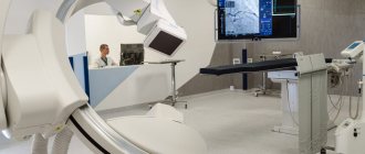
In some situations, it is necessary not only to examine the vascular bed, but also to evaluate the elasticity of the vessels, their elasticity, and ability to respond to changes in blood pressure and oxygen content in the atmosphere ( resistance ). For these purposes, the Carbonik , which is equipped in the ultrasound room at the Smart clinic. It has been clinically proven that with the help of a breath test using the Carbonik device, the most reliable information about changes in the resistivity of the vascular bed is obtained.
This photo shows the process of conducting a study of cerebral vessels with the Carbonik apparatus.
Who is this procedure indicated for?
As a measure to prevent serious complications of cerebrovascular accidents, such as stroke, BCA ultrasonography is recommended to be performed on individuals at risk at least once a year.
- People over 40–45 years of age.
- Patients with diabetes mellitus.
- Persons with a family history of stroke.
- If abnormalities are detected in the lipid profile.
- Patients with pathology of the heart and vascular system.
- Smoking and alcohol abuse.
- Patients with osteochondrosis of the cervical spine.
- Autoimmune vascular disease (hemorrhagic vasculitis)
This study is also indicated for all people with complaints indicating changes in the vessels of the neck.
- constant or recurrent headaches;
- migraine
- dizziness;
- sensation of noise or ringing in the ears;
- absent-minded attention;
- impaired coordination of movement;
- decreased vision;
- memory change;
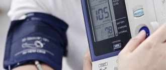
- blood pressure problems;
- fainting;
- neck injuries;
- condition after a heart attack or stroke.
Preparing for the study
BCA ultrasound does not require , but on the day of the examination it is better to exclude from the diet foods that can change the tone of the vascular wall, such as coffee, strong tea, alcohol, energy drinks, cola. It is advisable to refrain from smoking two hours before the test. If you are taking medications that affect the functioning of the cardiovascular system, be sure to tell your doctor.
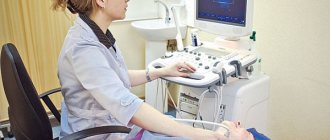
Carrying out the procedure
The procedure takes about 30-40 minutes, the patient lies on his back, the chin should be raised up and slightly turned in the direction opposite to the one being examined. The doctor may press a little on the patient’s neck during the examination, but in general this does not cause any discomfort. Our clinic has a “ platinum ” version of the device, including a device for heating the gel , which makes the procedure even more comfortable. The ultrasound scanner will record all the indicators, and the doctor will enter them into the study protocol.
Summary: Duplex ultrasound scanning of the brachiocephalic arteries is accessible and effective diagnostic method. In addition to high resolution, the method is absolutely safe . The Smart Clinic has an expert level ultrasound machine installed, and our doctor, Irina Alekseevna Romadova, is an experienced specialist, which together gives our patients timely and effective diagnosis, and therefore, subsequently, the correct treatment.
Indications for use
Ultrasound Doppler Doppler of the BCA – what kind of examination it is, it’s almost clear. You need to figure it out when it's needed.
Headache
There can be many reasons for a person to experience periodic headaches. To eliminate this symptom and the disease associated with it, it is important to correctly determine the cause of the pain. Quite often it turns out that the blood supply to the brain is impaired due to dysfunction of the brachiocephalic arteries and vessels. Using ultrasound scanning of veins and arteries
manages to find out.
Dizziness
Dizziness can be caused by various factors: disruption of the vestibular apparatus, inner ear, fasting, stress, poor diet. Often, this pathological condition, when the patient loses coordination (for example, during sudden movements, walking, and so on), is caused by a disruption in the functioning of the brain. It, in turn, can be caused by disruptions in the blood supply from the brachiocephalic artery (BCA).
Fainting
Fainting is a short-term loss of consciousness. Its main cause is a temporary disruption of cerebral blood flow, which in turn can occur due to disruption of blood flow in the brachiocephalic artery. Systematic fainting is an indication for urgent ultrasound examination of the BCA.
Sleep disorders
The lack of normal rest due to a sleep disorder has a very negative effect on a person’s general condition, his performance, mood, and so on. One of the reasons for this problem may be impaired hemodynamics in the BCA. Doppler ultrasound of BCA arteries for sleep disorders can be prescribed in the presence of other alarming symptoms (fainting, dizziness, and so on).
Permanent or transient visual disturbances
Deterioration of vision is another indication for ultrasound examination of the central nervous system (what it is is described above). The blood supply systems of the brain and eyes are adjacent, therefore, for an accurate diagnosis and treatment, it is important to study the blood flow in all interconnected systems.
Flashing “flies”, a veil before the eyes
“Floaters” and blurred vision can be associated both with purely eye problems (for example, retinal detachment) and with brain pathologies. The most dangerous and, unfortunately, quite common pathology is stroke. Ultrasound scanning of the brachiocephalic vessels will make it possible to find out exactly what is causing such discomfort in the eyes and, perhaps, will help prevent hemorrhage in the brain.
Tinnitus, ringing in the head
The sensation of noise and ringing in the ears, which interfere with a person’s normal life and work, can also be one of the symptoms of a violation of cerebral blood supply from the BCA. If such a symptom is observed in combination with dizziness, migraines, fainting, then the doctor must prescribe an ultrasound scan of the brachiocephalic arteries (vessels).
USDG BCA
Hello. The neurologist prescribed an ultrasound examination of the BCA. What is this procedure, how is it carried out and is special preparation required? Olga M.
Answered by Nadezhda Ivanovna, ultrasound diagnostics doctor at the MedMix Plus clinic.
Doppler ultrasound of the BCA is an ultrasound examination of the brachiocephalic vessels, that is, in other words, ultrasound of the vessels of the neck. This examination is very important in assessing the condition, anatomy and patency of arteries and veins. Using this ultrasound technology, it is possible to judge the state of the blood supply to the brain, determine the degree of stenosis, thereby enabling the attending physician to determine treatment tactics.
In medical practice, there are several methods for studying the vessels of the neck:
1.USDG (ultrasound Dopplerography ) is a method aimed at assessing the patency of blood vessels and their condition. This method gives a general idea of the vessels. Allows you to identify the internal cause of obstruction, for example, a blood clot or atherosclerotic plaque.
2. Duplex/triplex scanning , which is used by ultrasound diagnostic doctors at our MedMix Plus clinic. On the monitor screen, a specialist sees a vessel in color against a gray background and evaluates its blood flow. This method allows you to assess the cause of the disturbance in the blood supply to the vessels, whether it is external or internal.
Using BCA ultrasound, you can diagnose diseases such as:
- vascular atherosclerosis;
- abnormalities in the structure of blood vessels;
- delamination of the vessel wall;
- arterial aneurysms;
- vasculitis, etc.
Before the symptoms of the disease appear, BCA should be performed when:
- hypertension;
- suffered a stroke;
- lupus;
- diabetes mellitus;
- smoking.
Research is also necessary in the presence of conditions such as:
- dizziness;
- decreased visual function;
- memory impairment;
- headache;
- fainting, etc.
Preparing for the study.
Before performing an ultrasound examination, you need to stop taking foods that can affect vascular tone: tea, coffee, energy drinks, alcohol. You should not visit stuffy or smoky rooms, and you should also stop taking medications that improve memory and attention. It is better to discuss the discontinuation of vascular and cardiac medications with your doctor.
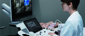
Conducting research.
Doppler ultrasound of the BCA is a standard ultrasound examination procedure. The patient lies on the couch, a cushion is placed under the head. A gel is applied to the neck area, and a specialist conducts a study with a special sensor. The procedure lasts on average 30–35 minutes.
Doppler ultrasound of the brachiocephalic vessels is a painless and accurate ultrasound examination method, which in less than an hour will give an answer about the condition of the neck vessels and the causes of discomfort. Performed routinely, ultrasound of the neck vessels will prevent stroke by 80%. Be healthy!
Preparation for the procedure
Preparation for the BCA ultrasound (BCA) procedure involves following nutritional rules.
Drinks containing caffeine (tea, coffee, energy drinks)
At least one day before the test, the patient should exclude tea, coffee, and energy drinks from the diet. These are the drinks that can affect the condition of blood vessels and the results of the examination.
Alcohol
Drinking alcohol the day before the procedure is absolutely excluded (it is even better to increase the period of abstinence).
Salty foods
Excessive salt content in the blood can somewhat affect the speed of its flow and the condition of blood vessels, therefore, the day before an ultrasound scan, consuming various pickles is also prohibited.
Order of conduct
Let's look at how an ultrasound scan of the brachiocephalic arteries (BCA) is done. To undergo Doppler ultrasound of the BCA, the patient needs to undress to the waist and lie down on the couch. The doctor will apply a special thick gel to the skin, which will ensure tight contact of the ultrasound sensor with the skin. During the study, the patient will need to change position - turn over on his side, on his stomach. Also, the doctor may suggest performing some functional tests: holding your breath or changing a horizontal body position to a vertical one.