Ultrasound diagnostics is one of the safest and most effective ways to detect various diseases of internal organs. This technique is used in various fields of medicine, including assessment of the condition and possible pathologies of blood vessels. In this case, Doppler ultrasound or ultrasound is performed, which is used to examine the neck and head. Early diagnosis allows you to choose gentle and effective treatment methods and prevent the development of complications. First you need to understand what an ultrasound scan of the vessels of the head and neck shows, why this method is effective in diagnosing many diseases.
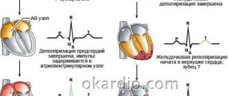
The Doppler ultrasound effect is based on the ability of ultrasound waves to be reflected from blood cells. As a result, electronic impulses appear that appear on the device screen in the form of images of veins and arteries with blood flow.
This ultrasound examination allows you to assess the condition of the large arteries that supply the brain, carotid and vertebral arteries, determine the intensity of blood flow, and also identify traces of obstruction.
How is Dopplerography performed?
This examination is carried out in the presence of certain symptoms. Preliminary preparation is necessary so that the results of the ultrasound examination give an accurate picture of the patient’s condition.
There are two types of procedure:
- Transcranial. The sensor is installed on the skull bones in those places where they are thinnest. The procedure gives an accurate picture of the vessels located directly in the brain.
- Extracranial. The technique is used to examine large vessels passing through the neck (carotid arteries, jugular veins, etc.). The sensor is located at each site separately.
General characteristics of the examination
To explain what kind of examination this is – Doppler ultrasound – and how it is done, you need to start by deciphering the abbreviation: Doppler ultrasound (you can also hear the name “duplex scanning”). In more detail, Doppler ultrasound is a combined study that combines standard ultrasound and the Doppler effect. Ultrasound allows you to visualize a patient’s specific organ on the screen, determine its size, structure, assess its integrity, and so on. Doppler allows you to assess the condition of the vessels of the examined organs, as well as the quality of blood flow in them.
An ultrasound examination, as practice shows, is informative for both assessing venous and arterial circulation. This procedure is safe, painless, non-invasive.
Indications for the procedure
Doppler sonography is indicated in the following cases:
- Problems with coordination and other symptoms indicating a malnutrition of the brain (the patient is dizzy, has tinnitus, etc.).
- Unstable heart rhythm.
- Hypertonic disease.
- Transient ischemic attacks.
- IOP.
- Previous stroke or heart attack.
- There are suspicions of atherosclerosis.
- Diabetes.
- Fatigue quickly for no apparent reason.
- Cervical osteochondrosis.
This procedure is also indicated if heart surgery is to be performed, if there are pulsating formations in the neck, or if thrombosis is suspected. In children, it is performed in case of serious postural problems and delayed development of the child.
Preparing for the study
No special preparation is required for ultrasound of the neck and head vessels. However, if the patient is taking medications that lower blood pressure or affect blood clotting, the doctor must be informed. He may recommend that you avoid taking medications in some cases.
On the day of the examination, it is recommended to refrain from smoking, drinking alcohol, energy drinks and physical activity. In general, it is not recommended to perform actions that may affect the tone of the vascular wall.
Are there any contraindications?
Ultrasound waves are absolutely safe for humans, so there are no special contraindications to this type of ultrasound. The only exception is a violation of the integrity of the skin. The procedure involves contact of sensors with the head or neck, so the examination is postponed until the wound heals.
However, there are a number of special cases that make duplex examination of blood vessels difficult:
- cardiac arrhythmia, which changes the speed of blood circulation through healthy arteries and veins;
- the area under study is hidden under bone tissue;
- slow blood flow in the patient.
These are not contraindications, but in this case changes in the location of the sensors are required. It will be necessary to navigate non-standard conditions, which requires professionalism on the part of the doctor.
What is the difference between duplex and triplex scanning?
These diagnostic methods are very similar to each other and have practically no differences, with the only difference being that with triplex scanning the vessels can be viewed in three-dimensional space. Duplex mode presents information in only two planes. In fact, triplex scanning is positioned as an additional procedure when conducting a duplex study.
And if we take into account the fact that the crystal transmitting and receiving the signal is the same, then the resolution of the triplex method is considered slightly worse than the duplex one. The quality of the work will depend entirely on the quality of the apparatus and the qualifications of the doctor conducting the research, and not on the diagnostic method.
Features of ultrasound examination
As a rule, Doppler sonography is planned in advance, but it can be performed urgently. The procedure itself is painless, so patients tolerate it calmly.
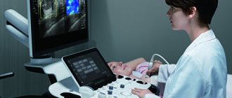
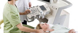
The examination is carried out according to the following scheme:
- The patient lies on his back. The head should be relaxed (a flat pillow is placed under it).
- You need to remain calm, breathe evenly and, if possible, not move.
- A special gel is applied to the area under study to prevent the penetration of air between the skin and the sensor.
- The movement of the sensors is controlled by a specialist. As a result, an image of the vessels appears on the monitor screen.
The study lasts no more than 1 hour. During the study, the doctor may ask the patient to do some action: turn his head, squeeze a vein in a certain place, breathe less often, etc.
Dopplerography of extracranial vessels begins with examination of the right lower segment of the carotid artery. The sensor is then moved up the neck. Next, the color mode is activated, which makes it possible to identify a number of pathologies: deformations of the walls of blood vessels, impaired blood flow in certain areas, etc.
Why is ultrasound examination of the vessels of the head and neck prescribed?
The main task of Doppler ultrasound is to determine the patency of the vascular bed and blood flow parameters. Depending on which vessels are being examined, there are two types of diagnostics:
| Type of ultrasound | Distinctive features |
| Doppler ultrasound of intracranial vessels | A complex branched network is subject to diagnosis, which is represented by:
|
| Doppler ultrasound of extracranial vessels | Vessels that are located outside the skull are examined. Their task is to deliver blood from the heart, and they themselves are represented by:
|
Both examination methods are usually used simultaneously, covering not only the head, but also the neck in order to provide an accurate and objective assessment. The goals of diagnostics are as follows:
- making a diagnosis and determining treatment;
- the need to prescribe additional diagnostic studies;
- control of the treatment and the need for its correction.
What can she show?
Duplex scanning allows you to determine areas of narrowing of the lumen of blood vessels, the direction of blood flow, its speed, as well as the condition of the vascular walls. This study can show the location of cholesterol plaques and the presence of blood clots, which is very important when diagnosing atherosclerosis.
A decrease in the elasticity of the walls of the arteries, as well as their thickening, indicate that the patient has hypertension (high blood pressure). If a change in blood flow is diagnosed, this indicates the presence of obstacles. For example, an aneurysm is a sac-like protrusion of a vessel.
Doppler ultrasound: what is it?
The abbreviation “UZDG” stands for “ultrasound triplex Dopplerography”.
This is one of the ultrasound diagnostic techniques based on the ability of moving blood cells to reflect acoustic ultrasound waves. Thanks to it, it is possible to determine the parameters of cerebral circulation and identify those areas where it is absent or its speed changes. The procedure is carried out in real time and is reliable. In the process, the ultrasound scanner sensor emits waves, and then picks up their modified reflections - and after they are converted, a corresponding image is displayed on the scanner screen.
What pathologies can be diagnosed?
Doppler ultrasound has certain limitations in diagnosing diseases. For example, she will not be able to study small veins - for this she needs to perform angiography. But there are a number of pathologies that can be identified:
- Atherosclerosis. By examining the head and neck using ultrasound, it is most often possible to diagnose atherosclerotic disorders. This condition is indicated by echogenicity, the presence of plaques, and thickening of the vascular walls. Moreover, if the lumen of the artery narrows by more than 20%, then the patient is diagnosed with “stenosis.”
- Thrombosis. Thrombosis is characterized by blocking of the arterial lumen by a blood clot. On Doppler ultrasound, inflammation of the vessel wall with a homogeneous formation inside is clearly visible.
- Compression of blood vessels. The examination allows you to see areas where blood flow is impaired due to the impact of swollen tissue on the channels. This allows you to diagnose cervical osteochondrosis and identify problems with blood circulation after injuries.
In addition, these ultrasound examinations can detect a number of other diseases, including vasculitis, post-traumatic dissections of the arterial walls and other pathologies.
Doppler ultrasound is a safe diagnostic method that can be performed in any case if there is a medical need for it. Timely implementation of the procedure allows you to identify the disease at an early stage of its development.
Interpretation of the results of ultrasound examination of the neck and head
In the process of studying the results obtained, the doctor compares them with normal indicators in a number of categories:
- lumen diameter and vessel wall thickness;
- the nature of blood flow and its quality;
- indications of blood flow in the arteries of the same name, synchronicity of the process;
- Vd, Vs and TAMX indicators (diastolic, peak systolic and maximum blood flow velocity, respectively);
- RI and PI (resistance index, pulsatility index, respectively).
We invite you to familiarize yourself with the average indicators of blood flow in certain arteries of the head, presented in our table below:
| Artery name | General sleepiness | Vertebrate | External carotid | Internal sleepy |
| D lumen | from 4.2 to 6.9 mm | from 2 to 4.4 mm | from 3 to 6 mm | from 3 to 6.3 mm |
| Vs | from 50 to 104 cm/s | from 20 to 61 cm/s | from 37 to 105 cm/s | from 32 to 100 cm/s |
| Vd | from 9 to 36 cm/s | from 6 to 27 cm/s | from 6 to 27 cm/s | from 9 to 35 cm/s |
| TAMX | from 15 to 51 cm/s | from 6 to 21 cm/s | from 5 to 26 cm/s | from 9 to 35 cm/s |
| R.I. | from 0.6 to 0.8 | from 0.6 to 0.8 | from 0.6 to 0.9 | from 0.6 to 0.8 |
| P.I. | from 1.1 to 3.5 | from 0.6 to 3 | from 1.1 to 3.9 | from 0.8 to 2.8 |
Correct decoding allows us to identify a number of pathological conditions of the patient’s vascular system.
What diseases does it detect?
Doppler ultrasound is a highly informative method used to diagnose various vascular pathologies:
- cerebrovascular accidents;
- phlebeurysm;
- vascular malformations;
- deep vein thrombosis;
- hypoplasia of the vertebral arteries;
- atherosclerosis;
- temporal arteritis;
- postthrombotic syndrome;
- vasculitis;
- aneurysms and other vascular pathologies.
The examination allows you to identify the presence of formations that impede or change blood flow (sclerotic plaques, blood clots), check the homogeneity and rhythm of blood flow, assess the compensatory capabilities of blood flow, identify defects in the structure and flow of blood vessels - narrowing, kinks, tortuosity, aneurysms, compression by scars, vertebrae or spasmodic muscles.