Leukoaraiosis is diagnosed when the white matter of the brain is affected. Additionally, changes are observed due to chronic ischemia and disruptions in blood flow. The disease is rare.
The pathology does not develop against the background of disturbances in blood circulation. The lesions may be located deep in the hemispheres or evenly surround them. The condition is dangerous and poses a real threat to the patient’s life. Changes in the functioning of the central nervous system cannot be reversed.
What is cerebral leukoaraiosis
The absence of an established etiology of the disease requires the identification of provoking factors. The condition is provoked by impaired blood supply, ischemia (insufficient oxygen supply), encephalopathy, Alzheimer's disease, multiple sclerosis. It is detected accidentally after magnetic resonance imaging of the primary nosological form.
The morphological substrate of the disease is destructive changes in nerve fibers with loss of myelin. Loss of the sheath results in a “short circuit” where nerve signal transmission is disrupted. The overlapping of unprotected nerve trunks on top of each other leads to a redistribution of the transmitting impulse, like exposed electrical wires.
Morphological changes in the zone of leukoaraiosis:
- Local intracerebral edema;
- Formation of cysts;
- Expansion of spaces around blood vessels;
- Single lacunar infarctions.
Clinical symptoms of white matter changes do not form. The absence of signs excludes earlier detection of pathology.
Manifestations of periventricular leukoaraiosis
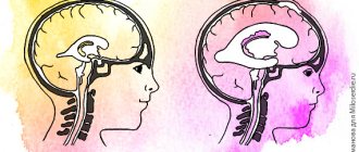
The cause of the disease is the expansion of the spaces around the ventricles due to excess fluid accumulation. The expansion of perivascular gaps leads to the formation of lesions, strokes, and heart attacks. The primary cause of the morphology is a violation of the blood supply to the perforating arteries of the brain. The diameter of the vessels is 100-300 microns. The anatomical structure includes many collateral anastomoses between arteries. Shunts provide additional microcirculation when one separate vessel is blocked. Instability of blood flow creates vulnerability of the centripental branches of the cerebral arteries.
An increase and decrease in intracranial pressure in the anterior horns of the lateral ventricles of both hemispheres of the brain forms the prerequisites for changes in the white matter.
Depending on the location of the zones of intracerebral foci, two types of pathology are distinguished:
- Subcortical;
- Periventricular.
The latter form is characterized by morphological changes in white matter located around the lateral ventricles, or domes over the ventricular segments.
The subcortical variety is characterized by the formation of foci inside the deep brain structures (semioval center).
MRI signs of periventricular leukoaraiosis:
- “Caps” along the posterior or anterior pole of the ventricle;
- A uniform stripe along the periventricular spaces.
MRI signs of subcortical leukoaraiosis:
- Scattered lesions within the cerebral hemispheres;
- Incomplete heart attacks;
- Demyelination – destruction of the myelin sheath of the neuron;
- Cysts are limited cavities with a clear contour and fluid inside;
- Angiectasias are local areas of arterial dilatation;
- Enlargement of perivascular spaces.
European scientists have conducted dozens of studies of patients with leukoaraiosis. Experts using magnetic resonance imaging have identified small areas of the brain deprived of blood supply. The lack of oxygen ensures gradual progression of the condition. Nosologies cannot be considered a natural sign of aging. In people over 60 years of age, minor vascular ischemia on magnetic resonance imaging is often observed, but pathologies also occur in young people.
Pathological areas of damage within the white matter lead to impaired communication and decreased speech functions. The information has been verified by scientists who have studied the brain of older and younger people. In response to performing mental tasks in patients with leukoaraiosis, the activity of the speech centers decreases, but the efficiency of the visual-spatial zones increases.
The more perivascular lesions, the more communication skills suffer.
Symptoms of leukoaraiosis
Destruction of white matter is characterized by disruption of brain functions. Clinical manifestations depend on the location and stage of the pathological process. The degree of damage to nervous tissue is determined using instrumental studies. When performing MRI, it is possible to diagnose leukoaraiosis at the initial stage.
The clinical picture of brain destruction is characterized by:
- motor dysfunction;
- cognitive impairment;
- psycho-emotional disorder;
- speech disorders;
- muscle weakness;
- headache;
- insomnia.
The first stage of leukoaraiosis is accompanied by slight dizziness, fatigue, and weakness. The patient notes:
- noise in ears;
- decreased concentration;
- general depression.
Speech disturbances and memory loss are possible. On examination, increased tendon reflexes are revealed.
The second stage occurs with a significant decrease in the functionality of the body. Note:
- impaired coordination of movements;
- loss of balance when walking;
- slowing of psychomotor functions;
- partial or complete loss of speech;
- decreased memory and attention.
The patient loses control of his actions. Apathy, irritability, and depression are noticeable. Frequent urination and night diuresis are possible.
The third stage is accompanied by an intensification of the listed symptoms. Observed:
- severe behavioral disorders;
- falling while walking;
- loss of speech, memory;
- urinary incontinence.
The patient is not capable of self-care.
Leukoaraiosis on MRI images
Destruction of white matter in the frontal lobe is characterized by a decrease in intellectual abilities against the background of satisfactory motor function.
The main causes of leukoaraiosis
The etiological components of the disease have not been identified. Main provoking factors:
- Infectious conditions with cerebral ischemia;
- Poor quality food;
- Age over fifty years;
- Adynamia (low mobility);
- Increased intracranial pressure;
- Alzheimer's disease;
- Dementia;
- Hypertonic disease;
- Ischemia, stroke;
- Disturbance of cerebral metabolism.
High blood pressure damages small arteries. Destruction of the periventricular vessels provides minor ischemia of the white matter.
Persistent hypertension significantly increases the likelihood of minor ischemia. Arterial congestion creates opportunities for increased arterial permeability.
In approximately 40% of people, the cause of the disease is a decrease in cerebral blood supply. Binswanger's disease develops due to a lack of oxygen supply.
The causes of occurrence after 50 years are abnormalities in the course of blood vessels, damage to the endothelial lining, and decreased elasticity of the arteries.
Metabolic disorders in middle age with increased formation of homocysteine create conditions for increased permeability of the arterial wall. Endothelial dysfunction of small capillaries creates the preconditions for the development of nosology.
Formed elements of blood responsible for the healing of lesions, platelets. Blood clots inside small capillaries obstruct blood flow.
Sleep disordered breathing (apnea) increases the risk of obstruction (narrowing of the bronchial tube). Snoring increases blood pressure. Weakness of the wall and impaired fiber elasticity during intracranial hypertension increases the risk of pathology.
Leukoaraiosis is observed in patients with type 1 or type 2 diabetes mellitus due to the development of metabolic syndrome and impaired mitochondrial functionality. Lack of glucose supply to brain tissue leads to hypoxia, ischemia, and circulatory pathology.
Degrees of leukoaraiosis at a young age
The formation of pathological foci and ischemic segments near the ventricles has different severity.
Classification of leukoaraiosis by morphology:
- 1st degree – slight loss of coordination, decreased walking speed;
- 2nd degree – development of clinical symptoms of movement disorders, loss of concentration and memory;
- Grade 3 is accompanied by a significant expansion of the ventricular spaces, increasing the manifestations of the disease. Patients require outside supervision;
- Grade 4 is characterized by uncontrolled urination, loss of balance, and suppression of psychomotor activity.
The progression of the pathology takes several years. Morphological changes are irreversible, so it is better to verify first-degree brain disorders, when supportive treatment can be selected.
The child has no clinical symptoms. Diagnosis is carried out due to the impossibility of treating speech disorders.
Stages of development
Leukoaraiosis of the head, as a disease, goes through three stages of development:
- With stage 1
of the disease, a medical examination can reveal revitalization of tendon reflexes, human instability, a reduction in step length when walking, and the ability to move only slowly. Neuropsychological examination reveals suppression of cognitive changes. The person complains of poor attention and memory deterioration, and cognitive activity is significantly reduced. - At stage 2,
the presence of clinical syndromes is clearly visible. The patient notices memory deterioration, and at the same time mental and psychomotor reactions slow down. A person cannot control his actions and notices uncertainty in his own walking. In some cases, the presence of apathy, depression, irritability and lethargy is noted. There is deterioration in the functioning of the genitourinary system - uncontrolled urination at night. A person loses social adaptation, which manifests itself in the form of deterioration in performance and the inability to care for oneself independently. - At stage 3
, the clinical picture worsens and leads the patient to disability. Behavioral disorders are clearly noticeable (severe lethargy, inability to walk independently, which sometimes ends in a fall, chronic urinary incontinence). At this stage, doctors note pronounced signs of a disorder in the functioning of the cerebellum.
Clinical picture of Binswanger's disease
The asymptomatic course of leukoaraiosis continues for several years until multiple lesions accumulate inside the brain. Cortical-subcortical disconnection syndrome develops during progression. The condition is accompanied by a disruption of the relationships between the cortical parts of the cortex and thalamic zones. Conductive fibers are located within the white matter. The formation of ischemic foci leads to difficulty in conducting a nerve signal along the conduction tracts.
The consequences of the condition are intellectual-mnestic disorders (pathology of memory, intelligence), emotional disorders, imbalance of muscle tone, vascular dementia.
People with stroke have severe leukoaraiosis, which puts them at high risk of developing post-stroke dementia in the future. Neuroimaging methods can visualize numerous changes, but it is impossible to prevent the rapid progression of the third stage.
The main symptoms of leukoaraiosis:
- Movement disorders;
- Speech problems;
- Defects in thinking, memory;
- Dysphoria, depression and other emotional disorders.
The type of symptoms is determined by the predominant localization of the area of leukoaraiosis. The subcortical form causes problems in the motor sphere, while the periventricular form causes speech disorders. In young people, the pathology is most often located along the ventricles.
First signs and development of symptoms
At the first stage of the development of the disease, it is difficult to identify the exact symptoms. Sometimes the clinical picture goes unnoticed. Most often they are paid attention to as they progress:
- disturbances in a person’s emotional state, which manifests itself in the form of constant depression and discomfort in society;
- memory deteriorates sharply, disturbances in the ability to think clearly are observed;
- changes in speech functions;
- disturbance of movement and functioning of the musculoskeletal system.
The further course of the disease can also occur without obvious symptoms until the moment of exacerbation or with a gradual deterioration of the condition. You can consider the most characteristic signs of leukoaraiosis:
- change in a person’s speech capabilities for the worse;
- frequent mood swings, which are accompanied by depression and chronic dystrophy;
- obvious disturbances in the functioning of the musculoskeletal system;
- deterioration of basic memory properties;
- a noticeable change in intellectual and thinking abilities.
MRI diagnosis of leukoaraiosis
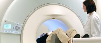
Before clinical symptoms occur, MRI detects lesions of leukoaraiosis by chance. Diagnosis of Alzheimer's disease, Parkinson's disease, verification of brain changes after a stroke are common situations in which nosology is identified.
MRI of the brain reveals a decrease in the thickness of the parenchyma, focal structures within the white matter.
Detection of Binswanger's disease of the first stage on magnetic resonance imaging helps to properly treat and delay the appearance of the second stage. Irreversible disorders of subcortical structures are revealed by T2-weighted imaging.
The nosological form is diagnosed using computed tomography, but the information content of the method is less. MR angiography, a method of contrasting blood vessels after injecting an enhancing drug (gadolinium), into a vein allows you to identify the provoking factor.
What does a brain CT scan show:
- Hypodense zones;
- Vascular dementia;
- Expansion of the ventricular system;
- Diffuse changes in white matter.
The information content of the examination is increased by using the “fat suppression” mode. The scan helps detect lesions within the cerebellum, brainstem, corolla radiata, and basal ganglia.
Degrees of expression
The severity of leukoaraiosis is divided into three degrees:
- Minor cognitive pathology of the subcortical region of the brain occurs. Such changes manifest themselves in the form of dizziness, headaches, and rapid loss of energy. Tendon reflexes are slightly activated. If you pay attention to the width of a person’s step, you will notice that it has become shorter.
- Leukoaraiosis of the second degree is actively progressing, which is expressed in a decrease in the patient’s cognitive functions. These include disorders of the neuropsyche and psychomotor skills, degradation of thought processes, restraint is replaced by the inability to keep oneself under control. Sometimes the patient begins to experience urinary incontinence, and frequent urination appears. Adaptation to society is lost, performance decreases. The symptoms are similar to Alzheimer's disease.
- The third degree is characterized by the same factors as the second, but they already have a pronounced character.
Prevention of leukoaraiosis
The causes of the disease have not been identified, so preventive procedures are aimed at eliminating provoking factors. After clarifying the pathogenetic mechanism of formation of areas of hypodense (low-intensity) MR signal, a list of procedures is prescribed:
- Organizing a healthy lifestyle (excluding smoking, alcohol abuse);
- Normalization of the diet (balanced diet of proteins, fats, carbohydrates);
- Moderate physical activity;
- Elimination of provoking factors.
Elderly people should periodically undergo MRI of the brain to identify pathology and monitor the dynamics of pathological processes.
Preventive measures
To date, there is no vaccine or medicine that could protect against the development of leukoaraiosis in the future. For preventive purposes, doctors may advise leading a healthy lifestyle.
A person should live in a mode of moderate activity and monitor nutrition. It is important to give up bad habits, because they have a detrimental effect on the functioning of the central nervous system.
If there is a cerebrovascular disease, the patient should receive timely treatment. Older people should have their functioning assessed regularly.
