Myocardial infarction is always a great risk to a person’s life. The cause of almost every third death in our country is cardiac arrest due to insufficient blood supply (ischemia) to the heart muscle (myocardium), as a result of which the organ tissue begins to die. Up to 40% of people with this diagnosis die in the first 15-20 minutes after the onset of the disease. That is why it is so important for patients with pathologies of the cardiovascular system to undergo examinations, to know the first signs of the development of myocardial infarction in women and men, and to always be ready to call an ambulance. Your health and life, as well as the lives of your loved ones, next to whom you find yourself at the right moment, may depend on the ability to correctly identify the symptoms of the disease, competent and prompt actions.
Symptoms of myocardial infarction at different stages of the disease
Signs of myocardial infarction differ at different stages of pathology development. During the disease there are 4 stages:
- stage of ischemia or acute period;
- stage of necrosis (acute period);
- stage of organization or subacute period;
- post-infarction period (scarring stage).
About 40% of all recorded heart attacks come as a surprise to the patient. Most often, doctors explain these by people’s inattention to their health. In the remaining 60% of cases, the disease is preceded for a long time by angina attacks.
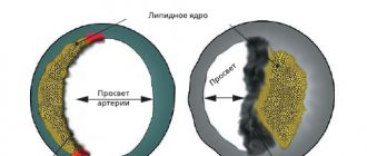
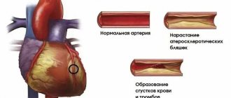
The first signal to show concern is the appearance of pain in the chest, back, left shoulder or forearm, sometimes radiating to the jaw, ear and, less often, to the stomach and lower back. The nature of the pain varies: from mild and sluggish to acute and cutting. Typically, such pain appears after playing sports or during everyday physical activity, during emotional experiences and stress. These are the first signs of coronary heart disease. In nine out of ten cases, IHD develops due to vascular atherosclerosis (deposits of cholesterol in the inner walls of blood vessels) that supply blood to the heart muscle.
Such attacks can be observed in a patient from several weeks to two, three or more years and often, if not intervened, end in myocardial infarction. Have you noticed these in yours? Contact specialists immediately! Timely measures taken, diagnosis and observation by doctors will help avoid the development of the disease.
If coronary heart disease is not treated, then the next stage may be the most acute period of a heart attack.
Five periods of illness
Each period has its own characteristics and characteristics:
- Pre-infarction. This period can last for several days or even weeks. The main symptom is an increase in angina attacks.
- The sharpest. It has a short duration - maximum 2 hours. In other words, little time passes from the development of ischemia to the occurrence of necrosis. This period can last only 20 minutes, so urgent hospitalization is necessary.
- Spicy. As a rule, several days pass from the onset of necrosis to myomalacia. Maximum duration – 2 weeks.
- Subacute. Scars are already beginning to form. The maximum duration of this period is 2 months.
- Post-infarction. The scar matures, and the myocardium has to adapt to new working conditions. It is very important here how the heart behaves during fundamental changes. If there is no adaptation, there is a high probability of a second attack.
Remember the following signs and symptoms of myocardial infarction:
- The first symptom of myocardial infarction is acute, squeezing or pressing pain in the chest, radiating to adjacent organs, the left ear, jaw, left arm, and sometimes the upper abdomen. The pain can last from half an hour to a day and, unlike angina, is not relieved by taking nitroglycerin;
- patients with the first signs of a heart attack may complain of toothache;
- the painful condition is accompanied by chills, weakness and dizziness, sometimes shortness of breath and cold sweat;
- arrhythmia develops, the pulse increases noticeably;
- the skin becomes pale gray due to lack of blood supply.
The nature and severity of pain during a heart attack depend on the location of the muscle damage. The larger it is, the stronger the pain. Sometimes the only symptom of a heart attack is sudden cardiac arrest.
The acute period is one of the most dangerous stages of the disease. Some of the cardiac muscle tissue is already dead, and the heart, finding itself in unusual conditions, may not withstand the load and stop. If you feel such symptoms and make sure that the usual remedies for angina pectoris do not help, immediately call an ambulance.
Symptoms during the acute stage of myocardial infarction become less pronounced. The pain subsides. As a result of tissue necrosis, the patient's temperature rises, which, depending on the focus of necrosis, may not subside for up to 7-10 days.
In the subacute period, the signs of myocardial infarction disappear, heartbeat and body temperature normalize. A week or two after a heart attack, the dead portion of the heart muscle begins to scar.
In the post-infarction period, there are no symptoms of the disease. But angina attacks will not simply disappear, so if measures are not taken, there is a high probability of developing a recurrent myocardial infarction. To avoid this, it is necessary to treat the root causes of the pathology: coronary disease and atherosclerosis. Due to damage to blood vessels by cholesterol plaques, not only the heart suffers, but also the limbs, digestive organs and brain (cerebral infarction).
Professional treatment. Why should you only contact experienced doctors?
As soon as myocardial infarction is suspected, the patient should be immediately admitted to the local cardiac intensive care unit. If an acute period is recorded, then the person first needs to remain in bed. Any psychological stress during this period is excluded! As for nutrition, it is severely limited in volume and calorie content. When the subacute period begins, the person can already be transferred from the intensive care unit to cardiology, where he will continue the recovery course after a severe attack. Treatment of myocardial infarction can take a long time, when the patient will systematically have to be checked by the attending physician, take various tests and undergo a bunch of examinations.
To relieve severe pain, the doctor prescribes narcotic analgesics. In this case, treatment is carried out in combination with antipsychotics, as well as the administration of nitroglycerin through an injection into a vein.
Therapy carried out by experienced specialists in case of a heart attack is necessary in order to prevent and eliminate interruptions in the heart and cardiogenic shock. As a rule, during this period the patient takes antiarrhythmic drugs, including thrombolytics, antispasmodics and nitrates.
It is extremely important that treatment for myocardial infarction is carried out by an experienced doctor. Only a prestigious clinic employs specialists who can correctly assess the patient’s condition. When a person survives an attack, this does not mean that the danger is over. There is a long course of treatment ahead, and the success of the process depends on the selection of medications, the experience of doctors and the technical equipment of the clinic.
Of course, when a person loses consciousness as a result of a heart attack, it is not advisable for family and friends to go to a private clinic. But the recovery period is better spent in a commercial medical institution. When the patient can walk normally, you can make an appointment with a specialist who will help minimize the consequences of a serious illness.
Symptoms and signs of myocardial infarction in women
Female heart attacks have a number of atypical features and are less studied compared to male ones. The thing is that women are less likely to suffer from coronary heart disease. They are helped in this by estrogen, a hormone that is produced by the female body almost throughout life. It prevents the development of atherosclerosis. After menopause, the amount of estrogen in the body decreases and the chances of developing coronary heart disease in women increase.
Precursors of myocardial infarction in women are often swelling of the extremities that appear at the end of the day, chronic fatigue, shortness of breath, and digestive problems.
Often, a heart attack in women occurs without severe chest pain and may be accompanied by nausea, high fever and high blood pressure. Sometimes a false toothache develops. There are also cases of asymptomatic heart attacks.
This course of the disease is much more dangerous, since the patient can endure the first signs of the disease without attaching much importance to them. Some patients who have experienced a heart attack find out about it only after a while, undergoing diagnostics.
Symptoms and signs of myocardial infarction in men
In men, the course of the disease has all the characteristic signs:
- sharp pain behind the sternum, in the left arm, shoulder and forearm;
- sticky cold sweat;
- weakness, dizziness, pale gray skin color;
- shortness of breath, rapid pulse;
- high blood pressure.
Despite the development of medicine, acute myocardial infarction is increasingly being diagnosed in both men and women under 40 years of age. The reason for this is a sedentary lifestyle; a diet with a predominance of unhealthy fats, which are rich in fast food products; obesity; smoking; diabetes. Young patients often have an asymptomatic form of heart attack.
Signs of atypical myocardial infarction in women and men
Atypical signs of the disease are observed in elderly patients who have experienced one or more myocardial infarctions. Based on the nature of the symptoms, several atypical forms are distinguished:
- the abdominal form is characterized by indigestion, nausea, vomiting, hiccups;
- in the asthmatic form, the patient feels suffocated and may develop a cough;
- the cerebral form is represented by dizziness, presyncope;
- atypical pain syndrome (toothache, pain in the neck, left ear, spine, left leg, left fingers).
Often, acute myocardial infarction occurs without signs or symptoms in both women and men and is detected only after a while, after the patient has done an ECG. A silent heart attack is typical for patients with diabetes mellitus because it dulls the pain syndrome. This course of the disease is much more dangerous: the absence of symptoms of myocardial infarction makes it difficult to provide first aid to the patient. The only way to avoid the development of coronary heart disease and prevent myocardial infarction is to undergo timely and regular diagnostics of the cardiovascular system. At the Center for Circulatory Pathology, our specialists work with the latest equipment from Europe, the USA and Asia, which is capable of diagnosing the disease long before the first signs and symptoms of myocardial infarction. Treatment in our center is based on the best experience of domestic and foreign medical practices.
For patients whose health does not allow them to come to our Center, there is a special on-site diagnostics department. Based on a special method of non-surgical treatment of cardiovascular diseases, we develop a unique set of therapy and diagnostics for everyone who comes to our clinic.
Clinical observation
Patient S., 28 years old, was admitted on June 25, 2019 to the State Clinical Hospital named after. S.S. Yudina DZM" to the emergency department of intensive care and intensive care with complaints of very strong pressing pain in the chest and in the heart area, radiating to the back, both arms, left collarbone, severe shortness of breath, weakness, cold sweat.
History of the disease.
The patient is an active athlete. Since 2011, he has been involved in martial arts with regular sports training, which was accompanied by weight lifting and taking anabolic steroids (methandrostenolone and testosterone) in courses every 3–4 months. Since 2011, he has been constantly taking the same energy drink (composition: alpine water, taurine (250 mg / 100 ml), caffeine (32 mg / 100 ml), B vitamins (B3, B5, B6, B12), sucrose , glucose, acidity regulators (citric acid, sodium bicarbonate, magnesium carbonate), dyes (simple sugar coloring, riboflavin), flavorings) 250 ml, 5-6 cans per day. Since 2021, he stopped training in martial arts, but continued regular physical training with weight lifting. In 2021, he started smoking 10 cigarettes a day, and since April 2021 he has been smoking 20 cigarettes a day. Since July 2021, after the death of his father from a traumatic brain injury with cerebral hemorrhage, the patient was in a state of psycho-emotional stress, slept 4–6 hours a day, and did not feel rested after sleep. During this time, he worked intensively and was actively involved in bodybuilding. At the end of May 2021, after bloodletting (according to a religious rite), the patient’s body temperature increased to 39 °C and frequent urination appeared. These manifestations were observed for 2 weeks, but he did not consult a doctor or take medications. From this time until hospitalization, I lost 10 kg.
On June 22, 2019, the patient began another course of anabolic hormones. On June 24, 2019 at 22:00, after drinking 1 can of energy drink, the patient began physical training, although he usually trains during the day. At the beginning of the training, very severe headaches, dizziness, then nausea and one-time vomiting suddenly appeared. I stopped training and returned home. At about midnight he went to the 24-hour medical care department, where he was examined by a therapist. An electrocardiogram (ECG) performed at the same time showed no changes. No treatment was prescribed. On June 25, 2019, at about 2 a.m., the patient woke up due to severe pain behind the sternum and in the heart area, radiating to the back, both arms and left collarbone. The patient endured pain throughout the day, did not take medications, and did not seek medical help. On June 25, 2019 at 7 p.m., due to weakness, cold sweat, severe shortness of breath and increasing pain up to the point of fear of death, the patient called an ambulance and was taken to the hospital.
Anamnesis of life.
As a child, he suffered an appendectomy and hepatitis A. He did not take medications. I didn’t notice any allergies. Doesn't drink alcohol. Habitual blood pressure (BP) 110/70 mmHg. Art. There is no heredity for cardiovascular diseases.
Results of physical examination upon admission.
The condition is serious. Consciousness is clear. Position active. The skin is of normal color, warm, moist. There is no swelling. Height 176 cm, body weight 81 kg, body mass index 26.15 kg/m2, waist circumference 88 cm, body temperature 37.1 °C. On percussion the sound is pulmonary, there is no dullness. On auscultation, breathing is vesicular, there are no wheezes, respiratory rate (RR) is 18 per minute, blood oxygen saturation is 96%. The area of the heart is not changed, the apical impulse is weakened. Percussion: the boundaries of the heart are not expanded. On auscultation, heart sounds are sonorous, rhythmic, and there is a weakening of the first sound at the apex. Heart rate 90 per minute, blood pressure 100/70 mm Hg. Art. Peripheral arteries and veins are not changed. The tongue is pink and moist. The abdomen is symmetrical, soft and painless on palpation. The edge of the liver is palpated along the edge of the costal arch. The kidney area is not changed. The symptom of effleurage is negative.
Results of clinical diagnostic examination.
Chest X-ray
(06/25/2019): without pathology.
ECG data on admission
(06/25/2019; Fig. 1, A): ST segment elevation was noted in leads I, AVL, V1–V6 with a maximum of 6 mm - the acute stage of anteriorly advanced left ventricular (LV) MI.
Coronary angiography
(06/25/2019): the type of blood supply to the heart is balanced. The trunk of the left coronary artery is usually developed and not changed. Occlusion of the anterior descending artery (ADA) in the proximal segment; distal segments are not visualized. The diagonal branch is not visualized (Fig. 2, A). The circumflex branch, the branch of the obtuse margin, and the right coronary artery are not changed. Conclusion: acute occlusion of the LAD.
On June 25, 2019, an operation was performed: mechanical recanalization, thrombus extraction, angioplasty and stenting of the LAD with a drug-eluting stent (1DES).
Progress of the operation.
A coronary guidewire was used to recanalize the occlusion of the proximal segment of the LAD, and the guidewire was passed into its distal segment. Thrombus extraction was performed and fragments of blood clots (with fibrin contents) were removed. During control coronary angiography, the result was considered suboptimal: thrombotic occlusion of the distal and apical segments of the LAD, occlusion of the distal segment of the diagonal branch. Angioplasty was performed in the occlusion zone with a 2.0×20 and 2.5×20 mm balloon catheter with a pressure of 14 atm. A solution of eptifibatide 6.2 ml (2 times) was connected, and in parallel, dosed administration of the drug was connected at a rate of 11 ml/h. A Promus PREMIER stent (Boston Scientific, USA) 3.5×28 mm (pressure 18 atm) was positioned and implanted in the area of residual stenosis of the proximal segment of the LAD. In the area of occlusion, thrombus extraction (with the removal of thrombus fragments) and angioplasty with balloon catheters 2.0×20 and 2.5×20 mm with a pressure of 4 and 6 atm were performed multiple times. Blood flow in the area of the stented segment TIMI (Thrombolysis In Myocardial Infarction) - 3 (normal blood flow); There are no dissection zones, extravasation of the contrast agent, or residual hemodynamically significant stenoses. Blood flow in the distal and apical segments of TIMI is 0 (no antegrade blood flow).
Immediate result.
Blood flow in the LAD: TIMI 3 (in the proximal segment), TIMI 0 (in the distal and apical segments; Fig. 2, B).
Echocardiography (EchoCG) in B- and M-mode with Doppler analysis
(06/26/2019). B- and M-mode: the aorta is not compacted, the root is 3.4 cm, the ascending section is 3.5 cm (normal is 2.0–3.7 cm). The valve apparatus is intact. Left atrium: anteroposterior size 3.6 cm (normal 2.0–4.0 cm), apical size 3.7×4.2 cm (normal <4.4×4.8 cm). Right atrium: 3.2x3.8 cm (normal <4.4x4.8 cm). Right ventricle: anteroposterior size 2.5 cm (normal 2.1–3.5 cm), interventricular septum 1.0 cm (normal 0.6–1.0 cm). The posterior wall of the left ventricle is 1.0 cm (normal 0.6–1.0 cm). LV: end-diastolic size 5.5 cm (normal in men 4.2–5.8 cm); end-systolic size 4.4 cm. LV ejection fraction 38% (normal in men >52%). Violation of myocardial contractility - akinesis of the basal-septal, mid-septal, upper-septal, superolateral, supero-inferior, supero-anterior, supero-posterior segments, basal anteroseptal, middle anteroseptal segments of the LV. There is no separation of the pericardial layers. The inferior vena cava is not dilated, collapses on inspiration >50%. Doppler analysis: aortic valve - no regurgitation detected, mitral valve - 1st degree regurgitation, tricuspid valve - 1st degree regurgitation. Systolic pressure in the pulmonary artery is 20 mm Hg. Art. Pulmonary valve: regurgitation degree I.
Conclusion.
The cavities of the heart are not dilated. Violation of local contractility of the LV. Decreased global LV contractility. Mitral valve insufficiency degree I. Tricuspid valve insufficiency degree I.
treatment was carried out in the Department of X-ray Endovascular Surgery
: heparin 5000 IU/ml subcutaneously 4 times/day, acetylsalicylic acid 100 mg 1 tablet in the evening, omeprazole 20 mg 1 tablet 1 time/day (06/25/2020–07/01/2020), warfarin 2.5 mg 2 tablets in the evening , bisoprolol 5 mg 1 tablet in the morning, atorvastatin 40 mg 1 tablet in the evening, clopidogrel 75 mg 1 tablet in the morning, enalapril 2.5 mg once a day, spironolactone 25 mg 1 tablet in the morning (07/01/2020–07/04/2020).
Data from laboratory research methods in the dynamics of observation. Clinical blood test:
06.26.2019 - hemoglobin 164.0 g/l, red blood cells 5.44×10¹²/l, platelets 193×109/l, leukocytes 17.2×10⁹/l, hematocrit 47.3; 06/02/2019 - hemoglobin 162.0 g/l, red blood cells 5.52×10¹²/l, platelets 198×109/l, leukocytes 7.51×10⁹/l, ESR 19 mm/h, hematocrit 47.5.
Blood chemistry:
06/26/2019 - glucose 6.94 mmol/l, cholesterol 4.07 mmol/l, triglycerides 1.94 mmol/l, total protein 61 g/l, urea 4.4 mmol/l, creatinine 63.53 µmol/l , total bilirubin 8.8 µmol/l, sodium 137 mmol/l, potassium 3.5 mmol/l, chlorine 98 mmol/l; 06/27/2019 - without pathology.
Whole blood glucose:
06/27/2019 - 5.5, 10.3, 7.1 mmol/l, 06/28/2019 and 07/02/2019 - within normal limits.
General urine analysis:
06/26/2019 - protein 0.5 g/l; 07/02/2019 - protein negative.
Coagulogram:
06/26/2019 and 06/29/2019 - within normal limits; 06/28/2019 - activated partial thromboplastin time (aPTT) >100 s; international normalized ratio (INR) 1.00, prothrombin time 13.3 s; 01, 02, 03 and 07/04/2019 - aPTT 24.0 s, INR 0.88, 2.10, 1.96 and 2.50, respectively; 03 and 04.07.2019 - prothrombin time 23.8 and 29.7 s, respectively.
Troponin (06/26/2019) 27.00–89.70 ng/ml.
The assessment of psycho-emotional stress according to the Reader test corresponded to 1.71 points - a high level of psycho-emotional stress.
24-hour Holter ECG monitoring
(07/02/2019): sinus rhythm. Maximum heart rate 112 per minute. Minimum heart rate 69 per minute. Average heart rate is 78 per minute. 1 single ventricular and 2 single supraventricular extrasystoles were recorded. No significant ischemic dynamics of the ST segment or clinically significant pauses were detected.
EchoCG in B- and M-mode with Doppler analysis
(07/04/2019). Conclusion: Violation of local contractility. Thrombus in the LV cavity (size 21×17 mm). Global contractility of the LV myocardium is reduced (ejection fraction 40%).
ECG data at discharge
(04.07.2019; Fig. 1, B): ST segment elevation remains in leads I, AVL, V1–V5 with a maximum of 2 mm; pronounced regression of R in leads I, AVL, V2, V5, V6; QS in V3, V4; the formation of negative T in leads I, AVL, V1–V6 is a subacute stage of anteriorly advanced transmural LV MI.
Clinical diagnosis.
Main disease:
transmural anteriorly advanced left ventricular myocardial infarction with ST segment elevation dated June 25, 2019.
Mechanical recanalization, thrombus extraction, transluminal balloon angioplasty and LAD stenting with a Promus Premier 3.5×28 mm stent from 06/25/2019. Complications of the underlying disease:
acute aneurysm of the apical anteroseptal region of the left ventricle. Thrombus in the cavity of the left ventricle.
The patient was discharged on 07/05/2019 under the supervision of a cardiologist at the local clinic.
Condition at discharge
: satisfactory. The skin is of normal color, there is no swelling. In the lungs there is vesicular breathing, no wheezing. NPV 16 per minute. Heart sounds are muffled and rhythmic. Heart rate 60 per minute. Blood pressure 110/70 mm Hg. Art. The abdomen is soft and painless. Coagulogram data: INR 3.20; prothrombin time 35.2 s.
Recommendations for discharge:
1) diet with limited fat, carbohydrates, salt; 2) drug therapy: clopidogrel 75 mg in the morning for 1 year, acetylsalicylic acid 75 mg in the evening for 1 month, warfarin 2.5 mg 1.5 tablets in the evening (INR control, target level 2.0–3.0), omeprazole 20 mg in the evening, atorvastatin 40 mg in the evening, spironolactone 25 mg in the morning, bisoprolol 2.5 mg in the morning, perindopril 2.5 mg in the afternoon; 3) echocardiography monitoring after 10 days (thromb in the LV); control of biochemical blood parameters, INR.