Diagnosing coronary heart disease is not always an easy task. In some cases, the diagnosis is beyond doubt. But there are also situations when the patient’s complaints are ambiguous, and studies performed at rest do not completely exclude this serious disease. Then the attending physician prescribes stress echocardiography.
During this study, the subject’s heart works under conditions of increased load:
- 1) Physical a
treadmill (treadmill) or bicycle (bicycle ergometer); - 2) Pharmacological ,
when a drug injected into a patient’s vein makes his heart beat stronger and faster.
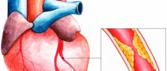
The essence of the method: the use of these loads increases oxygen consumption by the heart. At the same time, in a healthy person, the arteries that encircle the heart (coronary) bring him the necessary amount of oxygen and the contractility of the heart evenly increases. And in a patient with narrowed coronary arteries, the blood flow does not increase adequately to the load, the contractility of the heart is regionally impaired, which is recorded by the doctor on the monitor of the ultrasound machine.
Carrying out stress echo-CG
The total examination time is no more than 45 minutes. It takes place in several stages:
- The patient lies on the couch on his left side. A gel is applied to the chest area and a resting ultrasound is performed.
- A load is placed on the heart depending on the chosen technique. During a pharmacological test, drugs are administered. Physical activity is created on a treadmill or bicycle ergometer.
- During a pharmacological test, heart parameters are recorded during the period of exercise. When using a bicycle ergometer, ultrasound of the heart is performed immediately after completion of the movement.
- The intensity increases every 2-3 minutes.
- The duration of the test is 10-15 minutes.
The doctor compares the results of ultrasound at rest and movement. The analysis makes it possible to make a correct diagnosis of heart disease.
In our clinic, patients can get advice from experienced cardiologists and functional diagnostic doctors and undergo stress echo-CG. We employ leading specialists who regularly improve their skills. The latest Swiss and Italian equipment is used for diagnostics. By contacting us, the patient receives a guarantee of the quality of medical services. Call or schedule a consultation online at a convenient time.
Preparation
- 3 days before the test, you must stop taking certain medications, primarily beta blockers, under the supervision of your doctor.
- Patients with arterial hypertension should come for the test with a blood pressure below 180/110 mmHg. High blood pressure is a contraindication for the study.
- The day before the study, you should not smoke, drink alcohol or coffee.
- 3 hours in advance - you must stop eating and limit physical activity. You can drink water.
- You must bring comfortable sportswear and shoes.
- You must have with you: a referral from your attending physician, the latest ECG, an EchoCG report, and for a pharmacological test, a Holter monitoring report.
Types of load tests
The type of technique is chosen depending on the patient’s heart condition. If the patient has problems with the musculoskeletal system, or an angina attack occurs during physical activity, drug or electrical stimulation is performed. For allergies to drugs, asthma and lung diseases, stress echocardiography with physical activity is used.
The latter study is used more often because the test conditions are as close as possible to real life. A type of stress echocardiography is the treadmill test and bicycle ergometry. Despite the reliability of the information, due to shortness of breath it is not possible to obtain a clear image of the left ventricle.
The procedure for performing stress echocardiography:
- The patient changes clothes, electrodes for recording an ECG and a cuff for measuring blood pressure are attached to his body.
- At the first stage of the procedure, the doctor records an ultrasound of the heart at rest.
- Then the patient is given an ever-increasing physical or pharmacological load, the doctor monitors and records ECG changes, pulse rate and blood pressure, the absence or occurrence of complaints.
- As soon as the patient performs the amount of physical activity characteristic of his gender, age and health status, a repeat ultrasound of the heart is performed. With a pharmacological test, the doctor performs an ultrasound of the heart throughout the test. After stopping the test, the patient is monitored for about 10 minutes.
- All recorded indicators are saved on a computer, processed and compared. The total duration of the test is 40-60 minutes, after which the patient receives a conclusion.
Preparation for the procedure
Stress echocardiography of the heart measures parameters that depend on external factors. In this regard, the patient was required to comply with certain conditions to ensure the reliability of the test.
On the day of the study, the patient should not take medications that affect the results: beta blockers, nitrates, etc. They change vascular tone. During the day, the patient refuses foods that increase or decrease blood pressure and heart rate: tea, coffee, alcohol and others. It is recommended to stop smoking as it affects the functioning of the heart.
During the preparation stage, you should not play sports or experience increased physical activity the day before.
What stress tests will a cardiologist suggest?
The type and duration of EGC examinations differ. Depending on the indications and goals, the cardiologist may offer the following tests:
Load tests (Step test). A simple type of diagnostics that does not require special equipment. First, an electrocardiogram is taken from the patient at rest. Then he performs several exercises (running, walking in place for 2 minutes, 20-30 squats). After this, another cardiogram is recorded, the data of which is compared with the first results. In some cases, another ECG is performed, for example, 3 minutes after exercise.
Bicycle ergometry. A person exercises on a bicycle ergometer (a special bicycle connected to a computer). In this case, the intensity of the load may change. It all depends on the purpose of the examination. The device records indicators at rest, during the test and after exercise.
Treadmill test. It is carried out on a treadmill, which allows you to more accurately change the load and speed of the moving belt, adjusting it to each person being examined, even for a child.
Indications for stress echocardiography
- Complaints of chest pain.
- Complaints of shortness of breath during physical activity.
- Diagnosed heart diseases or the need for their diagnosis (ischemia, myocardial infarction, arterial hypertension, atherosclerosis, etc.).
- Assessing the need for coronography, bypass surgery, and other cardiac surgeries.
- Determination of myocardial viability after myocardial infarction or before surgical treatment of the heart.
- Assessing the patient's endurance in case of need for another complex non-cardiological operation.
Features of the method
An early sign of weakened blood circulation in the myocardium is a decrease in the number of heart contractions in response to additional load, whereas in a normal physiological state contractions remain unchanged or increase.
- deterioration in the contractility of the damaged area (visualized by ultrasound);
- pathological changes during ECG recording (determined by stress test);
- the appearance of pain behind the sternum.
Myocardial movements are preliminarily assessed before testing. After this, the patient is given a drug that increases the heart rate or is asked to do an exercise test. A pharmacological test causes more complications from the cardiovascular system than stress tests. In the case of a load test, it is recommended to rotate the pedals on a bicycle ergometer in a horizontal position. This makes it possible to quickly move the patient to the couch.
Evaluation of results
The results of the study are displayed in the form of a two-dimensional graph, which makes it possible to fully assess the quality of left ventricular functioning. Interpretation of the results includes an assessment of the degree of thickening and mobility of the heart muscle tissue in individual areas. A preliminary analysis of the graphs is carried out by a specialist immediately after their registration. After the screening is completed, the cardiologist can view a video recording of diagnostic indicators in slow motion. The obtained data is stored on disks, creating an information base for the patient with further assessment of the dynamics of heart performance. Thus, stress echocardiography is a modern method for diagnosing coronary artery disease. The study allows us to determine the initial stage of the disease, when other methods reveal low effectiveness. However, before the procedure, possible cardiac complications associated with excessive load on the organ should be taken into account.
How is cardiac ultrasound performed?
During echocardioscopy, the doctor uses various modes of the ultrasound machine:
- one-dimensional (M-mode),
- two-dimensional (B-mode),
- Doppler mode (assessment of the speed of blood flow in chambers and vessels),
- color Doppler - CDK (to determine the direction of blood flows and identify pathological ones),
- power doppler (records the very fact of the presence of blood flow in the vessels),
- tissue Doppler (a deeper assessment of myocardial contractility, based on a study of the nature of the movement of the walls from the sensor and to it),
- 3D echocardiography (it brings maximum benefit before operations on the valves - they are almost completely visualized before the intervention, which is important for the surgeon to determine tactics).
Transthoracic ultrasound
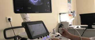
Echocardiography through the chest is performed in an ultrasound or functional diagnostics room. Sometimes in a hospital, due to the severity of the patient and the impossibility of transportation, a portable ultrasound machine is used to examine him. The nurse or doctor asks the patient to undress from top to waist, including women who need to remove their underwear. Next, the patient needs to lie on the couch on his left side and place his left hand under his head. In this case, the head end of the couch is slightly raised - this way, the maximum distance between the intercostal spaces is achieved for better visualization of the heart.
The position of the patient in relation to the doctor can be different, it all depends on the preferences of the latter and the arrangement of the office. The patient can be turned with his face or back towards the doctor, with his head towards or away from the device.
The specialist lubricates the sensor with ultrasound conductive gel for better contact with the skin, applies it to the left side of the chest, visualizes the heart, displaying its standard positions for measurements.
Transesophageal echocardiography
Transesophageal ECHO is performed strictly on an empty stomach in an ultrasound or functional diagnostics room. They take the patient’s consent to conduct the study, having first explained its entire essence and course of events. The throat is irrigated with lidocaine spray, the patient is asked to remove dentures and lie on the couch on his left side, bending his knees and placing his hands under his cheek or on his stomach. A mouthpiece is inserted into the mouth to prevent the patient from biting the probe. The doctor then inserts the endoscope. At the beginning, he asks the patient to make swallowing movements to move the device easier. Having reached a certain position, the doctor begins the examination itself. It lasts 10-20 minutes.
Echocardiography with contrast
When using a contrast agent, it is injected into one of the veins or arteries on the thigh - depending on the type of drug and the purpose of the study. In this case, the ultrasound sensor remains on the patient in order to register all data and take measurements in a timely manner.
Benefits of stress echocardiography
What is a stress ECG? This method was formed by merging two types of studies - cardiac ultrasound and stress ECG (bicycle ergometry). This is one of the most modern and sensitive methods of non-invasive diagnostics - it allows you to detect hidden disorders in the heart that are not noticeable at rest. Most often used for:
- detection of silent myocardial ischemia and angina pectoris;
- determining the viability of the myocardium in the area of post-infarction damage;
- assessment of disturbances in heart rate, blood flow and blood pressure at rest and during exercise;
Stress echocardiography is a safe research method that can be performed an unlimited number of times.
Decoding the results
Immediately after stress echocardiography, the cardiologist begins to decipher the data obtained. It assesses the condition of the walls of the heart, determines the number of affected areas of the left ventricle, and the ability of various segments to contract. The patient receives the results of the examination on the same day; the doctor describes the clinical picture in detail and gives a conclusion.
Make an appointment
You can see prices for services
Advantages and disadvantages
Among the significant advantages of stress echocardiography are:
- Ability to visualize each segment of the left ventricle;
- Large selection of echocardiographic indicators of cardiac contractility;
- Evaluation of research results in real time;
- Greater mobility of equipment;
- Does not cause any discomfort to the patient;
- Low cost of research;
- Safety and non-invasiveness of the procedure;
- No side effects.
Thanks to the study, the doctor will be able to identify patients with low and high risk of cardiovascular pathologies. It also helps to choose an effective treatment regimen. Despite this, stress echocardiography has disadvantages:
- High requirements for the qualifications of a doctor;
- Great importance of subjectivity when interpreting results;
- The duration of the procedure is from half an hour to several hours;
- Impossibility of carrying out in patients with poor visualization of cardiac structures.
Contraindications to stress echocardiography
Stress echocardiography should not be performed in patients with acute myocardial infarction, hepatic, respiratory, renal, heart failure, thromboembolism, acute myocarditis, or pericarditis. The study cannot be done if there is an uncontrolled disturbance of heart rhythm and cardiac conduction. In addition, the doctor always pre-assesses exactly what kind of load the patient can normally tolerate, and in which cases the load should not be given. Patients with severe obesity do not undergo the study due to its low information content. Significantly elevated baseline blood pressure, tachycardia and the presence of aneurysms are relative contraindications.
Types of loads during research
There are several types of load used in stress echocardiography. Among them:
- Physical: treadmill running, vertical and horizontal bicycle ergometry, work on a manual ergometer, isometric load;
- Pharmacological: tests are carried out with dipyridamole, ATP, dobutamine and other drugs;
- Transfood: direct electrical stimulation of the atria;
- Read the types: cold, mental, test with hyperventilation.
The doctor will decide what type of load will be applied during stress echocardiography. In patients at high risk of coronary heart disease, bicycle ergometry or dobutamine tests are used.
How to prepare for research?
At the preparatory stage of the study, the patient is prescribed medications containing nitrates, which can reduce the number of myocardial contractions and also lower blood pressure. The medication is prescribed to protect the heart muscle from the effects of adrenaline produced during stress, which can cause adverse reactions from various organs and systems. To fully prepare the body for the procedure, it is necessary to follow these recommendations:
- one day before the procedure, drinks containing caffeine and alcohol should be avoided;
- It is necessary to avoid physical activity for several hours;
- last meal – no less than 3-4 hours before the procedure;
- quit smoking immediately before screening.
On the day of diagnosis, patients are allowed to take nitroglycerin to stop a possible attack of angina. However, the use of funds must be agreed with a specialist.