| For a pregnant woman, the heartbeat of her unborn baby is a very important indicator and moral support. This knock can indicate whether the child is healthy or not. Of course, the mother will not understand this on her own, but doctors will identify pathologies if they are present. |
- Timing of detection of fetal heart rate
- Causes of fetal heart rhythm disturbances
- Why is the fetal heartbeat determined?
- Methods for listening to the fetal heartbeat
- Determining the sex of the baby by heartbeat
- How can you hear your child’s heartbeat?
Timing of detection of fetal heart rate
In the first months of gestation, the heart of the unborn child can already be heard.
With the help of ultrasound diagnostics, specialists hear the heart in the 5th week of gestation. Even earlier (at the 3rd or 4th week), the fetal heart rate can be heard by being diagnosed with a vaginal sensor. It is at these dates that you can hear the heart of your unborn child for the first time. With the period of gestation and with the activity of the fetus, the heart rate also changes. 110-130 beats per minute are recorded at 6-8 weeks. A maximum of 190 beats is heard at 8-11 weeks of gestation, and 140-160 beats can be heard starting from the 11th week of conception. The gynecologist must assess what phase the child’s activity is in when determining the heart rate, the time of the procedure, whether the unborn baby has any illnesses, and whether his mother has certain pathologies.
Obstetric stethoscope or phonendoscope
On the one hand, this method is available to everyone: you can buy this tube at any pharmacy for little money. On the other hand, it is not easy to count heartbeats using this tool at home. For this you need an assistant, because you can’t do it alone. At the same time, he must have skills that he most often does not have. Moreover, you need to listen to tones at certain points. Correctly counting the number of beats will be interfered with by extraneous noises (mother’s heartbeat, contraction of the muscles of the intestinal walls and other sounds), which you need to learn to distinguish from the fetal heartbeat.
Before 25 weeks, it is unlikely to be possible to hear the heart of an unborn child at home.
Causes of fetal heart rhythm disturbances
The reasons can be very different. If the heart rate is less than 80 beats/min, in the first weeks this may indicate that the pregnancy may be terminated. If the period is less than 4 weeks, then the heart rate may be less than 120 beats/min, this will not be a pathology. From the 12th week to the last day of gestation, a heart rate less than 120 beats/min may indicate compression of the umbilical cord or lack of air (chronic) in the fetus. The same heart rate during childbirth indicates that the baby is in a state of acute hypoxia, or that the umbilical cord is compressed by the mother’s muscles during contractions.
A fetal heart rate of 170 beats per minute in rare cases may indicate a dysfunction of the placenta, but in general this is a variant of the norm. At the 12th week and later, an increased fetal heart rate indicates chronic hypoxia, or may occur in response to the stress of the expectant mother. The reasons for increased heart rate during childbirth are similar to those described above.
If the tones are heard muffled, the doctor makes efforts to listen to the fetal heart rate, this may indicate the following:
- the child has cardiovascular system defects
- the mother is obese (significantly overweight)
- ultrasound sensor is outdated
- pregnancy is too early
Muffled tones, starting from the 12th week of gestation and until the last day of gestation, may have the following cause:
- defects of the cardiovascular system in a baby
- the fetus is positioned so that its heart is difficult to listen to
- oligohydramnios or polyhydramnios
- anterior placenta previa
- fetoplacental insufficiency
- obesity in the expectant mother
If the doctor cannot listen to the fetal heart sounds, do not be alarmed ahead of time. Again, the reason may be an old ultrasound sensor. But this may also indicate more serious problems, including an incipient abortion or a frozen pregnancy (the fetus stops developing and dies in the mother’s belly). The absence of fetal heart rate starting from the 12th week indicates that antenatal fetal death has occurred, or the doctor is using an old sensor, or listening to heart sounds incorrectly. During childbirth, the reasons are similar to the last two mentioned.
What problems are diagnosed using cardiotocography?
Cardiotocography is an additional examination method. Therefore, it is impossible to make an accurate diagnosis based only on its results. However, with the help of CTG it is possible to detect the development of various fetal pathologies, including the following disorders:
- heart rhythm disturbance;
- hypoxia;
- pressing or entwining the umbilical cord.
If CTG reveals abnormalities in the condition of the fetus, additional ultrasound and Doppler sonography are performed.
Why is the fetal heartbeat determined?
- to establish the fact of pregnancy
When a woman sees a positive result on a home pregnancy test, she should go to the gynecologist. He does an ultrasound to confirm or refute the test result. From the third week, as already noted, you can hear the heartbeat of the embryo. If at the first appointment you are told that there is no fertilized egg in the uterus, or that the embryo’s heartbeat cannot be heard, do not panic! After a week, most likely, your baby's heart will be sufficiently formed and the doctor will hear its beating.
In some cases, a return visit to the gynecologist reveals deformation of the ovum and lack of heart rate. Then the doctor speaks about a frozen pregnancy. Need a medical abortion. The doctor prescribes hormone-based medications. You can try to conceive again 3-6 months after an abortion for medical reasons.
- to assess the condition of the unborn baby
All environmental and maternal health factors affect the baby's heart. Heart rate depends on:
- the percentage of oxygen in the air that the mother is breathing at the moment
- fetal activity
- baby's sleep
- physical activity pregnancy
- mother's illnesses
- the stress that the mother is currently experiencing
But when exposed to these factors, the baby’s heartbeat changes temporarily. If the heart rate is not normal for a long time, the doctor may suspect that the baby is not getting enough blood. A diagnosis of fetoplacental insufficiency is made. It is almost always chronic. Sometimes the condition requires emergency delivery.
- to monitor the condition of the child during childbirth
Childbirth is a difficult stage in a baby’s life. There is a lack of oxygen; it is compressed as it passes through the mother’s birth canal. Basically, babies' hearts can withstand this stress. But in some cases there are emergency conditions, and the child will not survive without the help of qualified doctors. During the birth process, it is customary to check the baby’s heart rate after each contraction, otherwise you may miss the moment when acute hypoxia begins, which is life-threatening for the baby.
Methods for listening to the fetal heartbeat
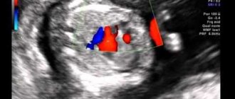
- Ultrasound (ultrasound)
With the help of ultrasound, you can not only hear the baby’s heart, but also see what condition the placenta is in, what size the fetus is, etc. The gynecologist-obstetrician pays special attention to the fetal heart rate if the pregnant woman has developmental defects or children born her in the past, had malformations of the heart and blood vessels.
- Auscultation
To implement this method, an obstetric stethoscope is needed. Relevant only from the 18th or even 20th week of gestation. But this method is not always easy to implement. In these cases, auscultation is difficult (and sometimes impossible):
- oligohydramnios
- polyhydramnios
- location of the placenta along the anterior wall of the uterus
- maternal obesity
- Cardiotocography (CTG)
The method helps to identify hypoxia and provide the necessary treatment. When measuring heart rate using this method, heartbeats are recorded on a special film. The activity of the uterus is also determined, which is very important during the birth process. The procedure usually takes about an hour, sometimes a little less. This duration is explained by the fact that the baby can sleep for some period, and then wakes up. This affects heart rate and indicators that are recorded during CTG.
Sometimes the doctor decides that the sensors should remain on the woman’s stomach for 24 hours. The first time cardiotocography is performed after the 32nd week of gestation. An earlier date is not advisable. In 9 months you need to undergo CTG 2 times. The procedure is absolutely safe for the fetus and the mother herself. What will be recorded on the tape will not be understood by the average person. The doctor must decipher it, taking into account the data of tests and ultrasound diagnostics.
CTG norms:
- Heart rate 120-160 beats per minute
- the fetal heartbeat does not become less frequent, or is recorded very rarely
- When the fetus moves, the heart rate increases
These three factors are important to determine the PSP index, which should be less than 1.
CTG may show abnormalities when:
- lack of oxygen baby
- pressing the umbilical cord to the fetal head or bones (but then changes will be recorded for a short period of time)
- sensors incorrectly attached to the mother's belly
- Echocardiography
This is another method of listening to the baby's heartbeat. The method is relevant for the 18th – 28th week of gestation if the doctor suspects that the child’s heart is not forming quite correctly. Echocardiography shows the structure of the heart and blood flow.
Advantages and disadvantages
The advantage of the study is the speed of obtaining results: take a measurement and immediately see the data on the screen and hear the rhythm with your ears. And this without leaving home. The examination procedure is painless. The devices are easy to use, with an intuitive interface.
The disadvantage is that home Doppler cannot replace consultations with an obstetrician-gynecologist or a full ultrasound scan in a clinic or hospital. And it does not serve as a basis for making a diagnosis, much less taking any medications on your own.
How can you hear your child’s heartbeat?
You don't have to go to a gynecologist to hear your child's heart. The first way to listen to your heart rate is with a stethoscope . It can be bought at a pharmacy at a low price. But you will need help. Before the 25th week of gestation, people without appropriate medical education can almost never hear the fetal heart. You can repeat listening with a stethoscope every week. It is important to remember that the sounds of the fetus moving can be confused with the beating of its heart, as well as with the sounds of processes that occur in the mother’s abdomen.
The second way to hear the baby’s heart yourself is fetal doppler . This is an ultrasound detector that you can buy yourself. It does not record data on tape, but you will hear the baby's heartbeat. The method is relevant from the 8th and later weeks of gestation. Duration of the procedure: maximum 10 minutes. The device is expensive, so not all pregnant women can afford it.
Third way: put your ear to the pregnant woman's belly . But this is only relevant after the 30th week of gestation. But if a woman is overweight, this method will not work. If the fetus is head down, it is better to listen to its heart below the pregnant woman's navel.
If you tend to worry about little things and independently diagnose yourself and your child, it is better to entrust listening to the fetal heartbeat and determining the norm to doctors. Enjoy motherhood and be in harmony with yourself.
Rules for using a fetal doppler
Immediately after purchase, you should study the instructions on how to use the fetal doppler. The procedure does not require any preparation. When using for the first time, sensors are attached to the device and turned on. It has a volume control button and speakers.
The examination is carried out lying on your back. A gel or cream is applied to the sensor and placed on the stomach. The first time you will have to look for the place where the rhythm is best heard, placing it on the area above the pubis and moving towards the navel. Next time you will immediately place the sensor in the desired area.
Depending on the stage of pregnancy, it will be:
- in the first trimester - immediately above the pubis;
- in the second trimester - on the left or right just below the navel;
- in the third trimester - below the navel with an occipital presentation (the head is below), and above the navel - with a gluteal presentation (the baby’s buttocks are below), at the level of the navel with a transverse position.
During multiple pregnancy, the rhythm is heard in different areas of the abdomen.
The duration of the study is no more than 5 - 7 minutes. Experts do not recommend taking measurements too often; once a week will be enough.
You will hear the sound of heart contractions through the speakers, and the device will calculate the frequency and display the result on the display.