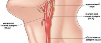
Origin of the internal carotid artery
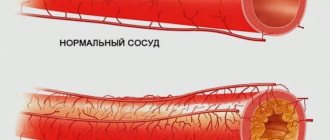
Atherosclerosis of the upper (proximal) part of the internal carotid artery is usually most pronounced during the first 2 cm and is localized on the posterior wall.
Often atherosclerosis spreads downwards (to the distal portion) of the common carotid artery. Often (in 50-80% of stroke cases), such damage by atherosclerosis makes itself felt by the development of a small stroke (micro-stroke) or transient ischemic attack (TIA), caused by a critical decrease in cerebral blood flow or embolism from the carotid artery into its intracerebral branches. Clinical experience and data from pathological studies suggest that strokes with damage to the carotid artery are more often caused by embolism than by blood flow deficiency. Emboli from an atherosclerotic plaque located at the beginning of the internal carotid artery can cause a transient ischemic attack (TIA, microstroke), but if they are repeated, short-lived and stereotyped, then their more likely cause is hemodynamic disorders.
How is ultrasound of neck vessels performed?
Fear in anticipation of the result is a traumatic factor for the psyche; we must remember that modern medicine is able to help many patients, provided that the pathology is detected early.
According to the appointed time (by appointment), the patient arrives at the diagnostic center. The initial position during the examination is lying on your back, with a pillow placed under your head for comfort. The neck and upper shoulder girdle must be freed from clothing. During the procedure, the doctor may ask you to hold your breath, tilt your head back, or change your position. There will be no pain. A special gel will first be applied to the skin to facilitate the sliding of the sensor and enhance visualization due to tighter contact. It is hypoallergenic and does not leave marks on clothes after drying.
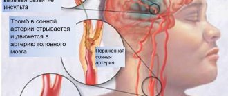
Cerebral ischemia caused by a lack of blood supply
Weak arterial blood flow can lead to the development of a stroke (infarction) of the brain or cause a transient ischemic attack (TIA, microstroke) in the border zone or in areas of adjacent blood supply. The development of stroke and micro-stroke (TIA) with a lack of blood supply to the brain is facilitated by two conditions:
- A decrease in blood pressure below (distal) stenosis in the lesion of the carotid artery when the diameter of its lumen decreases by more than 80% or, which is equivalent to this, when the diameter of its remaining lumen is less than 1.5-2 mm.
- Insufficiency of collateral blood flow to the ischemic area.
Cerebrovascular accident usually occurs when the circle of Willis is incompetent due to congenital atresia of the initial (A1) section of the anterior cerebral artery or the anterior or posterior communicating arteries. Less commonly, brain damage develops when there is blockage (occlusion) of the opposite carotid or basilar artery, limiting blood flow to the circle of Willis. Sometimes sufficient blood supply is provided through orbital collaterals from the external carotid artery or superficial cortical collaterals at the border of the arterial basins. In this case, the blood supply becomes adequate and the area of ischemia is limited even with the existing inferiority of the circle of Willis. It is the diversity of collateral blood supply that determines the diversity of localization of lesions during strokes and transient ischemic attacks (TIA, micro-strokes) in the carotid artery system against the background of insufficient cerebral blood flow.
There are other explanations for the development of transient ischemic attack (TIA, microstroke) with a decrease in cerebral blood flow. It is believed that pronounced narrowing (stenosis) in the area of the bifurcation of the common carotid artery can lead to transient blocking (occlusion) of the lumen of the vessel due to spasm. In rare cases, systemic factors of circulatory disorders lead to a decrease in blood flow through the area of gross narrowing of the lumen of the vessel to critical values. On the other hand, regional blood circulation in the cerebral hemisphere can change due to the maintenance of reduced blood flow in the carotid artery system, and transient decompensation of this mechanism can provoke a transient ischemic attack (TIA, microstroke). Other factors, including polycythemia vera, thrombocythemia, cardiac arrhythmia, sometimes cause repeated transient ischemic attacks (TIA, micro-strokes) against the background of insufficient blood flow in the vessels of the brain and other internal organs.
Preparation for ultrasound of neck vessels
Insomnia, stress, alcohol, smoking and strong coffee contribute to a rise in blood pressure, which can affect the result of an ultrasound scan of the vessels of the neck and head
. No special measures are required before an ultrasound scan of the vessels of the neck, but experts recommend giving up strong tea and coffee, smoking , alcohol, avoid stressful situations. If you plan to take any pills that affect vascular tone, inform your doctor in advance. A skinny stomach is not required to perform Doppler sonography, but you should not eat too much breakfast: blood will rush to the stomach to support digestion, and lying on your back for 30-40 minutes will be uncomfortable. It is necessary to ensure adequate fluid intake, as hypovolemia (decreased circulating blood volume) can lead to unreliable sonograms.
Take with you the results of previous studies (ultrasound, MRI, CT) of this area, a referral from a doctor with a suspected diagnosis, a passport, money or a health insurance policy (if the procedure is paid for by the fund), napkins. Before starting the procedure, remove jewelry: earrings, chain, etc.
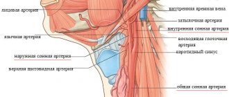
Embolism of the carotid artery and its branches
When emboli (blood clots, thrombi) originate from a vessel narrowed by stenosis or an ulcerated area with atherosclerosis at the beginning of the internal carotid artery (local or arterioarterial embolism), the symptoms that appear are usually associated with blockage (occlusion) of the ophthalmic artery, the trunk of the middle cerebral artery, one or more of it branches and sometimes the anterior cerebral artery or its branches. The size of the embolus corresponds to the caliber of the vessel that is being occluded.
The anatomy of the common carotid and internal carotid arteries provides insight into the mechanism of ischemic stroke.
Small platelet emboli (blood clots, thrombi) clog the most distant (distal) branches of the middle cerebral artery or ophthalmic artery, causing the patient to experience transient blindness in one eye or small asymptomatic infarctions (micro-strokes) in areas of adjacent blood supply to the cerebral arteries. Larger emboli, consisting of platelet-fibrin clots, can close the primary and secondary branches of the middle cerebral artery. At the same time, neurological syndromes make it possible to judge the involvement of certain areas of the brain in the pathological process.
Some emboli (blood clots, thrombi) are so large that they block the “trunk” of the middle cerebral artery at its very beginning, causing severe ischemia of the entire area of \u200b\u200bthe blood supply to the middle cerebral artery (deep white matter, lenticular nucleus, surface of the cortex). Other emboli (blood clots, thrombi) large enough to block the trunk of the middle cerebral artery can only cause a deep infarction (stroke) if there is sufficient collateral blood flow through the superficial cortical arteries.
Carotid artery stenting is a minimally invasive procedure that restores proper blood flow to the brain when there is an area of narrowing (stenosis) in one of the carotid arteries. A small metal tube (stent) is passed through the area of stenosis to keep the artery open.
A large embolus that occludes a large vessel can move, dissolve, and disintegrate. If the splitting of the embolus (blood clot, thrombus) occurs quickly, then the neurological deficit in the patient may be transient or disappear completely.
In some patients with neurological symptoms, vascular damage may be represented by a single plaque in the area of the bifurcation of the carotid artery, but much more often a narrowing (stenosis) is detected with a residual lumen diameter of the arterial vessel of less than 2 mm.
The frequency of massive embolic strokes (cerebral infarctions), which develop solely against the background of ulceration of the area of the vascular wall affected by atherosclerosis, remains unclear. It is possible that the frequency of such embolic strokes is low, and such cases are observed only with large ulcers of the vascular wall (4 mm or more). The development of a stroke or a single transient ischemic attack (TIA, microstroke) with long-lasting symptoms in the absence of narrowing (stenosis) of the lumen of the carotid artery or its insignificant severity suggests that the source of the embolus is the heart. Atheromatous lesions localized at the beginning of large branches of the aortic arch can also cause embolism in the cerebral vessels and cause transient ischemia or infarction, but the incidence of this mechanism has also not been established.
Computed tomography angiography of the neck arteries revealed occlusion of the proximal left internal carotid artery (black arrow).
Cerebral angiography demonstrates a filling defect (thread sign) in the proximal left internal carotid artery (black arrow).
Blockage (occlusion) of the internal carotid artery in its initial section can be completely asymptomatic for the brain with an adequate level of collateral blood flow of arterial blood through the circle of Willis. On the other hand, if the collateral blood supply is insufficient, a stroke or transient ischemic attack (TIA, micro-stroke) may develop. In addition, a blood clot or thrombus can spread upward from the site of blockage of the lumen of the vessel through its siphon to the initial sections of the middle and anterior cerebral arteries, leading to a stroke. But more often, fresh emboli break off from the thrombotic material and get stuck in the lumen of the middle, anterior arteries or in one of their branches. According to some authors, emboli can originate from the underlying stump of the internal carotid artery and be transported through the collaterals of the external carotid artery, reaching the intracranial part of the internal carotid artery and its branches. But such embolisms are probably quite rare.
The cause of delayed stroke, which develops several months after complete blockage of the lumen (occlusion) of the carotid artery, often remains unclear, and its frequency is unknown. According to one study, delayed stroke occurs in 5% of cases per year. But taking into account clinical experience, this figure seems overestimated. In most cases, embolism due to blockage of the lumen (occlusion) of the carotid artery occurs during the first year, although they can be observed within two years. Brain strokes caused by insufficient blood supply develop earlier, usually in the first few weeks after blockage of the lumen (occlusion) of the carotid artery.
Postoperative duplex scanning demonstrates normal blood flow and no recurrence of internal carotid artery stenosis.
Duration of the ultrasound procedure of neck vessels
The duration of the ultrasound procedure depends on a number of aspects, including the human factor: during a collegial examination, more time is required to assess an unclear situation.
Since there is no harmful effect on the body when performing ultrasound with Doppler sonography, the diagnostic procedure continues as long as the doctor needs to detect all pathological changes or make a statement absence of the latter. In municipal institutions, due to the large influx of patients, time is strictly regulated; in a private medical center, the doctor has the opportunity to examine the patient longer. On average, the duration of an ultrasound scan of the neck vessels is 30-40 minutes, and another quarter of an hour will be required to fill out the study protocol.
Intracranial (intracranial, intracerebral) section of the internal carotid artery
The siphon of the carotid artery is subject to atherosclerosis with thrombosis less frequently than the inferior portion of the internal carotid artery. Lesions in the area of the carotid artery siphon can cause strokes and transient ischemic attacks (TIA, micro-strokes), the pathophysiological and clinical manifestations of which repeat those given above. The patient's history of narrowing of the lumen (stenosis) of the carotid artery siphon is nonspecific. It should be taken into account that narrowing of the lumen (stenosis) of the carotid artery siphon is asymptomatic until the atheromatous process reduces the remaining lumen to 1.5 mm or less. Only angiography can accurately diagnose narrowing of the lumen (stenosis) of the carotid artery siphon above the ophthalmic artery. Collateral blood flow through the circle of Willis undoubtedly influences the pathogenesis of these lesions and the effectiveness of their medical or surgical treatment.
Atherosclerosis with thrombosis of the middle cerebral artery
Atherosclerosis with thrombosis of the trunk of the middle cerebral artery can cause symptoms of cerebral ischemia both due to a narrowing of the lumen of the artery, and as a result of blockage of the initial segments of one or more arteries of the lenticular nucleus and striatum, supplying blood to the deep white matter and subcortical nodes. Clinically manifested atherosclerotic plaque is rarely located below the first division (bifurcation) of the middle cerebral artery. Since the circle of Willis is located below the beginning of the middle cerebral artery, collateral blood supply in the middle cerebral artery basin should be carried out through small superficial cortical vessels of the adjacent blood supply zones and anastomoses from the anterior and posterior cerebral arteries.
The cause of ischemic stroke in the middle cerebral artery is atherosclerotic thrombosis.
Available data indicate that before the development of cerebral infarction (stroke), narrowing of the lumen of the vessel is usually “prevented” by transient ischemic attacks (TIA, microstrokes) in the middle cerebral artery basin. Their symptoms are similar to those of transient ischemic attacks (TIA, micro-stroke), associated with deterioration of blood circulation as a result of severe stenosis of the internal carotid artery. In contrast to the internal carotid artery, the trunk of the middle cerebral artery and one or more of its major branches are usually occluded by an embolus (arterioarterial, cardiac, or unknown source).
Atherosclerosis with thrombosis of the anterior cerebral artery
Atherosclerotic deposits in the lower segment of the anterior cerebral artery rarely give pronounced clinical symptoms of neurological deficit, since blockage of the lumen (occlusion) is compensated by collateral blood flow through the anterior communicating artery. The likelihood of a transient ischemic attack (TIA, microstroke) and stroke increases if the path of collateral blood flow is characterized by a congenital fusion (atresia) or if there are atherosclerotic changes in a higher section of the anterior cerebral artery.