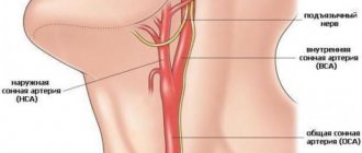
Carotid arteries
Diseases of the carotid arteries occur when developing atherosclerotic plaques partially or completely block their lumen. The carotid arteries are paired vessels that supply blood to the head and brain.
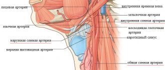
The carotid arteries are paired vessels that are located in the neck and provide blood flow to the brain.
Narrowing of the lumen of the carotid arteries due to the formation and growth of atherosclerotic plaques reduces the volume of blood flowing to the brain and increases the risk of stroke.
Symptoms
Since atherosclerotic lesions of the carotid arteries develop slowly and are often asymptomatic, the first clinical manifestations of this disease may be a stroke (acute cerebrovascular accident - stroke) or transient ischemic attack (TIA), sometimes called a microstroke.
Treatment of atherosclerotic lesions of the carotid arteries usually includes a set of measures, such as lifestyle changes, drug therapy and, in some cases, surgical treatment (open surgery or stenting).
In the early stages, atherosclerotic lesions of the carotid arteries are usually asymptomatic. You and your doctor may not be aware that you have a narrowing of the carotid artery until an acute disruption of cerebral blood supply develops as the first and very serious manifestation of the disease.
Clinical manifestations of stroke or transient ischemic attack may include:
- a sudden feeling of numbness or weakness in the face or limbs, usually on one side.
- Impaired speech or understanding
- sudden loss of vision in one or both eyes
- dizziness or loss of balance
- sudden, causeless severe headache
If you have risk factors for developing atherosclerotic lesions of the carotid arteries, you should consult your doctor. Your doctor may order an examination to determine the condition of these vessels. Even if you do not have clinical manifestations of the disease, your doctor may recommend a number of measures aimed at reducing the severity of risk factors and reducing the likelihood of developing a stroke.
Seek immediate medical attention when you experience symptoms of a transient ischemic attack or stroke.
Even if the duration of symptoms is short, usually less than an hour, but possibly longer, tell your doctor right away. The appearance of these symptoms indicates that you have suffered a transient ischemic attack - a short-term decrease in blood flow to the brain. The presence of transient ischemic attacks is the most important sign that you are at high risk of developing a stroke if preventive measures are not taken in time. A timely visit to the doctor increases your chances that atherosclerotic lesions of the carotid arteries will be identified and eliminated before an acute cerebrovascular accident develops.
A transient ischemic attack may also indicate a decrease in blood flow through other blood vessels in the brain. To clarify the diagnosis, your attending physician will prescribe the necessary examination.
Make sure your family and close friends are aware of the clinical manifestations of a stroke and that it is important to act quickly if they occur.
Dissection of the internal carotid and vertebral arteries: clinical picture, diagnosis, treatment
In recent years, interest in the dissection of arteries supplying the brain, a relatively new and insufficiently studied problem of cerebrovascular diseases, has been steadily growing in the world. Its main clinical manifestation is ischemic stroke (IS), which often develops at a young age. The study and intravital diagnosis of cerebral artery dissection has become possible thanks to the widespread introduction of magnetic resonance imaging (MRI) into the clinic. MRI makes it safe for the patient to carry out repeated angiographic examination, which is important for diagnosing dissection, since it represents a dynamic pathology, and also, using the T1 mode with signal suppression from adipose tissue (T1 fs), to directly visualize intramural (intrawall) hematoma (IMH) - direct sign of dissection. The use of MRI has shown that dissection is a very common pathology, and not a rarity, as previously thought. In addition, it has become apparent that cerebral artery dissection is only fatal in a small number of cases, whereas it was originally considered a fatal disease.
In Russia, targeted studies of cerebral artery dissection began in the late 90s of the last century at the Scientific Center of Neurology of the Russian Academy of Medical Sciences (until 2007, the Scientific Research Institute of Neurology of the Russian Academy of Medical Sciences) almost simultaneously with research carried out abroad. But the first morphological descriptions of individual cases of cerebral dissection, clinically, however, unrecognized, were made in the 80s of the 20th century. in our country, D. E. Matsko, A. A. Nikonov and L. V. Shishkina et al. Currently, this problem is being studied at the Scientific Center of Neurology of the Russian Academy of Medical Sciences, where more than 200 patients with intravital verified dissection of the cerebral arteries were examined, more than half of whom were patients with dissection of the internal carotid (ICA) and vertebral arteries (VA).
Dissection of the cerebral arteries is the penetration of blood from the lumen of the artery into its wall through an intimal tear. The IMG formed in this case, separating the layers of the arterial wall, spreads along the length of the artery to different distances, most often towards the intima, leading to narrowing or even occlusion of the arterial lumen, which causes cerebral ischemia. Minor stenosis caused by IMH may be clinically asymptomatic. The spread of IMH towards the outer membrane (adventitia) leads to the development of a pseudoaneurysm, which can cause isolated cervical headache, or to a true dissecting aneurysm. Thrombi formed in a dissecting aneurysm are a source of arterio-arterial embolism and IS. Dissection develops both in the main arteries of the head (ICA and VA) and in their branches (middle, posterior, anterior cerebral arteries, basilar artery). However, most researchers believe that dissection occurs more often in the ICA and VA than in their branches. At the same time, it cannot be excluded that dissection in the branches of the ICA and VA is often underestimated due to the difficulty of visualizing IMG in them and is mistakenly regarded as thrombosis. Dissection can develop at any age - from infancy to the elderly, but in most cases (according to the Scientific Center of Neurology of the Russian Academy of Medical Sciences - 75%) it is observed in young people (up to 45 years). It is noted that with intracranial lesions, the age of patients is, as a rule, less than with extracranial lesions, and with the involvement of the VA - less than with lesions of the carotid arteries. The distribution of patients by gender also depends on the location of the dissection: the ICA is more often affected in men, and the VA – in women. Dissection usually develops in individuals who consider themselves healthy, do not suffer from atherosclerosis, thrombophilia, diabetes mellitus, and rarely have moderate arterial hypertension.
Dissection of the internal carotid artery.
The main provoking factors for ICA dissection are head or neck trauma, usually mild; physical activity with tension in the muscles of the shoulder girdle and neck; bending, throwing back, turning the head; drinking alcohol; current or previous infection; taking contraceptives or the postpartum period in women. In conditions of previous weakness of the arterial wall, these factors and conditions play a provoking, rather than causal role, leading to intimal rupture and the development of dissection, which in these cases is considered spontaneous.
Clinically, dissection of the ICA most often manifests as IS, and less often as transient cerebrovascular accident (TCI). More rare (less than 5%) manifestations include isolated neck/headache, localized in most cases on the dissection side; isolated unilateral damage to the cranial nerves due to their ischemia, when the arteries supplying the nerve arise from the dissected ICA; isolated Horner's syndrome, caused by the effect of IMG on the periarterial sympathetic plexus, when the hematoma mainly extends towards the adventitia and does not significantly narrow the lumen of the ICA. Small IMHs may be asymptomatic and incidentally detected on MRI. A characteristic sign of cervical cerebrovascular accident during ICA dissection is headache/neck pain. Pain, usually dull, pressing, less often pulsating, shooting, appears several hours or days before AI on the side of dissection. Its cause is irritation of the sensitive receptors of the vascular wall by the hematoma developing in it. In approximately a third of patients with IS, it is preceded by a transient SMC in the cerebral territory of the ICA or ophthalmic artery in the form of a short-term decrease in vision on the side of the dissection. NMC, as a rule, develops in the middle cerebral artery (MCA) and is manifested by motor, sensory and aphasic disorders, which in half of the cases are detected in the morning upon awakening, in the other half of the cases - during active wakefulness.
The prognosis for life in most cases is favorable; death occurs in approximately 5% of cases. It usually occurs with extensive cerebral infarctions caused by dissection of the intracranial part of the ICA with transition to the MCA and anterior cerebral artery. In the majority of patients, especially with damage to the extracranial part of the ICA, the prognosis for life is favorable and good recovery of impaired functions is observed. When the intracranial part of the ICA is involved and the dissection extends to the MCA or with embolism of the latter, the recovery of impaired functions is much worse.
Relapses of dissection occur infrequently and are usually noted in the 1st month after the onset of the disease. They can appear both in an intact and in an artery that has already been dissected.
The main mechanism for the development of IS is hemodynamic under conditions of an increasing stenotic-occlusive process in the ICA caused by IMH. Less commonly, cerebrovascular accident develops through the mechanism of arterioarterial embolism. Its source is thrombi formed in a dissecting aneurysm, thrombosed fragments of IMH entering the bloodstream during a secondary intimal breakthrough, or thrombotic layers at the site of an intimal rupture.
Vertebral artery dissection.
Dissection of the VA, according to most authors, is observed somewhat less frequently than dissection of the ICA. However, it cannot be excluded that the VA is involved more often than indicated in the literature, since many cases of VA dissection manifesting as isolated cervicocephalgia are not clinically recognized and are not statistically taken into account.
The main clinical signs of VA dissection are ischemic cerebrovascular accidents and isolated neck/headache. This pain occurs in about a third of cases. Rare manifestations include circulatory disorders in the cervical spinal cord, isolated radiculopathy, and hearing impairment. In more than a third of patients, dissection is found in both VAs, and dissection of one VA may be the cause of UCI, and the second VA may be the cause of isolated neck/headache, or be clinically asymptomatic and detected only by neuroimaging. A characteristic feature of NMC during VA dissection, as well as during ICA dissection, is its association with neck/headache on the side of the dissected VA. Typically, pain is localized along the back of the neck and in the back of the head, appearing several days or 2-3 weeks before focal neurological symptoms. Pain often occurs after repeated bending, turning the head, or keeping the head in an uncomfortable position for a long time, and less often after a head/neck injury, usually mild. The tension of the VA observed in this case with weakness of the vascular wall causes intimal rupture and initiates dissection. The causes of neck/headache during VA dissection are irritation of the pain receptors of the arterial wall by the hematoma forming in it, as well as ischemia of the neck muscles, the blood supply of which involves the branches of the VA. Another feature of NMC is that often (about 80%) it develops at the moment of turning or tilting the head. Focal neurological symptoms are ataxia, vestibular disorders, less often - sensory disorder, dysarthria, dysphagia, dysphonia, paresis.
The most common mechanism for the development of CVA during VA dissection is arterio-arterial embolism. This is indicated by clinical manifestations (acute development of symptoms of cerebral ischemia, usually during active wakefulness, often when turning/tilting the head) and angiography results (the presence in most patients of hemodynamically insignificant stenoses, which do not significantly impair cerebral hemodynamics and ensure distal movement of emboli) . Many researchers consider the source of embolism to be fragments of IMH entering the bloodstream during a secondary intimal breakthrough. According to other researchers, these are intravascular mural thrombi formed at the site of intimal rupture. Neuroimaging studies, primarily MRI angiography (MRA) and MRI T1 fs, which allow identifying IMH, are of decisive importance in verifying the dissection of the ICA and VA. The most common characteristic angiographic sign of ICA/PA dissection is uneven, less often - uniform prolonged stenosis (“rosary symptom”, or “strings of beads”, “string symptom”), pre-occlusive cone-shaped narrowing of the ICA lumen (“candle flame symptom”). Such characteristic angiographic signs of dissection as dissecting aneurysm and double lumen are much less common. Dissection is a dynamic pathology: ICA/VA stenoses caused by IMG are completely or partially resolved in all cases after 2–3 months. Recanalization of the original occlusion caused by dissection is observed in only half of the cases. Characteristic MRI signs of dissection are IMG, which is visualized on T1 fs for ≥2 months, and an increase in the outer diameter of the artery. It should be borne in mind that during the 1st week of the disease, IMH is not detected by MRI in the T1 fs mode, so computed tomography (CT) and MRI in the T2 fs mode acquire diagnostic value.
In most cases, over time there is a good or complete restoration of impaired functions. VA dissection may recur. After 4–15 months, relapse was observed in 10% of patients.
Morphological examination of the cerebral arteries during dissection plays a fundamental role in elucidating the causes of weakness of the arterial wall leading to dissection. It makes it possible to identify dissection, thinning, and sometimes the absence of the internal elastic membrane, areas of fibrosis in the intima, and incorrect orientation of myocytes in the media. It is assumed that changes in the vascular wall are caused by genetically determined weakness of connective tissue, primarily collagen pathology. However, no mutations in the collagen gene were found. For the first time in the world, employees of the Scientific Center for Neurology of the Russian Academy of Medical Sciences have suggested that the cause of weakness of the arterial wall is mitochondrial cytopathy. This was confirmed by a study of muscle and skin biopsies. Histological and histochemical examination of the muscles revealed red ragged fibers, a change in the reaction to succinate dehydrogenase and cytochrome oxidase, and a subsarcolemmal type of staining in fibers with an intact reaction. Electron microscopic examination of the skin arteries revealed changes in mitochondria, vacuolization, deposition of fat, lipofuscin and glycogen in cells with altered mitochondria, and calcium deposits in the extracellular matrix. The complex of identified changes characteristic of mitochondrial cytopathy allowed Russian researchers to propose the term “mitochondrial arteriopathy” to designate arterial pathology that predisposes to dissection.
The treatment of IS caused by dissection has not been definitively determined, since there are no randomized placebo-controlled studies involving a large number of patients. In this regard, there are no clearly established treatment methods in the acute period of stroke. Most often, the introduction of direct anticoagulants is recommended, followed by a transition to indirect anticoagulants, which are used for 3–6 months. The purpose of their administration is to prevent arterio-arterial embolism and maintain IMG in a liquefied state, which contributes to its resolution. It should be borne in mind that the prescription of large doses of anticoagulants can lead to an increase in IMH and a deterioration in the blood supply to the brain. As an alternative to these drugs in the acute period of stroke, the use of antiplatelet agents is recommended, and, according to preliminary data, there is no difference in stroke outcomes. In order to assess the safety of treatment with low molecular weight heparin and aspirin in the acute period of dissection, French researchers measured the volume and extent of IMH during the 1st week of treatment. A slight increase in these parameters was observed in a third of patients, but in no case was there an increase in the degree of stenosis or the development of re-dissection. The use of anticoagulants and antiplatelet agents is limited to 2–3 months, during which the development of IMH occurs. Further prophylactic use of these drugs is not advisable, since the cause of IS during dissection is not hypercoagulation, but weakness of the arterial wall. Since the main reason predisposing to the development of dissection is weakness of the arterial wall, therapeutic measures both in the acute and long-term periods of stroke should be aimed at strengthening it. If we take into account the data on mitochondrial cytopathy, leading to energy deficiency of the cells of the arterial wall and its dysplasia, which contributes to the occurrence of dissection, the use of drugs with “trophic” and energy-tropic effects can be considered justified. One of these drugs is Actovegin, which is used in both acute and long-term periods of stroke caused by dissection. It is a biologically active substance of natural origin - a deproteinized derivative of calf blood. The main effect of Actovegin is to activate cellular metabolism by facilitating the flow of oxygen and glucose into the cell, which provides an additional influx of energy substrates and increases by 18 times the production of ATP, the universal donor of energy necessary for the life and functioning of the cell. Other drugs with neurometabolic effects are also used to restore functions impaired due to stroke: Cerebrolysin, piracetam, gliatilin, ceraxon.
In the acute period of dissection, in addition to drug treatment, compliance with the following rules is of great importance: it is necessary to avoid sudden head movements, injuries, physical stress, straining, which can lead to an increase in dissection.
Dissection of the ICA and VA is a common cause of IS at a young age, and less often – a cause of isolated neck/headache.
Knowledge of the clinical and angiographic features of this type of vascular pathology of the brain allows for its correct treatment and secondary prevention.
Causes
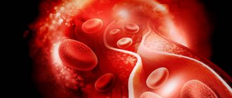
Over time, the walls of the carotid arteries can become thickened, their lumen narrows due to the formation of atherosclerotic plaques, which leads to a decrease in the volume of blood flowing through them. Atherosclerotic plaques consist of accumulations of cholesterol, calcium, fibrous tissue elements and cell debris that penetrate the artery wall through microscopic damage to the inner lining and form an atherosclerotic plaque in the area of which a blood clot (thrombus) can form.
Normal, healthy carotid arteries, like other normal arteries, are soft and flexible and allow blood to flow freely. If you place your finger on either side of the Adam's apple, you can feel the pulsation of the carotid arteries. The carotid arteries provide oxygen and nutrients to the cortex and other vital structures of the brain.
Clinic of the syndrome
The human brain is fed by two arteries – the carotid and the vertebral. The first provides up to 70% of blood flow, the second up to 30%. As a result of the progression of osteochondrosis, the vertebrae become deformed, increase in size, are destroyed or displaced; the specific pathology depends on the stage of the disease and the individual reaction of the patient’s body.
Against the background of progressive diseases of the spine, various symptoms develop that prevent a person from leading his previous lifestyle, limiting his mobility and causing him a lot of inconvenience. One of them is vertebral artery syndrome, the treatment of which is a very important task.
All unnatural changes negatively affect the condition of the arteries; they are unable to supply the required amount of blood to the brain. The lumen of blood vessels is reduced by external tissues or nerve endings react to stimuli with muscle spasms. Due to insufficient oxygen, the human brain cannot function normally and sends signals about an unfavorable state. The second name for vertebral artery syndrome is vertebrobasilar syndrome.
Vertebrobasilar syndrome
Risk factors
Factors that affect arteries and increase the risk of damage, plaque formation, and disease include:
- High blood pressure.
High blood pressure is the most important factor in the development of atherosclerotic lesions of the carotid arteries. Exposing the artery wall to high pressure weakens it and makes it more susceptible to damage. - Smoking.
Nicotine irritates the inner lining of blood vessels, and also increases heart rate and increases blood pressure. - Age.
With age, the arterial wall loses elasticity and becomes more susceptible to damage. - Violation of the blood lipid ratio.
Elevated levels of low-density lipoprotein cholesterol ("bad cholesterol") and high levels of triglycerides contribute to the formation of atherosclerotic plaques. - Diabetes.
Diabetes not only affects the ability to control blood sugar levels, but also lipid metabolism, increasing the risk of hypertension and the development of atherosclerosis. - Obesity.
Excess body weight increases the risk of arterial hypertension, atherosclerosis and diabetes. - Heredity.
The presence of atherosclerosis or coronary heart disease in relatives significantly increases the risk of developing atherosclerotic lesions. - Sedentary lifestyle.
Lack of physical activity contributes to the development of hypertension, obesity and diabetes.
Often, the listed risk factors are present in combination, thereby increasing the degree of risk.
Massage and preventive measures to minimize the negative effects of symptoms
Massage roller
Massage should be treated with great caution; such manipulations can only be performed by a medical professional after a full examination. Otherwise, excessive compression of the vessels, the formation of blood clots and their uncontrolled displacement, and the appearance of severe pain may occur. Preventive measures include the following.
- Healthy and active lifestyle . If work requires prolonged sitting in one position, straining the neck muscles, etc., then it is definitely recommended to take breaks for preventive exercises. You can do it right at your workplace for 10–15 minutes.
- Proper nutrition . It is necessary to reduce the amount of salt consumed to the values recommended by medicine, and do not abuse fatty and fried foods.
Proper nutrition
Complications
Atherosclerotic plaque
The most dangerous complication of atherosclerotic lesions of the carotid arteries is stroke. There are various mechanisms that increase the risk of stroke:
- Decreased blood flow.
The carotid arteries may become so narrowed by atherosclerotic lesions that they are unable to provide adequate blood supply to the brain. - Rupture of an atherosclerotic plaque.
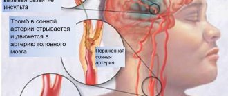
Particles of atherosclerotic plaque can break off and travel with the bloodstream into smaller vessels of the brain, blocking the lumen of these arteries and blocking the blood supply to the area of the brain that is fed by this vessel. - Blood clot formation.
The surface of some atherosclerotic plaques may rupture to form an uneven, ulcerated surface. When this happens, the body's response is to attract platelets to the site of the break and form a blood clot (thrombus). Large blood clots can partially or completely block the lumen of an artery, thereby impeding blood flow and causing a stroke.
The consequences of a stroke can be the formation of a focus of brain damage and dysfunction of organs, in particular paralysis of the limbs. In severe cases, a stroke can be fatal.
Stroke or transient ischemic attack are often the first manifestations of atherosclerotic lesions of the carotid arteries and are considered medical emergencies. If you or your loved ones have symptoms of impaired blood supply to the brain, you should urgently seek medical help. Do not try to get to the hospital on your own.
Signs of concern:
- sudden feeling of numbness or weakness in the face or limbs, usually on one side;
- impaired speech or understanding;
- sudden loss of vision in one or both eyes;
- dizziness or loss of balance;
- sudden causeless severe headache;
Signs of arterial stenosis
Long-term disruption of the blood supply to the brain causes the following phenomena.
- Numbness of facial muscles. If there is a one-sided pathology, then the face may become distorted. When it is bilateral, facial expressions become more complicated, speech becomes unclear, and the face becomes white.
Numbness of facial muscles - Dizziness, inability to hold a stable position . Such symptoms may indicate an approaching fainting state. The patient should be immediately laid down and the head should be placed in the most comfortable position.
Long-term painful conditions have a negative impact on the psyche of patients and serve as an impetus for the onset of depression. The earlier the diagnosis is made and treatment measures taken, the less likely there are negative consequences. In the most complex forms, pathologies cause strokes.
Diagnostic methods
After collecting anamnesis, identifying risk factors and characteristic symptoms, the doctor may conduct an additional examination, including:
- A physical examination,
during which the doctor, using a phonendoscope, can detect a noise above the carotid artery in the neck, which indicates a narrowing of the artery. The doctor may also conduct a neurological examination to assess your physical and mental status, such as strength in your limbs and ability to remember spoken language. - Ultrasonography.
The most common non-invasive research method to assess the condition of the carotid arteries is Doppler ultrasound. This is a variant of conventional ultrasound examination that evaluates blood flow speed, pressure and can detect narrowing of the lumen of the arteries due to changes in these indicators. - Computed angiography.
This procedure is performed using a contrast agent to highlight the arterial vessels. The drug is administered intravenously. When it reaches your carotid arteries, a series of X-ray images are taken of the neck and brain area in different views. - Magnetic resonance imaging.
Similar to CT scanning, this method can visualize brain tissue and detect early brain lesions or other pathology. - Angiography.
It is a more “sensitive” invasive research method. However, it is now used less and less due to the existing risk of stroke during the procedure. During the study, X-ray images of the arterial vessels supplying the brain are taken, with preliminary intra-arterial administration of a contrast agent.
Why does pathology appear?
Currently, medical science distinguishes two groups of pathologies that result in the formation of the syndrome.
Vertebral artery syndrome with cervical osteochondrosis
Table. Main causes of pathology.
| Reason type | Short description |
| Vertebrogenic | The functionality of the artery located in the vertebrae degenerates due to abnormalities in the structure of the cervical spine. Observed after acquired or other diseases and pathologies. Most often, this condition of the patient appears against the background of osteochondrosis or ankylosing spondylitis, with mechanical asphyxia of blood vessels caused by an increase in a benign or malignant tumor localized in the cervical spine. Unpleasant sensations can appear after complex mechanical injuries or as a result of prolonged muscle spasms. In children, vertebral artery syndrome appears as a consequence of varying severity of birth injuries. |
| Nonvertebrogenic | Such causes are not considered pathologies of the development of the cervical spine; the syndrome appears due to the adverse effects of scar tissue due to improper surgical treatment of other diseases. Often, blood circulation in the brain is disrupted by thrombosis, arteritis, and embolism. During development, the arteries have unnatural bends or greater tortuosity. Another reason is a deviation from the norm in the structure of the cervical vertebrae or a spasm of the muscle tissue surrounding the artery. |
Vertebral artery
Treatment
The goal of treatment of atherosclerotic lesions of the carotid arteries is to prevent the development of stroke. The choice of treatment method depends on the degree of narrowing of the artery lumen.
Slightly and moderately expressed narrowing.
In the presence of moderate narrowing of the carotid arteries, the following recommendations may be sufficient to prevent stroke:
- Lifestyle changes. Healthy lifestyle changes can help reduce damage to the carotid artery wall and slow the progression of atherosclerosis. Such changes include quitting smoking, losing weight, eating a healthy diet, reducing salt intake and regular exercise.
- Treatment of concomitant chronic diseases. At the same time, it is necessary to treat such concomitant diseases as arterial hypertension, obesity and diabetes mellitus.
- Prescribing medications. Your doctor may prescribe you to take aspirin or other antiplatelet medications daily to reduce the risk of blood clots. Therapy aimed at correcting blood pressure (ACE inhibitors, beta blockers) and correcting the lipid profile – taking statins – may also be recommended.
Therapeutic exercise to relieve the syndrome
Specific exercises are prescribed only by a specialist with a medical education. If you detect the slightest signs of deterioration, you should immediately stop exercising and consult your doctor again.
- Head tilts. You should start with a small amplitude; as the condition improves, the number of inclinations increases and the amplitude increases. The sensation of a slight painless cracking sensation during bending is not considered a reason to stop exercising.
Head tilts to the sides - Shrug . The head should always be in a strictly vertical position. The shoulders are raised to the maximum amount. The number of repetitions is not limited, the exercise does not have a direct effect on pathological areas, it only helps to improve blood flow in nearby areas.
- Head turns. During this exercise, cracking sounds may also be heard in the cervical vertebrae. Such crackling sounds indicate the presence of changes in bone tissue. If they are painful, then the amplitude should be reduced. In most cases, the cracking stops after a few days of exercise, which indicates an improvement in the condition of the spine.
Head turns - Tilts the head forward/backward with increasing effort . A more complex exercise, you need to counteract the tilt with your hands. A very effective exercise for improving the condition of the muscle tissue of the cervical region. Strong muscles are able to maintain the correct position of the head, due to which the load on the vertebrae is significantly reduced, they occupy a natural physiological position and free up pinched or curved arteries.
- Tilts of the head to the sides with increasing effort . The exercise is similar to the one described above, the only difference is in the direction of the inclinations.
Head tilts forward and backward