The description of aneurysmal dilatation of the carotid artery first appeared at the end of the 17th century. Since then, there are almost 1000 cases in the surgical literature. Most reports were of single cases or small series, each containing only seven reports of more than 25 patients. They show that the etiology, clinical presentation, and treatment of carotid aneurysm have changed significantly over the past 50 years. However, there are some common themes and important lessons that help the vascular surgeon who faces such a problem. This part will focus on the incidence, etiology, diagnosis, and treatment of both true and false extracranial carotid aneurysms. Aneurysms of the intracranial arteries are more common, but are usually the domain of neurosurgeons and neuroradiologists and will therefore not be considered.
Diagnosis of neck and brain disease
Examination algorithm:
- Survey and inspection . Complaints of bursting pain, fainting, decreased vision and hearing, sensory disturbances; history of vascular pathology. On examination, there is pastiness, facial hyperemia, and swelling of the veins of the neck.
- Objective research . Swelling and enlargement of the neck and thyroid gland, vascular murmur, hypertension.
- Laboratory data . Increased levels of platelets, fibrinogen, cholesterol.
- Angiography of neck vessels . Defect in the contour of the vascular wall, contrast leakage beyond the vessel.
- Ultrasound of neck vessels with duplex scanning . Limited vascular expansion, tortuosity of the branches of the carotid artery, atherosclerotic plaques, blood flow turbulence.
- CT (MRI) . Local edema, atrophy of cranial nerves 2, 3, 4, 6, hemorrhage, cidrocephaly, compression of the esophagus and trachea, foci of calcification.
- EEG . Disappearance of alpha waves, registration of theta and delta ranges.
You can read how to diagnose a cerebral aneurysm in a separate material.
Carotid artery aneurysm - characteristics depending on location
Carotid artery aneurysm (ICD-10 code - I72.0) is a local increase in its diameter by more than 2 times. It is a limited protrusion formed by a stretched vascular wall.
An aneurysm forms secondary to chronic diseases affecting the arterial bed. In the first stage, under the influence of a causative factor, nonspecific inflammation develops in the vessel wall: edema, destruction of endothelial cells and the muscle layer.
In the second stage, enzymes in the lesion lead to the destruction of collagen fibers. The artery wall becomes thinner and stretches. In the third stage, under the influence of a pulse wave, the thinned area is pressed and forms a protrusion.
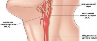
Common artery
The vessel extends from the aortic arch to the angle of the maxilla and is affected in 27-30% of cases . Causes: atherosclerosis, Takayasu syndrome. Symptoms are associated with compression of the tissues and organs of the neck, and cerebral ischemia on the affected side. Formation within 2-4 weeks. The prognosis is relatively unfavorable, with frequent relapses. Surgical treatment is indicated for rapid progression and increasing clinical symptoms.
In the area of the angle of the mandible, the common carotid artery is divided into external and internal branches.
External SA
The ESA does not penetrate the cranial cavity, but supplies blood to the soft tissues of the face, the auricle, and the tissues of the nose. Affected in 12-14% of cases . Causes: Takayasu disease, congenital anomalies. Symptoms are associated with ischemia of the facial muscles, orbit, nasal cartilage, and external ear. Formation is slow, over 2-4 months. The prognosis is relatively favorable. Treatment is symptomatic and surgical.
Internal carotid artery (ICA)
An aneurysm of the internal carotid artery of the brain can be in one of the sections of the ICA - the cavernous sinus, supraclinoid section and bifurcation.
Surgery as the only treatment option
None of the conservative methods can cure a carotid aneurysm or at least stop its growth. As the size increases, the arterial wall becomes thin; if time for surgery is missed, a rupture occurs, which has fatal consequences. There are several methods for disconnecting the enlarged part of the artery from blood circulation:
- complete excision and installation of a prosthesis at the site of the aneurysm;
- partial removal of the vascular sac, suturing and restoration of blood circulation;
- bypass bypass;
- placement of the prosthesis through a catheter inserted into an incision in the neck.
Preventive actions
Primary prevention:
- To give up smoking.
- Normalization of blood pressure.
- Low-salt diet.
- For persons over 50 years old - annual medical examinations with ultrasound.
- Laboratory screening for the detection of atherosclerosis, diabetes mellitus, hypertension.
Secondary prevention:
- Clinical examination.
- Dynamic monitoring of the condition of the aneurysm (ultrasound, CT).
- Treatment of concomitant diseases.
Aneurysm of the carotid artery and its branches is a common complication of chronic vascular diseases. The pathology is distinguished by a progressive course and an increasing clinical picture. Treatment is surgical. Prevention is aimed at early detection and treatment of cardiovascular pathology.
Most diseases associated with the condition of blood vessels are dangerous because they lead to death. Timely diagnosis and treatment are important in the early stages, before the disease affects vital organs.
Carotid artery aneurysm is a common anomaly, the victim of which can be not only an adult, but also a child .
Possible complications
Complications may include dissection and rupture, thrombosis, stroke, compression of the esophagus, loss of vision or hearing, memory loss, thyroiditis, paralysis of half the body.
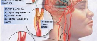
Rupture is a common complication of the natural course of the disease . It occurs due to overstretching of the carotid artery, which has undergone irreversible dystrophic changes.
Causes:
- Hypertensive crisis;
- Injuries;
- Detachment of an atherosclerotic plaque.
Risk factors are hypertension, smoking, diabetes, stress, physical overexertion.
Symptoms of a rupture:
- Anxiety;
- Dyspnea;
- Tachycardia;
- Increased pressure;
- Violation of visual, auditory, cognitive functions;
- Paralysis of half the body;
- Lack of response to external stimuli;
- Loss of consciousness.
Treatment in 100% of cases is surgical. The prognosis is unfavorable. Stroke occurs in a third of patients.
Symptoms of the disease
First symptoms:
- Bursting headaches and neck pains.
- Fatigue, tiredness.
- Increased pressure.
Symptoms depending on the location of the lesion:
- Common carotid artery - pain, feeling of a foreign body in the neck, hoarseness, pain when swallowing.
- NSA – loss of facial sensitivity, nosebleeds, pain in facial muscles.
- Cavernous sinus – decreased sensitivity of the tongue, facial skin, pain in the orbit.
- Supraclinoid region – paralysis of the eyeball, myopia.
- Bifurcation - flickering before the eyes, narrowing of the visual field.
Left and right side affected
Right-sided lesion appears:
- Loss of speech functions.
- Epilepsy.
- Impaired sensitivity (numbness, “crawling goosebumps”).
- Visual disorders.
Left-sided lesion is characterized:
- Migraine;
- Cramps;
- Fainting;
- Disorders of consciousness;
- Hypertension;
- Dizziness.
Signs of deterioration
As it progresses, symptoms of cerebral ischemia appear:
Therapy tactics
Treatment of carotid artery aneurysm is divided into conservative and surgical. Indications for conservative therapy:
- Internal carotid artery (segments according to Bouthillier)
- Preparation for surgery;
- No complaints;
- Size up to 1 cm;
- No progression.
Conservative therapy includes:
- Statins, antihypertensive, diuretic drugs;
- Symptomatic drugs (analgesics, NSAIDs);
- Vasodilators and nootropics.
Surgery
Indications for surgery:
- Growth more than 4 mm in 6 months;
- The appearance of the clinic;
- Hypertension;
- Increased thrombus formation;
- History of heart attack or stroke;
- Risk of complications.
Contraindications for surgery:
- Exacerbation of infectious diseases;
- Multiple organ failure;
- Sepsis;
- High risk of surgical complications.
Types of operations:
- Clipping is the removal of an aneurysm from the bloodstream by placing a clip on it. Selection operation for pouch shape;
- Resection – removal of the enlarged area and replacing it with a prosthesis. The prosthesis can be a segment of a peripheral artery or a synthetic tube;
- Endovascular embolization is the filling of an aneurysm with surgical glue through a balloon inserted from the femoral artery. The operation of choice in uncomplicated patients.
Conservative treatment is indicated for patients for whom surgery is contraindicated. In case of increased intracranial pressure, a palliative operation is performed - installation of drainage for the outflow of cerebrospinal fluid.
Emergency assistance in case of rupture
Algorithm of actions:
- Provide the patient with rest and fresh air;
- Offer an analgesic or sedative;
- Help the patient take a horizontal position with the head down.
When an ambulance appears, the following is carried out:
- Transportation;
- Stabilization of hemodynamics;
- Introduction of plasma substitutes;
- If clinical death develops, resuscitation measures are taken.
Emergency diagnostics (CT, ultrasound) and surgery are performed in the hospital. The intervention is carried out by a surgical team under angiography control.
Aneurysms of the vessels of the heart, brain, lungs, aorta, neck and splenic artery are no less dangerous. And when they rupture, professional, emergency care and prompt hospitalization are also very important.
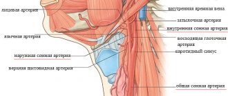
- Anatomy. Common carotid artery
Classification
Types of current:
- Acute - characterized by a sudden bright clinic, general cerebral symptoms, loss of consciousness. Observed during complications.
- Chronic - perennial, erased, wavy or asymptomatic.
Carotid aneurysms can be:
- Saccular - consist of a neck (through which they are connected to the vessel), a spherical body and a dome. This variety is more susceptible to rupture. The selection operation is clipping. The prognosis is relatively unfavorable.
- Fusiform - smoothly transition into a healthy vascular wall. Delamination is more typical for this variety. The operation of choice is resection. The prognosis is relatively favorable.
- Mixed - show signs of both forms. They are characterized by high vulnerability and rapid delamination. The operation of choice is resection. The prognosis is unfavorable.
Forms of the disease:
- Tumor-like – clinically resembles tumor growth. The course is chronic, the symptoms are associated with compression of anatomical structures and increasing compression syndrome.
- Apoplexy – clinically resembles apoplexy (convulsive syndrome). The course is acute, convulsions and loss of consciousness may be the first and only manifestation.
We wrote about the classification of cerebral aneurysm here.
You can read all the detailed information about this vascular lesion - what types there are, where they can occur, why saccular aneurysms, as well as true and false aneurysms, are dangerous - in separate articles.
Symptoms
An aneurysm the carotid artery is affected by pathology can be asymptomatic for quite a long time.
The brain does not react in any way to the gradual development of an arterial anomaly - until its blood circulation is disrupted.
If the saccular growth is small, the disease cannot be detected without the use of special diagnostic drugs. But it is very advisable to do this before it breaks.
The general symptom is an intermittent and systolic murmur that is heard in the area of a fairly large formation. You can also notice a pulsating swelling on the neck that has reached a significant size.
Additional symptoms:
- Noise in the head.
- Dizziness.
- Headache that occurs for no particular reason.
- Insomnia, sleep disturbance.
- Constant fatigue, even if the rest is complete and adequate.
The saccular formation filled with liquid blood has a tense consistency. Dense areas are palpated if blood clots have formed in it.
Another characteristic symptom is a gradual increase in frequency and intensification of headaches. The brain loses its ability to function normally.
Signs of pathology of the arterial wall are different:
- Pain in the heart area.
- Pain in the eyes, vision deteriorates.
- Dilated pupils.
- Hoarse voice.
- Numbness of the limbs.
- Loss of coordination.
- Pulsation radiating to the head.
- Painful sensations in the neck, back of the head, shoulder.
- Dyspnea.
An enlarged carotid artery can put pressure on:
- Trachea.
- Esophagus.
- Larynx.
- Throat.
This causes symptoms:
- Dysphonia.
- Dyspnea.
- Bleeding from the nose.
- Blue face.
- Paresis and paralysis.
Signs of a complicated aneurysm:
- Speech problems.
- Paresthesia.
- Bilateral blindness.
- Epileptiform seizures.
In addition, the affected left carotid artery gives a corresponding internal resonance:
- Disturbed consciousness.
- Difficulty digestion, stomach pain.
- Spasms in the head.
- Excited state.
- Convulsive syndrome.
Symptoms of carotid aneurysm
There are no initial signs of a small aneurysm. If the patient does not have factors that can provoke its growth, then the diagnosis is made only through a random examination.
Large protrusions are visible on the neck; they represent a swelling above which a murmur can be detected during the period of systolic contraction of the heart.
If there is blood in the cavity of the aneurysm, then its consistency is close to elastic, and if there are blood clots, then upon palpation it is hard.
Signs of aneurysm growth may include general cerebral symptoms:
- increased fatigue,
- chronic headache,
- sleep disorder,
- noise in ears,
- decreased vision,
- dizziness.
When the formation reaches a large size, the headache becomes stronger and worries almost constantly, sensations of pulsation in the temples are added, vision and hearing decrease, the voice becomes hoarse, and the gait becomes unsteady.
The pressure of the aneurysm on the nerve plexuses causes pain in the neck, shoulder girdle, and occipital region. When compression spreads to the jugular vein, the complexion becomes cyanotic, and sensitivity and motor function of the limbs are impaired.
The most dangerous complication is the rupture of an aneurysmal formation. Its symptoms:
- sharp, unbearable headache,
- double contours of objects,
- nausea and frequent vomiting,
- neck muscle tension,
- convulsions,
- paresis or paralysis,
- disturbance of speech and consciousness,
- anxiety,
- coma.
If the operation is not performed on time, death occurs from internal bleeding.
Saccular aneurysm
An aneurysm the carotid artery is affected , is a disease of the cardiovascular system and, unfortunately, often ends in the death of the patient. Often such an anomaly is formed in an area running in the neck .
Saccular type - this is the name given to the pathology that forms inside the vessel. For various reasons - internal and external - the carotid artery , which delivers blood to the head, loses its elasticity and gradually expands.
At the weakest area, fragility reaches such a degree that it stretches greatly and expands under blood pressure. The resulting protrusion resembles a bag. The aneurysm grows - its size and volume increase. Elastic fibers are replaced by connective tissue.
The carotid artery in this area is constantly filled with blood and blood clots. The danger of the situation is that the wall of the vessel becomes so fragile that it can easily burst when the pressure in the body increases. The location of the pathology can be the right or left branches. Bilateral formations almost never occur.
Types of arterial aneurysms in children and adults
- Saccular - is a blood sac that is attached to the artery using a neck.
- Fusiform - expansion of the vascular wall of a diffuse nature.
- Lateral – a tumor on the side of the artery.
The fusiform type has a second name - fusiform.
Extremely dangerous conditions develop when fusiform and other types of dilations rupture. Symptoms:
- Double vision.
- Nausea, vomiting.
- Paralysis - complete or partial.
- Speech impairment.
- Dark skin tone on the neck.
- Stiffness of the neck muscles.
- Falling into a coma.
The severity of the condition increases very quickly, and the person is in dire need of emergency medical care.
Diagnostics
Diagnostic measures that allow you to reliably determine the presence of a pathological formation:
- Angiography. It is an examination of cavities using x-rays.
- CT scan. Determination of possible hemorrhage.
- Magnetic resonance imaging. Gives a detailed picture of the state of the circulatory system.
- Cerebrospinal fluid analysis. It is done in case of suspected rupture of the aneurysmal sac.
Prevention
Measures to prevent carotid aneurysm involve eliminating risk factors for damage to the vessel wall. These include:
- getting rid of nicotine and alcohol addiction;
- treatment of hypertension and blood pressure control;
- review of nutrition with the exclusion of animal fats and sweets, flour products from the diet;
- maintaining a sufficient level of physical activity;
- reduction of excess body weight;
- compliance with occupational health recommendations;
- examination by a neurologist and cardiologist at least once every six months if there is a hereditary predisposition;
- taking vascular, antihypertensive drugs and blood thinners.
We recommend reading the article about atherosclerosis of the neck vessels. From it you will learn about the causes and symptoms of atherosclerosis, methods of diagnosis and treatment, and preventive measures.
And here is more information about cerebral vascular thrombosis.
A carotid aneurysm may be asymptomatic. If it is detected, patients should take precautions not to stimulate its growth. With large sizes, the wall becomes thinner, which can result in rupture and death.
For treatment, conservative therapy is not very effective; surgery is indicated. Prevention requires lifestyle modification and constant monitoring of blood pressure.
An aneurysm is a local expansion of the lumen of an artery due to changes or damage to its wall.
Cerebral aneurysms are the main cause of non-traumatic subarachnoid hemorrhage, accounting for up to 85% of all cases of intracranial hemorrhage.
Aneurysm rupture most often occurs between the ages of 30 and 50 years.
STORY. E. Moniz performed the first cerebral angiography for subarachnoid hemorrhage in 1927, and 10 years later WEDendy performed the first successful surgical intervention for a ruptured aneurysm.
Where do they “come” from? Currently, there is no single theory of the origin of aneurysms. Most authors believe that the origin of aneurysms depends on two reasons: the presence of degenerative changes in the vascular wall and the factors that cause them.
Defects of the arterial wall underlying the formation of an aneurysm:
- defect of the muscle layer,
- damage to the internal elastic membrane,
- intimal hyperplasia and atheroma of the arterial trunk,
- damage to collagen fibers of the artery,
- a combination of increasing rigidity of the artery wall with a decrease in its thickness.
Hemodynamic factors - aneurysms are often located in the area where the branches originate from the artery or at the artery bends, because these areas experience the greatest hemodynamic impact.
Aneurysms are often combined with malformations or diseases that lead to:
- arterial hypertension (hereditary arterial hypertension, coarctation of the aorta, polycystic kidney disease);
- damage to connective tissue (fibromuscular dysplasia, connective tissue syndrome);
- changes in cerebral hemodynamics (brain tumor, AVM, anomalies in the development of the arterial circle of the cerebrum)
Classification of aneurysms.
There are many classifications of cerebral aneurysms, the most common of which are classified according to shape, size and artery on which they are located.
Classification of aneurysm by shape. 1. Saccular (single- or multi-chamber). 2. Fusiform (fusiform).
3D-CT angiography – saccular aneurysm of the fork of the basilar artery
3D-CT angiography – fusiform dilatation of the basilar artery
Classification of aneurysms according to the artery on which they are located.
Classification of aneurysms by size. 1. Up to 3 mm – miliary. 2. 4 – 15 mm – ordinary. 3. 16 – 25 mm – large. 4. More than 25 mm – giant. Circle of Velisius (arterial circle of the brain) with a giant aneurysm of the internal carotid artery (left
The structure of aneurysms.
Circle of Velisius (circle of artery of the brain) with a giant aneurysm of the internal carotid artery (left)
An aneurysm consists of a neck, body and dome. The neck has a three-layer structure of the cerebral artery; it is the most durable part of the aneurysm. The body of the aneurysm is characterized by the absence of a three-layer vascular wall (primarily the muscular layer) and underdevelopment of the elastic membrane. The dome of the aneurysm is represented by one layer of intima, it is the thinnest, and bleeding occurs from here. In the area of the aneurysm there are almost always atheromatous changes and its rupture often occurs at the site of these changes.
Complications that develop in the acute period of aneurysm rupture are the following: 1. Repeated bleeding from the aneurysm. 2. Vascular spasm, which develops in 100% of cases (peak development of spasm on days 3-14), cerebral ischemia due to vasospasm develops in 64% of cases. 3. Intracerebral hematoma – 22%. 4. Intraventricular hemorrhage - 14%. Currently, leading neurosurgeons around the world are inclined to early operations for ruptured cerebral aneurysms. In this way, repeated hemorrhage, vascular spasm and hydrocephalus are prevented.
Examination of patients with cerebral aneurysm - see subarachnoid hemorrhage.
Treatment methods for cerebral aneurysms.
1. Surgical:
- aneurysm clipping (open interventions),
- endovascular intervention
2. Conservative.
Chief physician:
Prof. Doctor med. Thomas Gasser
Beta Clinic Bonn
location_on
Germany, Bonn
Department of Neurosurgery and Interventional Neuroradiology
Beta Clinic is a modern clinic consisting of 30 specialized departments. The clinic is equipped with state-of-the-art university-level equipment. The operating rooms, intensive care unit and rehabilitation center meet the highest standards. This also applies to highly qualified Diagnostics of aneurysm of the internal carotid artery 2081 Go to the program chevron_right
Open repair of internal carotid artery aneurysm 32189 Go to program
chevron_right
Reasons for the development of pathology
Aneurysm can be genetically determined or occurs during life. The causes of acquired vascular defects can be the following factors:
- atherosclerotic changes in the arterial wall;
- brain injuries and concussions;
- excessive physical activity;
- surgical interventions in the carotid artery area;
- blockage of the lumen by a thrombus or embolus;
- periarteritis nodosa or other systemic vasculitis;
- syphilis;
- tuberculosis;
- ischemic or hypertension;
- bacterial, parasitic infection.