Heart defects can be congenital or acquired.
When planning a pregnancy, a woman should identify (or rule out) a congenital heart defect. This will make it possible to take a conscious and balanced approach to family planning and, if pregnancy and childbirth are possible, to prepare for them in advance.
90% of acquired heart defects develop against the background of rheumatism; they can also occur during pregnancy (exacerbation of rheumatism in pregnant women is most often observed in the first three and last two months of pregnancy). Fortunately, there is now a wide range of methods for diagnosing and treating this disease. It is especially important for women suffering from rheumatism to plan a pregnancy. A favorable prognosis for pregnancy is possible if it occurs against the background of an inactive rheumatic process.
Thanks to the improvement of methods for diagnosing and treating heart diseases, many patients with similar diseases, previously doomed to infertility, have the opportunity to carry and give birth to a child.
How to plan a pregnancy with heart defects
Modern medicine has quite effective methods that allow us to calculate the degree of risk associated with pregnancy and childbirth in women with heart defects. With their help, doctors help a woman determine the optimal time for conception or decide the fate of an unplanned pregnancy.
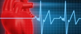
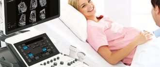
The most important method for assessing the state of the cardiovascular system in case of heart disease is ultrasound of the heart - echocardiography. It is harmless and helps to objectively assess the condition of the cavities, valves and openings of the heart. An auxiliary role in the diagnosis of heart defects is played by electrocardiography (ECG - graphic recording of the electrical activity of the heart), phonocardiography (PCG - graphic recording of cardiac sound phenomena) and Dopplerography (ultrasound, which allows assessing blood flow).
In pregnant women, heart defects account for 0.5 to 10% of all heart diseases. Most often, they have an atrial or ventricular septal defect or patent ductus arteriosus. Women with the above defects usually (with appropriate treatment that compensates for the defect) tolerate pregnancy and childbirth well.
Currently, many women who have undergone heart surgery have the opportunity to give birth. The recovery period after such an operation usually takes 1 year. Therefore, it is in a year that you can plan a pregnancy - of course, in the absence of contraindications (unfavorable results of the operation, the development of diseases that complicate postoperative rehabilitation and reduce the effect of the operation).
It is unnecessary to remind that the question of the possibility of pregnancy and the admissibility of childbirth should be decided individually before pregnancy, depending on the general condition of the woman, the nature of the disease, the severity of the operation, etc. After a comprehensive examination of the patient, the doctor can give a very definite conclusion.
However, even when a woman’s condition is stabilized after surgical (or therapeutic) treatment, pregnancy against the background of an increasing load on the heart increases the risk of recurrence of the underlying disease (a previously compensated defect may become decompensated) - this is another argument in favor of the need for consultation with a doctor and medical supervision before and during during pregnancy, even if the woman herself seems to be healthy and full of strength.
There are severe heart defects with significant circulatory disorders (stenosis of the pulmonary artery, tetralogy of Fallot, coarctation of the aorta, etc.), in the presence of which such dramatic disturbances in the functioning of the cardiovascular system can develop that in 40-70% of cases they lead to the death of the pregnant woman Therefore, with these defects, pregnancy is contraindicated .
Such defects can be inherited, and the likelihood of transmitting the disease to a child is determined in each specific case. (For example, if two or more family members have a heart defect, the likelihood of inheriting it increases.)
In general, the worse the prognosis for the expectant mother and child, the more pronounced the circulatory disorder and the activity of the rheumatic process. In severe heart failure and a high degree of activity of the rheumatic process, pregnancy is contraindicated. However, the issue of continuing pregnancy is decided by the patient and the doctor in each specific case.
Treatment methods
Treatment of congenital heart disease is carried out only by surgery . Cardiac surgeons perform complex reconstructive interventions, successfully curing those forms of congenital heart disease that were previously considered inoperable. If the diagnosis is made early and treatment is carried out promptly, heart function can be restored to 100%.
Non-invasive therapy (medication, physiotherapy, etc.) may be prescribed as an auxiliary measure. Such treatment is also carried out in cases where it is possible to postpone the operation to a later time or there is a “white” congenital heart disease that has little impact on the child’s well-being during growth and development.
Pregnancy management
During pregnancy, the load on the cardiovascular system increases significantly. By the end of the second trimester of pregnancy, the blood circulation rate increases by almost 80%. The volume of circulating blood also increases (by 30-50% by the eighth month of pregnancy). This is understandable - after all, the fetal blood flow also joins the maternal circulatory system.
With such an additional load, a third of pregnant women with a healthy heart may experience disturbances in heart rhythm (arrhythmias) and the functioning of the heart valves, what can we say about women with heart defects.
If necessary, drug treatment for heart defects is carried out throughout pregnancy. The goal of treatment is to normalize blood circulation and create normal conditions for fetal development. The question of prescribing drugs and their doses is decided individually, in accordance with the duration of pregnancy and the severity of circulatory disorders.
If therapy is ineffective, surgical treatment is resorted to, preferably at 18-26 weeks of pregnancy.
Echocardiotocography (ultrasound of the fetal heart) is performed periodically throughout pregnancy. Using Doppler ultrasound, uteroplacental and fetal (fetal) blood flow is examined to exclude hypoxia (oxygen starvation) of the fetus.
Naturally, the condition of the mother’s heart is constantly monitored (its methods were described in the previous section).
Often, even with an initially compensated defect, complications are possible during pregnancy, so every pregnant woman suffering from a heart defect should be examined at least three times during pregnancy in a cardiology hospital.
The first time is up to 12 weeks of pregnancy , when after a thorough cardiological and, if necessary, rheumatological examination, the question of the possibility of continuing the pregnancy is decided.
The second time - in the period from 28 to 32 weeks , when the load on a woman’s heart is especially high and it is very important to carry out preventive treatment. After all, a large load on the heart at this time can lead to the development of:
- chronic heart failure, characterized by fatigue, edema, shortness of breath, liver enlargement;
- heart rhythm disturbances (arrhythmias);
- acute heart failure and its extreme manifestation - pulmonary edema and thromboembolism (that is, blockage of the arteries of the lungs with blood clots) in the systemic circulation and pulmonary artery (these conditions pose an immediate threat to life, they must be immediately eliminated in an intensive care unit).
These complications can occur not only during pregnancy, but also during childbirth and in the early postpartum period.
For a child, such maternal circulatory disorders are fraught with a lack of oxygen (hypoxia). If timely measures are not taken, intrauterine growth retardation and insufficient body weight (hypotrophy) of the fetus may occur.
The third hospitalization takes place 2 weeks before birth . At this time, a repeated cardiac examination is carried out and a birth plan is developed and preparations are made for them.
What is heart disease
Heart disease is a pathology characterized by anatomical disorders of the structures of the heart muscle, valves, septa or large vessels that supply it with blood. The heart cannot cope with its job of supplying the organs with oxygen. They experience oxygen starvation and are in serious danger. There are acquired and congenital heart defects.
Congenital defect
Congenital heart disease is an anomaly in the structure of blood vessels and the heart, which, for various reasons, arose during the intrauterine development of the fetus. Pathology occupies one of the first places among congenital deformations of organs that can cause the death of newborns before they reach one year of age.
Often, congenital heart disease does not manifest itself in the prenatal phase. It happens that the pathology remains unnoticed during the first years of the baby’s life. But over time, she will definitely remind herself.
Responsibility for the occurrence of pathology lies primarily with the child’s parents. Their diseases, heredity and lifestyle directly affect the health of the unborn baby. The development of heart disease can be triggered by:
- infectious diseases;
- taking certain medications;
- addiction to alcohol;
- drug use;
- exposure to radiation;
- pathologies of the endocrine system;
- severe toxicosis during pregnancy;
- advanced age of the mother;
- bad heredity;
- chromosomal disorders.
There are several types of congenital heart defects:
- open holes in the heart muscle;
- blood flow difficulties;
- pathologies of blood vessels;
- heart valve defects;
- tetralogy of Fallot;
- aortic stenosis;
- common trunk of arteries;
- Ebstein's anomaly;
- simultaneous manifestation of several types.
An anomaly detected immediately after birth will allow timely treatment to begin and reduce the risk of death of the baby during the first days of life. Before planning offspring, it is necessary to find out how healthy the future parents are. You should ask your other “halves” whether they have genetic problems or cases of congenital heart disease in their immediate relatives.
- Treatment of heart disease with folk remedies
If a heart pathology is detected in the fetus during development, the mother is prescribed appropriate therapy during pregnancy. It should support the baby's cardiac functions until birth.
Heart disease in children
Timely treatment of pathology prevents the occurrence of complications. Children can grow and develop on par with healthy peers. Not all heart defects require emergency surgery. If so, specialists take a wait-and-see approach, keeping the cardiac activity of their patients under control. But in any case, a child with a heart defect needs special conditions for growing up.
Visually, symptoms of pathology usually appear when the baby turns three years old. At this time, attentive parents may notice:
- slow physical development of the baby;
- pallor of the skin, sometimes cyanosis;
- the appearance of shortness of breath during habitual movements.
Children with congenital heart disease are characterized by psycho-emotional experiences due to problems with development and learning. Typically, sick children begin to walk, talk, read and write later than their healthy peers. Over time, the situation can be aggravated by the appearance of excess weight, although initially, children with congenital heart disease are registered as underweight. A sick child's immunity is low, so he is at risk of infectious diseases.
But it's not just congenital heart defects that affect children. Adolescents are often diagnosed with acquired defects. This type of pathology can occur during exacerbation of various ailments. Harmful bacteria can enter the bloodstream:
- through infection during injection (contaminated syringes and needles);
- in case of violations of sanitation during medical procedures (including dental procedures);
- when abscesses occur.
Blue and white vices
There are blue and white heart defects. When blue, there is a reflux of venous blood into the arterial bed. In this case, the heart muscle “pumps” blood depleted of oxygen. The pathology is characterized by the early manifestation of symptoms of heart failure:
- cyanosis (blue color);
- dyspnea;
- nervous overexcitation;
- fainting.
With white defects, venous and arterial blood do not mix, oxygen enters the organs in the required quantity. The pathology is characterized by the same attacks that are observed with blue defects, but they appear later - at 8-12 years.
Medical practice shows that people with heart defects often live full lives without experiencing suffering or discomfort.
Acquired vice
Acquired heart defects affect the heart valves. Serious pathologies become the “trigger” for their development:
- Causes of heart murmurs in a newborn baby. Methods of medical intervention
- chronic vascular diseases (atherosclerosis);
- systemic connective tissue lesions (rheumatism, dermatomyositis, scleroderma);
- inflammation of the endocardium (infectious endocarditis);
- systemic joint diseases (Bechterew's disease);
- systemic venereal diseases (syphilis).
The cause of acquired heart defects is often the death of cells in the heart valves. Injuries can provoke the course of pathology.
There are compensated and decompensated acquired defects. In the first case there are no obvious symptoms of circulatory failure, in the second these symptoms are present.
The symptoms of the pathology are similar to those of other vascular and heart diseases. Therefore, the diagnosis is made only based on the results of an examination, including echo and electrocardiography. Some acquired heart defects include:
- Mitral - manifested by prolapse (sagging of the leaflets) of the mitral valve. Treatment is symptomatic. In parallel with it, drug therapy is carried out for the pathology that caused the heart defect. In case of serious damage to the valve, surgical correction is indicated;
- Aortic – the aortic valve is affected. The main pathology is treated with medication. Treatment of heart disease may require surgery, including valve transplantation;
- Combined - two or more valves of the heart muscle are affected. The mitral, tricuspid and aortic valves may become deformed, which will cause difficulties in diagnosing and treating the pathology. Most often, mitral valve insufficiency and mitral stenosis occur simultaneously. Under such circumstances, cyanosis and severe shortness of breath appear;
- Combined - one valve is subject to several violations. This is usually stenosis and failure. When diagnosing this type of heart defect, the severity of the lesions and the predominance of one of them are determined. This is necessary to prescribe adequate treatment and the type of possible surgical intervention;
- Compensated – difficult to diagnose, asymptomatic pathology. Dysfunctions of some parts of the heart muscle are fully compensated by the increased load on other parts of the heart. Only an experienced cardiologist, who has high-tech special equipment at his disposal, can diagnose this defect.
“Simple”, isolated heart defects are much less common than “complex”, combined ones. Infectious ailments haunt patients for years, affecting muscle tissue. As a result, another is added to one vice.
Childbirth
The question of the method of delivery is decided individually, depending on how much the defect is compensated by the due date. This could be a vaginal birth with or without pushing (see below) or a caesarean section.
Often, several weeks before giving birth, the increasing stress on the heart worsens the pregnant woman's condition so much that early delivery may be required. It is best if this happens at 37-38 weeks.
The birth plan is drawn up jointly by the obstetrician, cardiologist and resuscitator. Attempts - the period of expulsion of the fetus - represent a particularly difficult moment for the heart of the woman in labor, therefore they try to shorten this period of labor by performing a dissection of the perineum (perineotomy or episiotomy), and in case of stenosis of the mitral valve opening, circulatory failure of any degree, complications associated with impaired cardiac function -vascular system in previous births, - applying exit obstetric forceps.
Caesarean section is performed in the following cases:
- combination of the defect with obstetric complications (narrow pelvis, abnormal position of the fetus in the uterus, placenta previa);
- mitral valve insufficiency with significant circulatory disorders (severe regurgitation - backflow of blood from the ventricle into the atrium);
- mitral valve stenosis that cannot be corrected surgically;
- aortic valve defects with circulatory disorders.
After childbirth
Immediately after the birth of the child and the placenta, blood rushes to the internal organs, primarily to the abdominal organs. The volume of circulating blood in the vessels of the heart decreases. Therefore, immediately after childbirth, the woman is given drugs that support heart function (cardiotonics).
Women with heart defects are discharged from the maternity hospital no earlier than two weeks after birth, and only under the supervision of a cardiologist at their place of residence.
If a woman needs to take medications for a heart defect after childbirth, breastfeeding is excluded, since many of these drugs pass into milk. If after childbirth the heart defect remains compensated and treatment is not required, the woman can breastfeed.
Women suffering from rheumatism should especially carefully monitor their health in the first year after childbirth, when, according to statistics, exacerbations of this disease are quite common.
Symptoms of heart failure
In the clinical picture of the disease, there are general symptoms and specific ones, determined by the anatomical location of the defect. Common symptoms include:
- weakness and decreased ability to work;
- dizziness and fainting;
- shortness of breath and cyanosis (blue discoloration) of the skin;
- feeling of heartbeat;
- increase or decrease in blood pressure.
All these signs are the first signals and can only indirectly indicate the presence of heart disease.
Recommendations for women suffering from heart defects
Remember that the main reason for the unfavorable outcome of pregnancy and childbirth in those women with heart defects, for whom pregnancy is not contraindicated in principle, is insufficient or irregular examination in the antenatal clinic, the lack of comprehensive pregnancy management by an obstetrician and cardiologist and, as a consequence, the insufficient effectiveness of treatment measures and errors in the management of childbirth and the postpartum period.
Recommended:
- try to prevent unplanned pregnancy;
- consult your cardiologist before pregnancy; find out whether you are able to bear a child and what method of delivery you should prepare for;
- if you suffer from congenital heart disease, be sure (preferably before pregnancy) to consult a geneticist;
- find out what regime you should follow so as not to put yourself and your unborn child at risk, how to eat properly, what physical therapy exercises could help you bear and give birth to a child;
- do not miss your appointments with antenatal clinics and appointments with a cardiologist, complete all prescribed examinations on time;
- do not refuse hospitalization and taking medications - because not only your well-being, but also the health and life of your baby depends on how effectively your heart functions.
Answers to frequently asked questions
Is there accurate data on how many years people with congenital heart defects live?
As medical care and treatment for infants expands, patients with congenital heart disease live longer and healthier lives. Many children with congenital heart disease were able to transition into adulthood. It is estimated that more than two million people in the United States live with congenital heart disease.
Survival statistics:
- The survival of infants with congenital heart disease depends on how severe the defect is, when it is diagnosed, and how it is treated.
- About 97% of children born with non-critical congenital heart disease survive to one year of age, while 95% of children born with non-critical congenital heart disease survive to 18 years of age.
- About 75% of infants born with critical congenital heart disease survive to one year of age, while 69% of infants born with critical congenital heart disease survive to age 18. Thus, the population of people with congenital heart disease is increasing.
- Survival and medical care for children with critical congenital heart disease are improving. Between 1979 and 1993, about 67% of infants with critical congenital heart disease survived to one year. About 83% of children with critical congenital heart defects in the period from 1994 to 2005 were able to survive to the same age. [3 - Matthew E. Oster, Kyung A. Lee. Temporal Trends in Survival Among Infants With Critical Congenital Heart Defects. Pediatrics, May 2013.]
There are the following options for the development of congenital heart disease:
- Many people with uncomplicated forms of defects live without any problems.
- In other cases, the disease may develop over time.
- Some patients with congenital heart disease have genetic disorders or other health changes that increase the risk of disability or even death.
Even with improved treatment, many people with congenital heart disease are not completely cured, even if their heart defect has been repaired. With congenital heart disease, other health problems may develop over time. It all depends on the specific cardiac defect of the patient, the magnitude of the disorder and the severity of the pathology.
Against the background of congenital heart disease, even after treatment, various health problems may arise:
- irregular heart rhythm (arrhythmias);
- increased risk of infection of the heart muscle (infective endocarditis);
- weakening of the heart due to the development of cardiomyopathy.
The presence of congenital heart disease forces you to undergo regular examinations with a cardiologist, which allows you to maintain your health at an acceptable level. Repeat surgery may also be required at an older age, even after primary childhood surgery.
Sources
- Pereira SP., Tavares LC., Duarte AI., Baldeiras I., Cunha-Oliveira T., Martins JD., Santos MS., Maloyan A., Moreno AJ., Cox LA., Li C., Nathanielsz PW., Nijland MJ., Oliveira PJ. Sex dependent Vulnerability of Fetal Nonhuman Primate Cardiac Mitochondria to Moderate Maternal Nutrient Reduction. // Clin Sci (Lond) - 2021 - Vol - NNULL - p.; PMID:33899910
- Ben Abdelaziz A., Bchir A., Ben Salah A., Ben Salem K., Mansour N., Hsairi M., Ennigrou S., Nacef T., Dhidah L., Bellaj R., Mehdi F. Professor Mohamed Soussi Soltani : Leader, Innovator and Researcher in Public Health. // Tunis Med - 2021 - Vol99 - N1 - p.5-11; PMID:33899170
- Saavedra LPJ., Prates KV., Gonçalves GD., Piovan S., Matafome P., Mathias PCF. COVID-19 During Development: A Matter of Concern. // Front Cell Dev Biol - 2021 - Vol9 - NNULL - p.659032; PMID:33898461
- Alexanderson-Rosas E., Antonio-Villa NE., Sanchez-Favela M., Carvajal-Juarez I., Oregel-Camacho D., Gopar-Nieto R., Flores-Garcia AN., Keirns C., Hernandez-Sandoval S. ., Espinola-Zavaleta N. Assessment of Atypical Cardiovascular Risk Factors Using Single Photon Emission Computed Tomography in Mexican Women. // Arch Med Res - 2021 - Vol - NNULL - p.; PMID:33896676
- Bhasin D., Arora GK., Isser HS. Young woman with recurrent pregnancy loss. // Heart - 2021 - Vol107 - N10 - p.821-854; PMID:33893215
- Lee K., Laviolette SR., Hardy DB. Exposure to Δ9-tetrahydrocannabinol during rat pregnancy leads to impaired cardiac dysfunction in postnatal life. // Pediatr Res - 2021 - Vol - NNULL - p.; PMID:33879850
- Richards C., Sesperez K., Chhor M., Ghorbanpour S., Rennie C., Ming CLC., Evenhuis C., Nikolic V., Orlic NK., Mikovic Z., Stefanovic M., Cakic Z., McGrath K ., Gentile C., Bubb K., McClements L. Characterization of cardiac health in the reduced uterine perfusion pressure model and a 3D cardiac spheroid model, of preeclampsia. // Biol Sex Differ - 2021 - Vol12 - N1 - p.31; PMID:33879252
- Song Y., Xu J., Li H., Gao J., Wu L., He G., Liu W., Hu Y., Peng Y., Yang F., Jiang X., Wang J. Application of Copy Number Variation Detection to Fetal Diagnosis of Echogenic Intracardiac Focus During Pregnancy. // Front Genet - 2021 - Vol12 - NNULL - p.626044; PMID:33868367
- Khanna R., Chandra D., Yadav S., Sahu A., Singh N., Kumar S., Garg N., Tewari S., Kapoor A., Pradhan M., Goel PK. Maternal and fetal outcomes in pregnant females with rheumatic heart disease. // Indian Heart J - 2021 - Vol73 - N2 - p.185-189; PMID:33865516
- Kirby A., Curtis E., Hlohovsky S., Brown A., O'Donnell C. Pregnancy Outcomes and Risk Evaluation in a Contemporary Adult Congenital Heart Disease Cohort. // Heart Lung Circ - 2021 - Vol - NNULL - p.; PMID:33863666