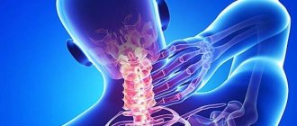
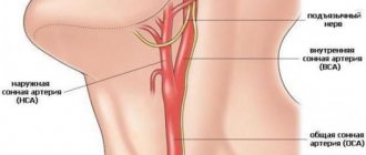
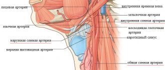
The carotid artery is a paired large blood vessel that rises to the head along the anterolateral surface of the neck on the right and left. It originates in the chest cavity, branching from the aorta, the main artery of the human body that supplies blood to all internal organs. The division of the carotid artery into external and internal trunks (bifurcation) occurs in the area of the thyroid cartilage of the larynx, hyoid bone or under the lower jaw. The outer part provides blood supply to the face, neck and scalp, while the inner part supplies blood to the brain.
Preventive measures
Prevention of the disease involves the following measures:
- reducing stress on the body and the cervical spine if there is a predisposition or primary symptoms of the disease;
- timely treatment of pathologies that provoke dilatation of the jugular vein;
- systematic planned examination by a specialist;
- proper lifestyle, physical activity, balanced diet.
People who have a hereditary predisposition to this pathology need to be especially careful.
It should be understood that vein abnormalities are very difficult to prevent, but the disease can be stopped and eliminated in the early stages. For this reason, a regular visit to the hospital will avoid serious health problems in the future.
S and C shaped tortuosity of the internal carotid artery
There are several types of pathology.
More common is the s-shaped tortuosity of the ICA with a smooth transition from one dislocation site to another. Usually with this type there is a deviation at two points. Most often, s-tortuosity of both ICAs occurs with deformities of the cervical spine. Straightening the cervical lordosis and static overstrain of the neck muscles entails a change in the position of the bed of this cerebral vessel. S-shaped tortuosity of the internal carotid artery in most cases does not give pronounced clinical manifestations. It is discovered during a study for another disease. If left untreated for 2-3 years, the disease will progress. In this case, the clinical picture worsens and emergency treatment of vascular pathology is required.
C-shaped tortuosity of the internal carotid artery develops in people suffering from high blood pressure. This type is typical for elderly patients suffering from atherosclerosis of cerebral blood vessels.
Children and adolescents are characterized by this type of pathology called kinking. This is a bend of the cerebral blood vessel at an angle of 45 degrees. It is a congenital vascular pathology. Gives attacks of acute cerebrovascular accident during excessive physical exertion. Such children and adolescents often complain of attacks of dizziness and fainting during physical education lessons. Parents should pay attention to the appearance of such complaints and conduct an examination.
Diagnostics
All about left side stroke
Some people are not even aware of their special structure of the internal carotid artery. In most cases, anterior trifurcation of the ICA is discovered by chance when a person decides to conduct a brain examination in connection with completely different diseases. Magnetic resonance imaging helps to identify this feature. If necessary, contrast angiography is used to obtain a clearer and clearer vascular picture.
In the resulting 3D-TOF images, trifurcation can be clearly identified. Clinical symptoms are determined by the absence of the right vertebral artery in the image and the origin of the anterior cerebral vessels from the right internal carotid artery. Such patients subsequently need consultation with a neurologist, who will prescribe a specialized course of therapy.
If a posterior trifurcation or anterior internal carotid artery is detected, you should not fall into despair and look for ways to solve this problem. This special structure of the Circle of Willis does not require special therapy. The structure of these arterial vessels cannot cause serious pathologies, since they are an additional source of nutrients for the brain and increase blood circulation during thrombosis.
Treatment in a medical facility may only be required if complications develop, such as an aneurysm. To eliminate it, you will need surgical intervention, which consists of ligating the vessels at the site of complications. When opening an aneurysm, conservative treatment will be required, which is generally accepted for any pathology with cerebral hemorrhage.
Diagnostics: identifying structural variants of the Circle of Willis
Consequences and recovery after a stroke on the left side
In most people, open VC does not manifest itself in any way. The development of the circle of Willis and its variants are most often detected accidentally, during examination of cerebral vessels for other reasons.
If the patient has symptoms of an unruptured cerebral aneurysm, the following examinations are performed:
- CT angiography is a non-invasive (that is, without penetration into the body) X-ray examination, during which a contrast agent is injected into the bloodstream to visualize the blood vessels of the brain, and then a computed tomography is performed.
- Magnetic resonance imaging is a non-invasive technique that uses a magnetic field and radiofrequency waves to produce detailed images of the blood vessels in the brain.
- Cerebral angiography is an invasive examination during which a special catheter is inserted into the brain artery. After this, contrast is injected through the catheter and an x-ray is taken.
Review of all options for the development of the Circle of Willis, what this means in practice
Causes and degrees of hydronephrosis of the left kidney: treatment and prognosis
From this article you will learn: what options exist for the development of the Circle of Willis, what it is, what arteries are included in its composition. What diseases can be caused by congenital or acquired pathology of the circle of Willis.
Author of the article: Nivelichuk Taras, head of the department of anesthesiology and intensive care, work experience 8 years. Higher education in the specialty “General Medicine”.
The circle of Willis (abbreviated as VC) is a system of anastomoses (connections between vessels) of blood vessels located at the base of the brain
It provides an important connection between the internal carotid artery systems and the vertebrobasilar territory
The VC consists of several arteries, which, connecting with each other, form a circle. In most cases, this circle is closed, but in some people one of the vessels may be missing, causing it to become open. These possible structural features of the VC are called its variants. Some of these developments can lead to an increased risk of dangerous brain diseases such as an aneurysm or stroke.
Nevertheless, for most people, various variants of the structure of the VC are the physiological norm, that is, they do not cause any symptoms or consequences.
Since complications of improper development of the circle of Willis arise in the brain, they are dealt with by neurosurgeons and neurologists.
The brain is, without exaggeration, the most important organ in the human body. Therefore, it is not surprising that its blood supply is one of the priority goals of the cardiovascular system. The brain receives blood from two sources - from the internal carotid artery system and from the vertebrobasilar system.
To avoid catastrophic consequences when one of the large vessels is blocked, there are anastomoses between these two blood supply systems that form the Circle of Willis at the base of the brain.
The VC consists of three pairs of main arteries:
- Anterior cerebral arteries (ACA) – arise from the internal carotid arteries.
- Internal carotid arteries (ICA) – the IC includes their terminal part, before the origin of the middle cerebral arteries (MCA).
- Posterior cerebral arteries (PCA) are the terminal branches of the basilar artery (BA), which is formed as a result of the fusion of the vertebral arteries (VA).
To complete the circle, two connecting blood vessels are also present:
- Anterior communicating artery (ACA) – connects the two ACAs.
- Posterior communicating arteries (PCA) are branches from the ICA that connect them to the PCA.
If the circle of Willis is closed, blood can, if necessary, pass through anastomoses from one artery to another.
Completely closed VC, in which there are no missing or underdeveloped (hypoplastic) components, occurs in only 20–25% of people.
There are a huge number of possible variants of the anatomical structure and development of the VC. The most common ones are:
- hypoplasia of one or two PCAs;
- hypoplasia or absence of the ACA segment;
- PSA hypoplasia;
- absence of one of the SSAs.
Causes and types of trifurcation
At the base of the brain lies a vascular formation in the shape of a polygon, the functions of which include the redistribution of blood flow from different pools when a deficiency occurs in one or another branch of the cerebral blood supply. In honor of the English physician Thomas Willis, who discovered and described this formation more than three hundred years ago, it is called the Circle of Willis.
Normally, the vessels of the circle should form a closed system, but sometimes there is a splitting of the internal carotid arteries included in its composition into three smaller vessels, which is called trifurcation. She may be:
- anterior - divided into anterior, posterior and basilar trunks;
- posterior - divided into anterior, middle and posterior cerebral arteries.
This feature of the cerebral vascular system is characterized as atavism of intrauterine development. In the first half of the period of fetal formation, such branching is the norm, but as it develops, the vessels unite and enlarge. However, sometimes this does not happen; triple cleft of the carotid artery remains with the person for life. This feature is found in all inhabitants of the planet with a frequency of 14 to 25% of the total population.
Depending on which of the paired vessels is subject to trifurcation, it is called right- or left-sided. Trifurcation can be complete when the branching of the internal artery, present in the prenatal state, is completely preserved, and incomplete when the vessels connect to a certain level and then divide into three branches.
On the one hand, the phenomenon is not considered a pathology, but only one of the variants of the structure of the circle of Willis; on the other hand, both complete and partial posterior or anterior trifurcation of the internal carotid artery can create favorable conditions for the occurrence of various vascular diseases, or aggravate their course.
Aplasia of the anterior cerebral artery
Hypoplasia, which is found in the area of the left cerebral artery, is an underdevelopment with a violation of the structure of the cerebral arteries, also at the stage of their formation. The consequences are a cerebral aneurysm or stroke. That is why anomalies in the development of the left cerebral arteries are of great importance in neurosurgical and neurological practice. Changes in the structure of the cerebral arteries influence the nature, localization and severity of pathological processes during the development of stroke. Factors that influence the occurrence of this pathology affect the fetus’s body even in the womb, which can be said about many other congenital pathologies.
Hemodynamically significant tortuosity of the ICA with local disruption
Any significant tortuosity of the ICA gives pronounced clinical manifestations and significantly reduces the patient’s quality of life. Decreased mental performance, progression of dementia, and inability to successfully complete training are only part of the potential problems.
Hemodynamically significant tortuosity of the ICA can provoke the appearance of attacks of cerebrovascular accident. With prolonged ischemia of areas of the brain, a focus of necrosis is formed. This is a cerebral infarction, which can lead to disability due to the appearance of persistent paralysis of the facial muscles, lower and upper extremities.
The least dangerous is the tortuosity of the ICA with a local disturbance of hemodynamics - with this pathology, the patient feels constant drowsiness, decreased performance, and deterioration in the quality of night sleep. Mild paresis of the muscles of the upper and lower extremities on the side opposite the affected vessel may occur.
1.1. Variants of origin (discharge) of vessels
1.1.1. Common origin: two different vessels can have the same origin (departure)
SCA/PCA: 2-22% Fig.3:
The common trunk arises from the basilar artery, then branches into the posterior cerebral artery (PCA) and the superior cerebellar artery (SCA).
PICA / AICA: common option Fig. 4:
The anterior inferior cerebellar artery (AICA) shares a common trunk with the posterior inferior cerebellar artery (PICA).
1.1.2. Funnel 7-15% Fig.5:
It is a funnel-shaped dilatation of the vessel at the origin. Its diameter should be no more than 3mm. It is most common at the origin of the posterior communicating artery (Pcom). A similar variant has also been described in the anterior communicating artery (Acom), ophthalmic artery, and anterior choroidal artery.
1.1.3. Abnormal origin (discharge) due to persistent fetal circulation:
Fetal type PCA:
The posterior communicating arteries are the terminal branches of the basilar artery. During development, RCAs originate from the internal carotid artery (ICA). This variant, if it persists into the postnatal period, is called “fetal origin.” This option can be classified into two subtypes: Fig. 6
Complete fetal PCA: 4-26% unilateral, bilateral 2-4%. PCA is entirely derived from the ICA. The P1 segment is absent, i.e., only the ICA supplies the occipital lobes. In bilateral complete fetal type PCA, the basilar artery may be hypoplastic Fig. 7.
Partial fetal PCA: 11-29% unilateral, 1-9% bilateral. The P1 segment is still present, but is smaller or equal in diameter to Pcom, i.e. Most of the blood supply to the occipital lobes comes from the ICA.
Persistent dorsal ophthalmic artery (PDOA): 1.1% Fig.8, Fig.46:
During embryonic development, the orbit is supplied with blood through the anterior and posterior rami, which originate from the ICA. Typically, the posterior branch is obliterated, while the anterior branch continues to supply the orbit. However, with this option the opposite happens. The PDOA enters the orbit through the superior orbital fissure.
The ophthalmic artery can also arise from other parts of the ICA, including the cavernous segment, in 8% of the population Fig. 9.
MMA from the orbital artery: 16% Fig. 10:
During embryogenesis, the middle meningeal artery (MMA) arises from the stapedial artery. The stapedial artery gives off branches to the ECA. One of these branches is the supraorbital artery, which forms an anastomosis with the developing ophthalmic artery. Along this anastomosis, failure of segmental regression or persistence of segments that should regress leads to a number of anomalies, such as the origin of the MMA from the ophthalmic artery. In this case, the foramen spinosum will be absent.
Fig. 3 TOF MRA, common trunk of PCA and SCA (red arrow) and ipsilateral dominant vertebral artery (white arrow)
Fig. 4 3D TOF MRA of the AICA (white arrow) extending caudally, supplying the PICA territory. Note the absence of PICA ipsilaterally. Contralateral PICA present (green arrow)
Fig. 5 3D TOF MRA, funnel-shaped expansion (funnel) at the origin of Pcom (arrow).
Fig. 6 3D TOF MRA, complete fetal PCA (white arrow) with P1 segment absence on one side and partial fetal PCA (red arrow) with P1 segment hypoplasia (green arrow) on the other side. Note fenestration of the proximal part of the P1 segment (blue arrow).
Fig. 7 3D TOF, complete fetal PCAs on both sides and hypoplastic basilar artery
Fig. 8 Persistent dorsal ophthalmic artery: MIP (a) and 3D TOF MRA (b) show the ophthalmic artery arising from the posterior surface of the ICA (arrow in a) and marked in red (b).
Fig. 9 Cavernous origin of the ophthalmic artery: MIP TOF lateral view (a), 3D TOF MRA ventral view (b), and 3D rotational DSA lateral view (c) showing the ophthalmic artery (white arrow) arising from the lateral surface of the cavernous segment of the ICA . Note the incidentally discovered Pcom aneurysm (red arrow)
Fig. 10 DSA (a) showing the MMA (red arrow) arising from the ophthalmic artery (white arrow). MIP reconstruction in the CT bone window (b), showing the absence of the foramen spinosum on the left side. Foramen spinosum on the right side is marked for comparison (blue arrow).
Treatment
If a posterior trifurcation or anterior internal carotid artery is detected, you should not fall into despair and look for ways to solve this problem. This special structure of the Circle of Willis does not require special therapy. The structure of these arterial vessels cannot cause serious pathologies, since they are an additional source of nutrients for the brain and increase blood circulation during thrombosis.
Treatment in a medical facility may only be required if complications develop, such as an aneurysm. To eliminate it, you will need surgical intervention, which consists of ligating the vessels at the site of complications. When opening an aneurysm, conservative treatment will be required, which is generally accepted for any pathology with cerebral hemorrhage.
Complications
Complications of trifurcation include the appearance of an aneurysm on any vessel. This pathology is determined immediately using an MRI or CT scanner. A feature of the formation of an aneurysm is the complete absence of signs of neurological diseases. No special treatment is provided in this case. Many concomitant diseases (hypertension, atherosclerosis, etc.) can also complicate the course of trifurcation.
Complications present with such a non-standard structure of blood vessels also include their increased length and the adoption of various unnatural shapes:
- Vascular pathology is very difficult to detect while they have soft bends at an obtuse angle. The clearly defined vessel has an ornate bend that can compress the main artery;
- Kinking. The direction of the artery forms an incomplete acute angle;
- Coiling. The artery takes the form of a loop, which significantly impairs blood flow. Symptoms of cerebral ischemia may also be present.
Features of the vessels, namely trifurcation of the left internal carotid artery, can lead to a narrowing of its lumen. This pathology occurs mainly in older people. Due to the formation of atherosclerotic plaques on the inner walls of the vessel, the lumen significantly narrows, which impairs blood circulation.
Such plaques are formed by fibrous tissue, cholesterol and calcium. When plaques become overgrown with platelets, complete blockage of arteries and vessels with a non-standard structure can occur.
The development of this complication can be prevented by using the following measures:
- Quitting bad habits (smoking and drinking alcoholic beverages);
- Regular exercise;
- Cholesterol-free diet;
- Body weight control.
If you detect a right posterior non-standard trifurcation of the internal carotid artery, you should not rush to search for specialists to eliminate this problem. This congenital feature of blood vessels is considered normal and does not require treatment. Regular periodic monitoring of their condition and diagnostics will be sufficient to avoid sudden complications.
Vessels
Anterior (Posterior) Atlanto Nuchal Membrane
Feb 06, 2021 Kokh V. A.
5935
Vessels
Arteries of the Head and Neck: Anatomy, Diagram, Atherosclerosis
Feb 06, 2021 Kokh V. A.
19266
Vessels
Carotid artery aneurysm
Head of the Institute: “You will be amazed at how easy it is to cure hypertension by taking it every day.
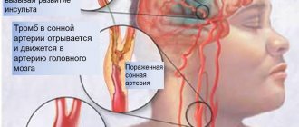
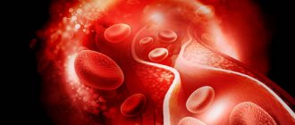
Diseases and ailments of the cardiovascular system in the body inevitably lead to irreversible consequences. This is due to the fact that the work of the heart and blood vessels directly affects the condition of all internal organs. Impaired blood supply leads to insufficient oxygen supply to all body systems.
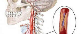
One of the most common and dangerous diseases of this type is carotid aneurysm. The picture of this disease is very sad: quite often it leads to death. A distinctive feature is that not only adults, but also children are exposed to the disease.
Enlargement and expansion of the carotid artery leads to the formation of a so-called pouch. Its cavity is mostly filled with plasma. The shape and size of the formation depends entirely on how severe the damage to the carotid artery is.
The formation of this sac and the enlargement of the artery leads to significant thinning of the tissue. They tear and become damaged at the slightest vibrations and other impacts. This leads to death.
This disease is also dangerous because the restriction of oxygen access to the brain is accompanied by various disorders and abnormalities in its functioning. This, in turn, affects other systems of the body.
As mentioned above, this type of aneurysm occurs due to a sudden and significant dilation of the carotid artery. A change in its size by more than 50% of the original can become a great threat to the brain and, consequently, other internal organs.
There are a number of reasons for this disease. These include:
- Atherosclerosis.
- Unsuccessful operations on the carotid artery.
- Hypertension.
- Previously suffered a stroke.
- Syphilis.
- Infectious diseases.
- Myocardial infarction.
- Heart disease.
- Other ischemic diseases of the cardiovascular system.
- Tuberculosis.
- Embolism.
- Hereditary diseases that manifest themselves in disruption of vascular function.
- Cervical injuries.
- Bad heredity.
- Thrombophlebitis, etc.
The disease can be caused by one of the reasons listed above, or by a combination of reasons.
Features of the pathology
This pathological process cannot be called a disease. Trifurcation is a structural feature of the Circle of Willis. This unusual structure of the arteries occurs quite often, in more than 20% of people. The pathology is still not normal, since some branches of the divided arteries may deliver insufficient blood to the brain due to the development of an aneurysm.
This structure of the Circle of Willis is considered the norm for an embryo located inside the womb in the first half of its development. During the growth process, the structure of the blood vessels changes, and they take on their usual state. But in some cases, their fusion does not occur, they retain the same size, and trifurcation of the internal carotid artery develops.
With anterior trifurcation, the artery delivers only 50% of the blood to the brain, and the posterior trifurcation only 10%, which is the result of hypoplasia of the proximal segment. Most scientists in this field argue that the non-classical structure of the circle of Willis can cause the formation of an aneurysm in any of its parts.
Cerebral circulatory disorders: trifurcation of the internal carotid artery
Anatomy
28.10.2016
26.3 thousand
17.6 thousand
4 min.
Trifurcation of the left internal carotid artery can occur in its two main parts - posterior and anterior. In the first option, there is a division into the posterior, anterior and middle arteries of the brain structures.
In the second option, the basal, posterior and anterior arterial structures of the brain are distinguished on the internal carotid artery.
This type of division can occur in the carotid arteries located on both the left and right.
Trifurcation is a term meaning the splitting of one artery into three parts. It is not considered a disease or any pathology. Doctors recognize this situation as one of the variants of the structure of the so-called Circle of Willis in humans. This pathology occurs in almost 28% of the total population of the planet.
It does not pose a great danger, but it can provoke a slight deterioration in the supply of blood plasma, oxygen and nutrients to various brain structures.
In some cases, an aneurysm may develop due to heterogeneity in the quantitative volume of blood transported through the three arteries.
In some cases, it happens that the anterior artery carries almost half of all flowing blood, and the basilar artery can pass through only 1/10 of the total volume.
This type of construction of the Circle of Willis is considered quite normal during the first half of fetal development inside the womb.
In this case, a person remains with three vessels coming from the carotid arteries, which supply the brain with blood, which in some cases can be considered as a special reserve.
This formation consists of many vessels located in front of the base of the brain. This is a backup system in case of insufficient supply of blood plasma, oxygen and nutrients to the brain.
It is designed to compensate for the resulting deficiency, as it promotes the pumping of blood from other sources to the brain. With a normal structure, a closed system of arteries is formed that feeds the brain structures.
In the presence of trifurcation, some phenomena are possible that arise due to uneven blood flow through different types of arteries. The symptoms are expressed as follows:
- 1. A person complains of feeling dizzy.
- 2. The carrier of this pathology may have a headache.
- 3. Doctors often detect migraine in such a patient, which sometimes has significant, very expressive symptoms.
If such a patient develops an aneurysm, the symptoms of the lesion can only appear when the vessel ruptures. At such a moment, blood transfusion occurs into the so-called subarachnoid region.
Then the patient begins to complain of unbearable pain in the head area. Very often, such pain in the patient is accompanied by a nauseating process, and then vomiting.
A person’s muscle structures at the back of the head become very tense, and then severe photophobia develops.
Sometimes, when a complication of trifurcation (aneurysm on a vessel) appears, it is almost immediately detected using MRI or CT.
But such cases are almost always an exception to the rule, since there are usually no signs of the disease from the neurological side. Therefore, no special treatment process is required in such cases.
Another concomitant disease, for example, the occurrence of symptoms of atherosclerosis or hypertension, can also complicate the course of trifurcation.
Typically, the described option for constructing the Circle of Willis does not require any special treatment.
We must take into account the fact that this is practically a reserve compensation system for such undesirable processes as thrombosis or disruption of the normal flow of blood plasma, oxygen and nutrients into the brain structures.
If the patient has complications in the form of an aneurysm, then after diagnosis it is recommended to perform only surgery, since such a disease cannot be cured by other methods.
When an aneurysm is opened, conservative treatment may be required using medications and other drugs that doctors use to treat cerebral hemorrhages.
Trifurcation of the carotid arteries is not considered a serious pathology or any disease.
It is one of the probable variants of the norm, which occurs during the growth of the fetus in the womb. This is simply incomplete development of the circulatory system in the brain, when some vessels do not enlarge.
The only complication of trifucation may be the development of an aneurysm on the vessels, which is eliminated surgically.
Structure of the Circle of Willis
The branching of the carotid artery consists of many vessels that are located in front of the base of the brain. Such a formation can be considered a reserve system that will work if the brain is insufficiently supplied with plasma, oxygen and nutrients. This special system is designed to compensate for problems with blood flow, since it is able to pump blood to the brain from other sources in the body. The usual structure of the Circle of Willis has a closed system of arteries that feed the brain structures.
During trifurcation, due to the uneven flow of blood through the vessels and arteries, some unpleasant phenomena may occur.
Such pathological conditions will have the following symptoms:
- Dizziness;
- Pain in the temples and back of the head;
- The manifestation of significant and pronounced symptoms of migraine.
If a patient with trifurcation develops an aneurysm, then symptoms may appear only when the vessels rupture. At this moment, the blood will flow into the subarachnoid area and the patient will begin to feel intense headaches. Unbearable pain is often accompanied by nausea and vomiting. The patient feels strong tension in the muscle structures in the back of the head and becomes sensitive to bright light.
Features of the pathology
This pathological process cannot be called a disease. Trifurcation is a structural feature of the Circle of Willis. This unusual structure of the arteries occurs quite often, in more than 20% of people. The pathology is still not normal, since some branches of the divided arteries may deliver insufficient blood to the brain due to the development of an aneurysm.
This structure of the Circle of Willis is considered the norm for an embryo located inside the womb in the first half of its development. During the growth process, the structure of the blood vessels changes, and they take on their usual state. But in some cases, their fusion does not occur, they retain the same size, and trifurcation of the internal carotid artery develops.
With anterior trifurcation, the artery delivers only 50% of the blood to the brain, and the posterior trifurcation only 10%, which is the result of hypoplasia of the proximal segment. Most scientists in this field argue that the non-classical structure of the circle of Willis can cause the formation of an aneurysm in any of its parts.
Symptomatic picture
In youth, trifurcation of the carotid artery in itself does not affect human health in any way. The lack of blood flow in the ICA is compensated by the high patency of other arteries and good elasticity of the vessel walls. Symptoms associated with this feature of the structure of blood vessels most often begin to appear in old age, when, as pathological signs accumulate, the condition of the vascular system as a whole worsens and it becomes unstable. But if clefting is accompanied by other abnormalities, it can make itself felt in a young person.
Trifurcation of the left or right internal carotid artery causes uneven blood flow to the brain. Hypoplasia (underdevelopment) of vascular branches leads to the fact that the intensity of blood flow through them varies significantly. With anterior trifurcation, the anterior cerebral artery accounts for 50% of the total volume of blood flowing through this artery, and the basilar trunk accounts for only 10%. Against the background of hypoxia of certain areas of the brain, the following symptoms may develop:
- Headache - usually in the temples and back of the head.
- Attacks of dizziness and nausea.
- Migraine with pronounced aura.
When complete posterior trifurcation of the right or left internal carotid artery accompanies a vascular aneurysm in the cerebral region, against the background of uneven blood pressure in different branches, the risk of its rupture and hemorrhage in the subarachnoid cerebral zone increases. In this case, the level of pain increases sharply, severe vomiting may begin, and the reaction to bright light worsens. Another symptom of a ruptured cerebral aneurysm is spasmodic tension in the back muscles of the neck and back of the head. In such a situation, it is necessary to call an ambulance as quickly as possible.
Failure to provide medical care in a timely manner leads to the development of severe complications that cause disability, and in the most difficult cases it can result in death.