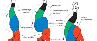
The process of formation and development of the heart in an embryo
The formation of the heart and its development is one of the most complex processes occurring in the fetus’s body and mother’s abdomen. 2 weeks after fertilization, the process of heart formation begins. After another 2 weeks, a hollow, curved tube had already formed. At the 5th week of pregnancy, the first contractions of the tube occur, the intensity of which increases.
At week 5, using ultrasound, you can already see the rudiments of the heart. At this time, the formation of the heart continues, transverse and internal septa appear, thanks to which the organ becomes two-chambered. Gradually, the formation of longitudinal partitions occurs.
By week 9, the baby’s heart becomes an “adult”, having two atria, two ventricles, valves separating them and vessels to ensure blood flow.
However, the heart of the embryo has its own characteristics related to the supply of oxygen from the mother: between the atria there is an oval window, which is connected to the ductus arteriosus. After birth, this window closes and the ductus arteriosus collapses.
The process of heart development ends only by the 22nd week of pregnancy, then only an increase in its muscle mass and growth of the entire blood supply system occurs.
The fetal heartbeat is monitored using ultrasound (US), cardiotocography (CTG), auscultation (listening with a special tube) and echocardiography (EchoCG).
How does the embryo develop in the period from 3 to 5 weeks?
The period of pregnancy from the 3rd to the 5th week is considered very important for the development of the fetus. This is due to the fact that it is at this time that embryogenesis occurs. The embryo has the appearance of the auricle, as it is curved in the shape of the letter C. At this time, when performing an ultrasound, specialists can already examine the rudiments of the head, back, upper and lower extremities.
In the period from 3 to 5 weeks of development, the spinal cord is released in the embryo (the development of the spinal cord and spine begins). The correct formation of the brain can be indicated by the process of flattening (at the wide end of the embryo) of the neural tube, which should occur gradually. During an ultrasound examination, specialists can see how tissue segments called somites are formed and subsequently rapidly increase, which are responsible not only for the development of muscles, but also of all tissue structures present in the human body.
Gynecologists pay close attention to this period also because the fetus begins to develop vascular and cardiac systems, which develop very quickly. Using a highly sensitive ultrasound machine, it is possible to examine the process of formation of large blood vessels. They are located in the central part of the embryo and are closely connected with a glomerulus of tissue, which at this time is only a prototype of the future heart. This tangle is a very important substance that takes an active part in the process of organogenesis.
If pregnancy is successful, it is from these tissues that the respiratory tract (upper) will begin to develop. In this case we are talking about the trachea and larynx. They also take an active part in the process of laying the pancreas, liver, gonoblasts (germ cells), which determine the sex of the child.
A photo of an embryo at the 3rd – 4th week is not informative, since the size of the embryo does not reach 0.2 mm
In the period from 3 to 5 weeks of pregnancy, gynecologists and uzologists are unable to hear the baby’s heartbeat (even when using ultra-sensitive equipment), but they confidently claim that the embryos already have all the necessary rudiments of the vascular and cardiac systems.
Heartbeat monitoring using ultrasound
So, when is a fetal heartbeat visible on an ultrasound? Since the first contractions occur already in the fifth or sixth weeks of pregnancy, the first beats can be heard using ultrasound.
To do this, use a special sensor that is inserted into the vagina. Using a sensor with which the doctor examines the fetus through the abdomen, the heartbeat is heard in the seventh week.
Each stage of pregnancy has its own heart rate:
- Between the sixth and eighth weeks, the fetal heart beats 110-130 times per minute.
- In the ninth and tenth weeks, the heart rate is 170-190 beats.
- Starting from the eleventh week and until birth, the baby’s heart in the womb makes 140-160 beats per minute.
The difference in indicators is affected by the process of formation and development of the fetal nervous system.
With the help of ultrasound examination of the fetal heart, not only the number of contractions is monitored, but also its location, size, nature of contractions, and their rhythm.
At what age does a baby's heart begin to beat?
Even during the process of heart formation, a small part of the tissue begins to contract (later it is transformed into full-fledged cardiac ventricles). It is worth noting that such contractile movements are in no way connected with the nervous system of the embryo. Modern medicine has proven that nervous tissue does not regulate the heartbeat process.
If, during an ultrasound examination of the patient (using heavy-duty devices), specialists are able to determine heart contractions in the 5th week of fetal development, then pregnant women will experience these amazing sensations between the 6th and 8th weeks.
Deviations from the norm
During ultrasound examination, there are cases of identifying deviations from the norm, which may be an indicator of deterioration in the condition of the fetus and its development process. So, deviations are considered:
- The number of beats per minute is less than 100.
- Increase in the number of heartbeats over 200.
- Complete absence of heartbeat.
During the first ultrasound, the doctor may not detect a fetal heartbeat. When its size exceeds 0.8 cm, there is a possibility of a non-developing pregnancy. However, this is not a definitive diagnosis. After five to seven days, a control ultrasound examination is performed, which confirms or refutes the diagnosis.
Also, the reason for the absence of heartbeat in the early stages may indicate a short period of time. After all, no one knows exactly when conception occurred and how many days the embryo is. It is necessary to undergo a re-test.
There is also a third reason for deviations in the number of heartbeats from the norm - the presence of pathological processes in the development of the internal organs of the embryo. To make such a diagnosis, the doctor examines the “four-chamber section.” An ultrasound image shows all four chambers of the heart at once. Ultrasound can detect more than 70% of heart defects. To clarify the diagnosis, echocardiography is performed.
Having discovered pathologies of the fetal cardiovascular system, doctors do not always suggest terminating the pregnancy. Today, surgery is well developed: highly qualified pediatric surgeons, high-tech equipment, innovative methods of treating pathologies of heart formation. The operation is carried out only with the consent of the parents and after a thorough examination. In practice, doctors have more than a dozen cases of saving babies born with heart defects.
In later stages, deviations from the norm depend on:
- Motor activity of the child.
- Physical activity of the mother.
- Presence of diseases during pregnancy.
What will an ultrasound show at 6 weeks of pregnancy?
What an ultrasound scan shows at the 6th week of pregnancy is very important, since it determines how the pregnancy will proceed in the future and whether its continuation is possible in principle.
Ultrasound at 6 weeks
shows:
- Is there a pregnancy?
- Location of the fertilized egg. The norm is the uterine location.
- Number of embryos. At the 6th week it will be possible to determine a multiple pregnancy.
- Dimensions of the fertilized egg. This indicator is necessary for doctors to clarify the gestational age.
- Fetal heartbeat. Only modern equipment can allow you to listen to the heart of an unborn baby at the 6th week. If the ultrasound machine does not have this capability, you will have to wait another 1-2 weeks to accurately assess the beating frequency of the small heart.
At the 6th week, it is too early to talk about identifying any developmental defects, since not a single system or organ in the fetus has yet formed.
If the embryo in the cavity of the ovum is not visualized, this may signal the fading of pregnancy (that is, the death of the fetus). After the ultrasound, you can take a photo in which the outlines of the future baby will be visible.
Heartbeat indicators and causes of deviations
When the heart rate is less than 120 per minute, this indicates: in the first weeks, a short period of time, a period of more than 12 weeks of fetal hypoxia and possible compression by the umbilical cord.
The number of beats is more than 170. In the early stages, it indicates a placentation disorder, however, in most cases this is the norm. From the 12th week, this indicator is affected by the motor activity of the fetus and mother, stressful situations of the mother and hypoxia. During labor, it indicates contraction or hypoxia.
When heartbeat sounds are difficult to hear: in the early stages, this indicates a short period of time, old equipment, excess weight in the mother, or heart defects. Muffled tones, starting from the 12th week, are a sign of: maternal obesity, placental insufficiency, presentation, high or low water, uncomfortable fetal position for examination or defects of the cardiovascular system.
During childbirth, dull sounds occur when contractions are active or the baby is starved of oxygen.
The absence of heart contractions indicates a short term, frozen pregnancy and spontaneous incipient abortion. At 12 weeks and during labor, the cause of this may be fetal death or incorrect examination.
The fetal heart goes through a difficult stage of formation, which is carefully examined by doctors at every appointment with the pregnant woman. When you can hear the fetal heartbeat on an ultrasound depends on the timing, quality of the equipment and the absence of pathologies.
How is an ultrasound performed at 6 weeks of pregnancy?
There are two options for how doctors do an ultrasound in the 6th week of pregnancy:
- The transvaginal method
is the insertion of a vaginal ultrasound sensor protected by a special condom into the vaginal cavity of a pregnant woman. - Transabdominal method
- through the skin of the abdomen. Before the scan, women apply a special gel to their abdomen.
The first method may cause some discomfort to the woman, but its results will be more accurate and complete. Contraindications to transvaginal ultrasound for a pregnant woman may include bleeding or abdominal pain.
How to prepare for research
We found out at what stage an ultrasound shows pregnancy. Now let's talk about how the expectant mother can prepare for the procedure. Since it is quick and completely safe for mother and baby, preparation will be simple and will not take much time.
- It is advisable to give up foods that contribute to gas formation two days before the examination: legumes, raw vegetables and fruits, dried fruits, dairy products, etc. The accumulation of gases in the intestines will interfere with the procedure.
- Depending on the type of ultrasound examination, the drinking regime may change. Thus, it is forbidden to drink water before a transvaginal ultrasound; the bladder must remain empty during the examination. When performing a transabdominal examination, in some cases it is necessary to drink water: the doctor will warn you about this in advance.