During pregnancy, a woman experiences a special state of her body. And the joy of upcoming motherhood should not be overshadowed by problems with the cardiovascular system. What should you do if you are carrying a baby and have been diagnosed with sinus tachycardia? Let's look at this diagnosis.
Pregnancy is not only a pleasant period in a girl’s life. This is also a responsible time when the expectant mother should pay special attention to her health. Due to the increased load on the body, women may develop various disorders of the cardiovascular system.
Attacks of tachycardia during pregnancy are a common occurrence that accompanies the pregnancy process.
Causes
Why does sinus tachycardia occur in an expectant mother and what to do? Factors that can trigger such problems:
- Changes in the body and all its systems;
- Increased load on the heart muscle due to the common circulatory system in the woman and fetus;
- Enhanced functioning of all organs;
- Compression of the abdominal and chest organs, which intensifies as the fetus grows;
- Changes in the hormonal levels of a pregnant woman.
Sinus tachycardia during pregnancy is an important symptom that requires attention from the woman. After all, a rapid heartbeat when a woman is at absolute rest indicates dysfunction of the sinus department, the main purpose of which is the formation of conductive impulses. As a result, a pregnant woman experiences an excessively rapid heartbeat (more than 90 beats per minute) against the background of normal temperature and other indicators of the body’s functioning.
We note the fact that the causes of tachycardia during early pregnancy must be identified and monitored with special attention. After all, the course of pregnancy, the health of the fetus and the expectant mother largely depend on the correct diagnosis of a woman.
Mild sinus tachycardia in pregnant women is considered normal and does not require treatment. Attacks of too fast heart rate occur and disappear suddenly. What to do at this moment?
At this time, you just need to try to calm down and relax as much as possible, and not react to nervous stimuli. Try to stay alone and calm down. The heart of the expectant mother works “for two”, so it contracts somewhat more intensely than usual, therefore, in the last trimester, sinus tachycardia is quite common in women and is not a cause for concern.
But if a patient is diagnosed with a pathological form of the disease during pregnancy, then this condition requires attention from doctors.
Pregnancy and childbirth in women with damage to the heart and blood vessels
The combination of pregnancy and CVD is fraught with increased stress on the body of the expectant mother. Dangerous symptoms may occur for the first time in those who have not previously consulted a cardiologist.
Arterial hypertension
Arterial hypertension (or hypertension, hypertension) is diagnosed in 7-30% of pregnant women, and complications associated with it occupy 4th place in the structure of maternal mortality. This is a condition in which systolic blood pressure rises to 140 mm. rt. Art. and higher, and diastolic - up to 90 mm. rt. Art. or higher. To confirm, pressure is measured at least 2 times on one arm with a minimum interval of 15 minutes; with self-assessment or daily monitoring, the diagnosis is established at a blood pressure level of 135/85 mm. rt. Art.
Frequent complications of pregnancy due to hypertension include:
- fetoplacental insufficiency;
- DIC syndrome;
- premature detachment of a normally located placenta;
- cerebrovascular accident;
- pulmonary edema;
- eclampsia;
- delayed fetal development;
- fetal death during pregnancy;
- obstetric hemorrhage;
- delayed fetal development;
- fetal death during pregnancy;
- retinal detachment, retinal hemorrhages.
Based on the time of onset of symptoms, arterial hypertension that developed before pregnancy, symptomatic (gestational) hypertension, hypertension complicated by preeclampsia, and the condition of eclampsia are distinguished. A special place is occupied by the so-called “white coat hypertension”, which is recorded when measuring blood pressure in a medical institution.
The course of the disease is complicated by hypertensive crises, in which a rapid rise in blood pressure occurs to significant values. Crises can be triggered by stress, refusal of therapy, or errors in diet. Increase in blood pressure to 170/110 mm Hg. - indication for urgent hospitalization and drug therapy.
Preeclampsia
Previously, this pathological condition was called late gestosis. This is an acute complication in which pathological changes affect the inner lining (or endothelium) of small arteries. The properties of the vascular wall and the properties of the blood change. This leads to disruption of microcirculation and damage to arterial walls. Preeclampsia is caused by pregnancy itself and can develop from 21 weeks. Typical symptoms include:
- increased blood pressure;
- proteinuria;
- sudden appearance of edema.
Often the condition is complicated by multiple organ failure, in which the mother experiences headaches, sensory disturbances, visual hallucinations, and blindness. Possible cerebrovascular accident, stroke. The gastrointestinal tract “reacts” with abdominal pain, nausea, vomiting, and the level of liver enzymes increases. Hypoxia and pulmonary edema develop. DIC syndrome, in which blood clotting is disrupted: blood clots form in the vessels, red blood cells disintegrate, poses a great danger. Lack of timely assistance can cause death of the mother and fetus.
Congenital and acquired heart defects
Congenital heart defects (or congenital heart defects) are various defects in the structure of the heart structures and large vessels extending from it. As a rule, such patients are operated on in childhood and are observed by a cardiologist, and undergo examination before planning pregnancy. Girls with suspected hereditary diseases associated with connective tissue deficiency - Marfan and Ehlers-Danlos syndromes - deserve special attention.
The most common options are:
- ventricular septal defect – up to 40% of all congenital heart defects;
- atrial septal defect (ASD) up to 15% of all congenital heart defects;
- patent ductus arteriosus – 15%
- coarctation of the aorta – up to 7%.
Less commonly - stenosis of the pulmonary artery, aorta and defects of the Fallot group.
Acquired heart defects are divided into 2 groups: rheumatic and non-rheumatic. The former arise as a complication of streptococcal tonsillitis, and the latter in diseases such as systemic lupus erythematosus, syphilis, infective endocarditis, scleroderma. They are characterized by the formation of stenoses and damage to the heart valves. Features of this group of CVDs during pregnancy include:
- the likelihood of rheumatic carditis in the 1st trimester;
- difficulty in pulmonary circulation in the 3rd semester;
- increase in blood pressure during childbirth;
- collaptoid conditions, obstetric hemorrhages in the postpartum period.
Symptoms for any heart defect depend on the size of the defect, its location, and the duration of the lesion. Patients in this group are concerned about nonspecific complaints: fatigue, muscle weakness, increased drowsiness, palpitations and shortness of breath that appear during physical exertion. Heaviness in the legs and drowsiness may occur. If the pregnant woman’s condition worsens, shortness of breath bothers her even at rest, swelling appears and increases, and arrhythmias are possible.
In addition to worsening heart failure, the following complications may be possible during such a pregnancy:
- fetoplacental insufficiency;
- preeclampsia;
- threat of premature birth.
Pregnancy with Marfan syndrome is associated with a high risk for the mother and baby: the formation and dissection of an aortic aneurysm and acute heart failure are possible. Due to systemic inferiority of connective tissue, premature birth, uterine rupture and prolapse are common. The probability of having a child with a hereditary disease is 50%.
Varicose veins
Varicose veins occur when collagen protein synthesis is disrupted and manifests itself in the presence of provoking factors. Pregnancy is the time when a woman is most vulnerable. Varicose veins in pregnant women develop and progress due to physiological characteristics: progesterone relaxes blood vessels, the volume of circulating blood increases, and body weight increases. The growing uterus puts pressure on the vena cava, and the outflow from the lower extremities and pelvic veins is disrupted.
A woman may be planning a pregnancy with an already diagnosed disease or discovering the problem for the first time. She may be concerned about:
- swelling of the legs in the evening;
- pain and cramps in the calf muscles;
- the appearance of spider veins, enlarged veins on the surface of the skin.
In some cases, damage to the veins of the lower extremities is combined with varicose veins of the small pelvis. Complications of the disease include placental insufficiency, preeclampsia and the threat of premature termination.
Cardiomyopathy
Damage to the heart muscle with changes in structure and impaired contractile function occurs with cardiomyopathy (or CMP). There are 2 types of disease: dilated and hypertrophic.
Dilated cardiomyopathy is a disorder of the myocardial structure with the proliferation of connective tissue. The contractility of the heart decreases, its wall becomes thinner, and circulatory failure occurs. Typical symptoms include shortness of breath, tachycardia, swelling of the extremities, pulmonary edema and hemoptysis.
Hypertrophic cardiomyopathy is often characterized by the formation of compacted chambers of the heart that do not relax well and do not fill during diastole. Hyperplasia of the inner layer of the coronary arteries is possible, causing myocardial nutrition to suffer. Characteristic symptoms include heart pain, poor exercise tolerance, and fainting. High risk of sudden death.
Most often, CMP is hereditary in nature. Pregnancy leads to decompensation of the disease and poses a danger to mother and child. Common complications include:
- delayed fetal development;
- premature birth;
- death of mother and child.
Symptoms
If a woman begins to feel severe nausea and dizziness, and also has bouts of vomiting, she should definitely tell her doctor about this. Such symptoms may indicate a pathological form of the disease, which is characterized by longer and more severe attacks of increased heart rate.
During pregnancy, a woman is under no circumstances recommended to expose herself to excessive physical exertion or experience any kind of nervous shock. If a patient experiences tachycardia while pregnant, doctors strongly advise her to protect herself from all kinds of activity and to rest and relax more.
Such measures in most cases help to avoid increased heart rate (heart rate) and attacks of tachycardia. But if after the patient’s rest this does not happen, the symptoms remain, she should consult a doctor for help.
If tachycardia during pregnancy is pathological, then this problem must be solved only with the help of qualified doctors in modern clinics. Therefore, it is so important to promptly diagnose and begin to treat such a disease.
Management of pregnancy in women with heart disease
Patients with a significant cardiovascular history should take a responsible approach to planning pregnancy. Even in the absence of any complaints, examination by a cardiologist is recommended. It is this doctor who decides the issue of prolonging pregnancy, choosing management tactics and method of delivery.
Preparing for pregnancy
The expectant mother should undergo examination to identify possible diseases. You should not be afraid of visiting a doctor and assume that pregnancy is a condition that cures all diseases, and the very fact of its occurrence automatically recognizes the woman as healthy. To assess the risk you must go through:
- ECG;
- EchoCS;
- stress tests to assess heart function;
- MRI for aortic lesions.
If you suspect hereditary diseases, you should contact a medical geneticist. A full examination will show whether it is possible to plan a pregnancy.
Pregnancy due to CVD
If pregnancy occurs in a woman with damage to the heart or blood vessels, the cardiologist conducts an ongoing examination to decide whether it can be saved. Thus, indications for interruption are conditions with severe circulatory disorders and an active rheumatic process:
- atrial fibrillation due to heart disease;
- aortic valve insufficiency;
- aortic valve stenosis with an increase in heart size and impaired myocardial function;
- mitral valve insufficiency with the development of circulatory failure, arrhythmia, active rheumatic process;
- previous heart surgery (individual);
- exacerbation of rheumatism with severe manifestations;
- severe cardiomyopathy;
- severe congenital defects (patent ductus arteriosus, pulmonary stenosis, ventricular septal defect).
If a council of specialists allows a woman to bear a child, she is subject to joint supervision by an obstetrician-gynecologist, a therapist and a cardiologist. During pregnancy, it is necessary to regularly evaluate the condition of the heart, and if the condition worsens, prescribe drug therapy. Treatment is carried out on an outpatient basis or in a hospital setting. Cardiologists recommend a minimum of 3 planned hospitalizations:
- in the 1st trimester to decide whether to continue the pregnancy;
- at a period of 228-32 weeks for preventive treatment;
- at 36 weeks to choose the method of delivery.
Possible complications
What complications can sinus tachycardia cause for a pregnant woman? Pregnancy is a period in a woman’s life when the risks of many diseases increase or existing health problems become more complicated.
If this disease is not treated, allowing its development to take its course, disruption of the nervous system may occur, the emotional background may change, mood swings may intensify, sleep disturbances and a state of lethargy may appear. As a result, the woman’s immunity will decrease, and the already weakened body will constantly remain in a weakened state.
Such problems in a pregnant woman can negatively affect the condition of her body and her quality of life. In addition, with such a disease, the risk of complications during pregnancy and delivery increases. That is why heart problems in the expectant mother must be identified early for the success of their treatment.
Relevance of the problem
The problem of admission to pregnancy and management of women with heart and vascular lesions is becoming increasingly urgent. The desire to move higher on the social ladder, to acquire a financial “safety cushion”, and a later age of marriage have led to an increase in the age of first-time women. Motherhood in adulthood is more often associated with the risk of endocrine and cardiovascular diseases (or CVD).
Also, the high level of development of medicine has become the reason that more and more women with congenital heart defects survive to reproductive age and are allowed to become pregnant, while the increased load on the body becomes the main cause of maternal mortality. Among all CVDs, the greatest risk of adverse outcome of pregnancy and childbirth is borne by:
- congenital heart defects;
- rheumatic heart valve lesions;
- conditions with increased blood pressure.
Cardiomyopathies are extremely rare, but pose the greatest danger to women.
Timely detection of CVD allows for preventive monitoring, hospitalization and timely treatment.
Treatment and its features
Tachycardia in a pregnant woman requires careful diagnosis and the correct treatment regimen. First of all, the patient is prescribed blood tests and an ECG. In some cases, at the doctor’s insistence, additional procedures may be performed.
Treatment of tachycardia during pregnancy cannot be carried out inconsistently. If the doctor has drawn up a treatment regimen using medications, consultations with a psychoanalyst or neurologist, this regimen must be strictly adhered to.
If you are worried about your health and the health of your unborn baby, if symptoms of tachycardia appear, consult a gynecologist or visit a cardiologist. Health-safe diagnostics will help to accurately establish a diagnosis or reassure the expectant mother if fears are not confirmed.
37th week of pregnancy for baby
At the 37th week of pregnancy, the baby’s height is approximately 48 cm, and his weight is 2,600 g. Externally, the fetus is almost no different from a newborn; all facial features are developed, and cartilaginous tissue is clearly visible. The accumulation of subcutaneous fat at this stage of pregnancy makes the body contours softer and rounder. The baby’s skin gradually smoothes out, it is no longer as pink as in the previous weeks of intrauterine development, and the integument gradually becomes lighter. The baby's body is still abundantly covered with lubricant, but the amount of vellus hair decreases noticeably, vellus hair remains only on the shoulders and back, and in some babies it disappears almost completely.
This week the accumulation of fatty tissue continues. It reaches a maximum of 15% of the child’s total body weight. It is difficult to overestimate the importance of adipose tissue for newborns; it is this tissue that protects the child from overheating or hypothermia, since the baby’s thermoregulation system after birth is not yet sufficiently formed and continues to develop in the first months of the little person’s life.
At this stage, not only the volume of subcutaneous fat increases, but also the muscles and skeleton intensively develop. The child constantly moves his arms and legs. These unique workouts help increase muscle mass. The baby also makes rhythmic breathing movements that strengthen the intercostal muscles and diaphragm, preparing the respiratory organs for childbirth.
Our doctors
Kedrinskaya Larisa Ivanovna
Experience 18 years / Doctor of the highest category, Candidate of Medical Sciences / General practitioner, cardiologist
Çà ïèñà òüñÿ ê âðà ÷ó
Kruzhalova Olga Sergeevna
25 years of experience / Leading specialist / Obstetrician-gynecologist
Çà ïèñà òüñÿ ê âðà ÷ó
Vershuta Elena Vasilievna
More than 23 years of experience / Highest category, Candidate of Medical Sciences / Pediatric cardiologist, cardiologist, therapist
Çà ïèñà òüñÿ ê âðà ÷ó
+
Pregnant woman at 37 weeks
As the due date approaches, pregnant women begin to notice the appearance of their precursors, that is, certain signs and changes that occur under the influence of hormones. A woman’s body is preparing to give birth to a child, progesterone gives way to the dominant role of the birth hormone estrogen, and the pregnant woman’s well-being changes.
From the 37th week, expectant mothers may observe the following changes:
- slight loss of body weight;
- reduction in abdominal volume;
- the appearance of training or “false” contractions and an increase in their intensity;
- discharge of mucus from the cervix.
The nature of the stool changes, it becomes weaker, aching pain in the lower back of varying intensity may appear, and the fundus of the uterus descends. The woman notes some signs on her own, others are observed by the gynecologist during a routine examination.
Precursors do not appear in all women. Some expectant mothers notice only some of the symptoms listed above, while others observe signs of impending labor not two or three weeks before the due date, but just a few hours. Both the appearance of precursors at the 37th week and their absence are normal and depend on the individual characteristics of the woman’s body.
This week, the female body is intensively preparing for the birth of a child. If the fetus is positioned correctly, head down, it gradually lowers, goes to the lower part of the uterus, presses it to the body and bends its limbs, intuitively taking the most comfortable position for passing the birth canal. The consequence of fetal movement is prolapse of the uterine fundus. The abdomen lowers, the pressure on the diaphragm decreases significantly, the pregnant woman can breathe easily, and the shortness of breath that plagued her in the previous weeks disappears. The pressure on the stomach also decreases, heartburn, a feeling of heaviness after eating and other unpleasant sensations disappear. Moving your baby can put pressure on your bowels and bladder. A pregnant woman at this stage often experiences the urge to urinate and may suffer from frequent loose stools. The reason for frequent bowel movements is not only the mechanical effect of the uterus on it, but also an increase in the content of estrogen in the body, hormones that promote the excretion of fluid. At the 37th week, the expectant mother can have bowel movements up to 3-4 times a day and at the same time observe significant dilution of the stool.
The appearance of acne.
This symptom is also associated with hormonal changes in the body. Acne can appear even in women who have never suffered from it before. Acne during pregnancy usually disappears when hormonal levels stabilize and no longer bothers the woman. You can often miss this phenomenon, mistaking it for a component of premenstrual syndrome, especially if you have previously been treated for acne. Topical, and especially systemic, retinoids can adversely affect fetal development, so you should stop using these medications if there is a delay.
40th week of pregnancy: how is the baby developing?
40 weeks – full-term pregnancy. The weight of a child born at this period ranges from 2,600 g to 4,400 g, and his body length is 48-53 cm. These indicators are very arbitrary, since at 40 weeks miniature babies weighing 2,600 g and real heroes are born, whose body weight approaches 5,000 g. The body length of newborns can also vary from 45 to 55 cm.
Most women give birth at 40 weeks. At this stage, the baby is completely ready for birth; it meets all the parameters of a full-term baby. Before birth, the baby presses its arms and legs closely to the body, bends its head as much as possible and presses against the exit of the uterus. This position makes it possible to pass the birth canal with the narrowest part of the skull. During labor, with each contraction, the child gradually moves downward; he does not move in a straight line, but makes helical-translational movements, as if screwing into the mother’s birth canal. As the newborn moves forward and his head descends completely, the cervix opens completely. This is followed by pushing, that is, contractions of the uterus that move the baby along the birth canal. The baby's head gradually appears, followed by his body. Childbirth is a complex mechanism that is aimed not only at the safe passage of the birth canal by the child, protecting him from accidental injuries due to increased pressure, but also at preventing ruptures of the woman’s soft tissues.
Sudden mood swings.
You woke up in a good mood, but a minute later absolutely everything irritates you? This is not a mental disorder. Until hormonal levels stabilize, constantly changing emotions, a sudden transition from negative to positive, can accompany a woman throughout pregnancy. Explain to your loved ones that now you are more sensitive to everything that happens around you. When your hormonal levels return to normal, you will feel better. This usually happens in the second trimester of pregnancy. If emotional problems bother you too much, be sure to ask your doctor to prescribe you a mild sedative that is not contraindicated for pregnant women.
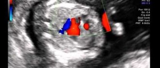
How can you hear your child’s heartbeat?
You don't have to go to a gynecologist to hear your child's heart. The first way to listen to your heart rate is with a stethoscope . It can be bought at a pharmacy at a low price. But you will need help. Before the 25th week of gestation, people without appropriate medical education can almost never hear the fetal heart. You can repeat listening with a stethoscope every week. It is important to remember that the sounds of the fetus moving can be confused with the beating of its heart, as well as with the sounds of processes that occur in the mother’s abdomen.
The second way to hear the baby’s heart yourself is fetal doppler . This is an ultrasound detector that you can buy yourself. It does not record data on tape, but you will hear the baby's heartbeat. The method is relevant from the 8th and later weeks of gestation. Duration of the procedure: maximum 10 minutes. The device is expensive, so not all pregnant women can afford it.
Third way: put your ear to the pregnant woman's belly . But this is only relevant after the 30th week of gestation. But if a woman is overweight, this method will not work. If the fetus is head down, it is better to listen to its heart below the pregnant woman's navel.
If you tend to worry about little things and independently diagnose yourself and your child, it is better to entrust listening to the fetal heartbeat and determining the norm to doctors. Enjoy motherhood and be in harmony with yourself.