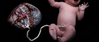
The umbilical cord is the main highway that provides blood flow with substances, microelements and oxygen vital for the fetus from the mother and removes waste blood. It connects the placenta (baby place) with the anterior abdominal wall of the fetus and consists of three vessels covered with a special protective membrane (Wharton's jelly).
Through the umbilical vein, the fetus receives oxygen and all the nutrients necessary for its normal development. Two umbilical arteries carry out the outflow of blood saturated with carbon dioxide and metabolic products back to the placenta. Therefore, structural features or anomalies of the umbilical cord have a great influence on the viability and dynamics of fetal development, as well as on the outcome of pregnancy.
What is the single artery of the fetal umbilical cord?
An anomaly in the structure of the umbilical cord, when instead of two arteries one is formed, is called a single umbilical cord artery.
The absence of one artery in the umbilical cord can be initial (congenital aplasia) or develop during pregnancy (artery atrophy, which occurs as a result of the complete cessation of its functioning).
A single fetal umbilical cord artery is considered a fairly common pathology: 1 in 200 singleton pregnancies – 0.5% and 1 in 20 multiple pregnancies – 5%.
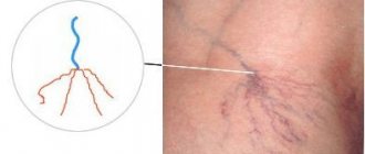
Normally, the umbilical cord contains 2 arteries and 1 vein
How many vessels are there in the umbilical cord, what are their functions?
How many vessels should the umbilical cord have? Normally, the umbilical cord vessels are represented in the amount of 3 pieces. Each of these tubular organs performs a specific function:
- Arteries. The umbilical cord contains two arteries that arise from the internal iliac veins. They exist exclusively during the gestational period. These blood vessels move embryonic blood, saturated with carbon dioxide and metabolic products, to the child's place. At the end of pregnancy, the umbilical arteries are emptied, turning into scar formations in the form of the medial umbilical ligaments. They pass along the anterior wall of the peritoneum under the parietal peritoneum, forming an equilateral triangle along the sides of the bladder to the umbilical cavity.
- Vein. Initially, 2 vessels are formed, subsequently one of them becomes clogged. This tubular organ delivers blood enriched with oxygen and nutrients from the baby's place to the embryo. 80% of the liquid connective tissue enters the systemic circulation through the vessel connecting the umbilical vein with the inferior vena cava, which passes along the surface of the liver and flows into the inferior vena cava. The remaining volume flows into the portal circulation through the opening between the umbilical vein and the left branch of the portal vein. These organs contribute to the blood supply to liver cells.
The causes of this pathology
In obstetrics, this umbilical cord anomaly is defined as single umbilical artery syndrome. The likelihood of its occurrence increases with diabetes mellitus, multiple pregnancies and other complex diseases in the expectant mother (pathology of the kidneys, liver, heart and blood vessels).
The reasons for the development of single umbilical cord artery syndrome are conditions and factors that negatively affect the formation of blood vessels and tissue differentiation in the early stages of intrauterine development.
Bad habits in a pregnant woman can cause pathology
The causes of umbilical cord vascular anomalies are not fully understood, but the most common predisposing and provoking factors are:
- chromosomal abnormalities (aberrations);
- severe somatic and infectious diseases of the expectant mother in the first trimester of pregnancy;
- intoxication;
- diabetes;
- bad habits (smoking, alcohol abuse, taking narcotic and other potent drugs);
- exposure to radiation, unfavorable environmental conditions, occupational hazards at work;
- multiple pregnancy.
The most dangerous period of intrauterine development with a high risk for the occurrence of congenital vascular anomalies of the placenta is considered to be 6-9 weeks of gestation - the period of formation of the future placenta.
Atrophy of a functioning umbilical cord artery can occur due to thrombosis, compression by a tumor (teratoma, hemangioma) or hematoma, or when nodes appear in one artery. These conditions occur extremely rarely under the influence of several pathological factors and require constant dynamic monitoring of the condition of the fetus and the expectant mother.
Where is the norm and where is the pathology?
Around the 21st week of gestation, all pregnant women are sent for a Doppler study, which allows one to evaluate not only the anatomical features of the fetus, placenta, and umbilical cord, but also the characteristics of blood flow (speed, number of vessels, anomalies). Often doctors do not bother to explain the results of the analysis to the patient, even briefly, so expectant mothers scour the literature and the Internet in search of answers.
Normally, at any stage of pregnancy, the umbilical cord has 3 vessels. Having received such a conclusion, the woman can calm down - the blood flow is fine (of course, if no other anomalies are found). In some cases, the ultrasound diagnostic doctor does not detect one artery in the umbilical cord, then the conclusion will indicate that there are only two vessels - one vein and one artery.
If there is an insufficient number of umbilical cord vessels, specialists will have to figure out whether the anomaly is isolated or combined with other defects, which is often found in this type of pathology. Some women note that the number of vessels during ultrasound differs at different times, and this creates even more questions, misunderstanding and unnecessary anxiety for the expectant mother.
Note that the number of vessels should not change throughout pregnancy, so there are two conclusions: either one of the arteries stopped functioning for some reason, or an error was made in one of the studies, and it is necessary to “recount” the vessels again, preferably – from a competent and experienced doctor who will dispel all doubts.
Symptoms and diagnosis of a single umbilical cord artery
In most cases, there are no clinical signs of the presence of this anomaly, and diagnosis is carried out by instrumental examination methods:
- screening (routine) or urgent ultrasound;
- Dopplerometry - determination of the main indicators of blood flow in the umbilical cord.
Ultrasound will help diagnose pathology
The presence of this pathology can be determined at 20-21 weeks of pregnancy by ultrasound examination of the umbilical cord in cross section. Diagnosis is made with a full bladder.
If a pathology is detected, an additional examination is prescribed - Doppler ultrasound to clarify the absence of weakening of blood flow.
After identifying a vascular anomaly of the umbilical cord in the fetus, constant monitoring of the course of pregnancy is necessary until the moment of birth. Repeated regular Doppler examinations are carried out in order to promptly diagnose changes in blood flow in the umbilical cord artery and avoid complications of the fetal condition.
Consequences and complications when identifying this anomaly
In 70% of cases, this condition does not have a negative effect on the intrauterine and postpartum condition of the child - a single artery completely copes with the increased load. Diagnosing this defect in most cases is not a cause for concern.
And yet, in 25-30% of cases, this anomaly of umbilical cord development can be combined with other developmental defects and genetic disorders:
- Chromosomal diseases.
- Congenital malformations: heart and blood vessels; urinary system; abdominal and thoracic organs.
Therefore, when diagnosing a single umbilical cord artery, it is necessary to conduct a comprehensive examination of the fetus to exclude the presence of other anomalies.
It is important to remember that isolated single umbilical cord artery syndrome is not an indication for termination of pregnancy and is not considered a marker of Down syndrome and other chromosomal diseases.
But the combination of this pathology with other developmental defects has a high risk for the life and further development of the child.
Consequences of combined developmental defects:
- intrauterine fetal death (frozen pregnancy);
- intrauterine growth retardation (IUGR);
- chronic intrauterine fetal hypoxia;
- intrauterine hypotrophy.
A single umbilical cord artery may be the cause of chronic fetal hypoxia and related consequences
Umbilical cord diseases
There is a connection between the fetal place and the fetus through the umbilical cord or umbilical cord (funiculus umbilicalis), which normally consists of three vessels: one vein , through which blood saturated with oxygen and nutrients enters the fetus, and two arteries , through which venous blood returns from fetus to placenta.
In the early stages of embryonic development, the umbilical cord has two veins, but at 4 weeks of pregnancy the right umbilical vein becomes blocked and disappears by 7 weeks of pregnancy. In very rare cases, the left umbilical vein becomes obstructed, and the right one supplies the fetus with the necessary blood, while the development of the fetus is not impaired.
About 80% of the blood from the umbilical vein enters the fetal systemic circulation through a special ductus venosus, which drains into the inferior vena cava. The remaining 20% of the blood enters the portal (hepatic) circulation of the fetus.
The volume of blood flow in the umbilical cord increases with the duration of pregnancy - from 35 ml/min at 20 weeks to 240 ml/min at 40 weeks.
Types of pathological conditions of the umbilical cord
All disorders and pathological conditions of the umbilical cord can be divided into the following:
- pathological set of umbilical cord vessels
- short and long umbilical cord
- pathological torsion of the umbilical cord
- compression and blockage of the umbilical cord
- disruption of blood flow in the umbilical cord
- umbilical cord nodes
- umbilical cord hernia
- pathological attachment of the umbilical cord
- umbilical cord entanglement
- umbilical cord tumors
- damage (trauma) to the umbilical cord
- prolapse of the umbilical cord
Any deviation in the structure of the umbilical cord found on an ultrasound requires a detailed examination of the structure of all fetal organs, as it may be associated with other fetal malformations.
Single umbilical artery
In 1% of all pregnancies, instead of two arteries, the umbilical cord contains one, which in 75% of cases is an isolated deviation that does not affect the development of the fetus and ends in the birth of a healthy child. However, in 25% of cases, a single umbilical artery is associated with other abnormalities in fetal development, in particular the presence of defects of the heart, kidneys, intestines and skeletal system. Also, this type of umbilical cord pathology is more common with trimosomy 18 (Edwards syndrome).
If a single umbilical artery is detected, it is recommended to perform fetal karyotyping through chorionic villus sampling or amniotic fluid sampling, detailed anatomical ultrasound, and echocardiography. In the absence of other abnormalities in the development of the fetus, the prognosis and outcome of pregnancy are positive, as in cases of a healthy pregnancy.
Short or long umbilical cord
The normal size of the umbilical cord at the time of birth is 55-61 cm, although it may be slightly smaller or larger. Determining the true length of the fetal umbilical cord using ultrasound is not always possible due to the compact position of the fetus and its constant movements.
Leonardo da Vinci, who was not only a famous artist and mathematician, but also an excellent anatomist, argued that the length of the umbilical cord usually corresponds to the length of the fetus, and he was right. At 8 weeks of pregnancy, the umbilical cord is 0.5 cm long, at 20 weeks it is about 16-18 cm. The growth rate of the umbilical cord slows down after 28 weeks of pregnancy.
One of the first signs of a long umbilical cord is the presence of umbilical cord entanglements or umbilical cord knots. The average length of the umbilical cord when such abnormalities are detected is from 75 to 95 cm.
A short umbilical cord limits fetal movements and is often a secondary sign of fetal pathology (against the background of existing abnormalities in fetal development). The umbilical cord is considered short if its size is less than 32-40 cm at 38-40 weeks. This type of umbilical cord deviation is observed in 2% of births and placental examinations.
Most often, a short umbilical cord is accompanied by lesions of the fetal nervous system, skeletal dysplasia and other developmental defects, as well as inhibited mental development of children. The shorter the umbilical cord, the greater the risk of serious developmental defects in the fetus. When the umbilical cord length is less than 15 cm, malformations of the anterior wall of the abdomen, spine, and limbs are often found.
Natural childbirth with a short umbilical cord is impossible if the fetus is born with a head, and may be accompanied by placental abruption during childbirth, severe bleeding and other negative consequences of such childbirth complications.
Genetic factors (heredity) can play a role in the occurrence of a long umbilical cord, since it has been noted that such an umbilical cord can be observed in the same woman during subsequent pregnancies. The evidence that the longer the umbilical cord, the more active the fetus and the more active the child in the future, is contradictory and not confirmed by clinical studies.
While diagnosing a short umbilical cord is not usually difficult, there is no consensus among physicians regarding the diagnosis of a long umbilical cord. The standard minimum permissible maximum size of the umbilical cord has not yet been adopted - it fluctuates around 70-90 cm.
Measuring the length of the umbilical cord after a normal birth is not usually done, so most cases of long umbilical cord remain undiagnosed. After a pathological birth or the birth of children with developmental defects, the placenta is usually examined and attention is rarely paid to the size of the umbilical cord, except in cases where there is pathological torsion of the umbilical cord, umbilical cord nodes, or a single umbilical artery.
A long umbilical cord is often accompanied by intrauterine fetal hypoxia, fetal growth retardation, intrauterine fetal death, a large number of brain development abnormalities, neurological disorders, neurological development disorders in children and blood clotting disorders in newborns.
Pathological torsion of the umbilical cord
Normally, the umbilical cord vessels are evenly twisted counterclockwise to the left (like a serpentine) with a certain frequency of torsion from left to right (7: 1.4). The average number of twists of the umbilical arteries and veins along the entire length of the umbilical cord is 40. The umbilical cord twist index is the number of twists per 1 cm, which is normally 0.2.
Umbilical cord torsion appears early in pregnancy and can be detected on ultrasound at 9 weeks. The cause and mechanism of twisting is unknown, but it is thought to be related to the turning and movement of the fetus. The torsion index can characterize low or high fetal motility.
Also, twisting of the umbilical cord may be due to different growth rates of the umbilical cord vessels.
Changes in the direction of torsion do not affect the development of the fetus, but are more often accompanied by placenta previa and bleeding during pregnancy.
Absence of torsion or low torsion index is associated with chromosomal abnormalities, fetal malformations, fetal distress, and increased fetal and neonatal mortality. Increased twisting or pathological torsion of the umbilical cord is often accompanied by premature birth, death of the fetus and newborn due to asphyxia, as it can lead to disruption of blood flow in the umbilical cord, which in turn can result in thrombosis, rupture, dilation (coarctation) of the umbilical cord vessels. Pathological umbilical cord torsion is more often observed with long umbilical cords.
Compression and blockage of the umbilical cord
The umbilical cord is located outside the fetal body in the uterine cavity and is surrounded by amniotic fluid, and also has a specific structure: the vessels of the umbilical cord are covered with a membrane and have a layer of gelatinous mass, somewhat reminiscent of jelly ( Wharton's jelly ), a derivative of connective tissue. This not only improves the strength of the umbilical cord, making it hard to the touch (like rubber), but also protects it from being completely crushed. The fetus often compresses the umbilical cord, but due to the constant movement and turns, such compression is very short-lived. If the umbilical cord has pathological changes, any compression can lead to serious negative consequences, primarily blockage of the lumen of blood vessels and disruption of blood flow in the umbilical cord.
All causes of umbilical cord compression can be divided into external (fetus, large uterine tumor) and internal (structural abnormalities of the umbilical cord, nodes, torsion, hernia, umbilical cord tumors, etc.). If compression develops gradually and does not completely block the lumen of the vessels, a state of chronic obstruction (blockage) of blood flow occurs. The fetus may develop normally, but most often there is a delay in fetal growth.
Acute obstruction occurs more often closer to childbirth or during childbirth, especially with the presenting part - the nascent head or body of the fetus. It is observed in the presence of another pathology of the umbilical cord (true umbilical cord nodes). Acute compression of the umbilical cord can result in the formation of umbilical cord thrombosis and rupture. Compression of the umbilical cord during childbirth can lead to fetal hypoxia and asphyxia of the newborn, which will result in brain damage due to oxygen deprivation. Such children suffer from neurological diseases of varying degrees. The risk of fetal death during childbirth also increases.
Since the umbilical vein carries “fresh” blood from the placenta to the fetus, its compression is characterized by more serious complications than compression of the umbilical arteries. There is blood stagnation in the placenta, and anemia and hypoxia in the fetus.
Diagnosing umbilical cord compression using ultrasound is not easy, but studying the blood flow in the fetal vessels and its biophysical profile allows us to suspect fetal hypoxia and conduct a more thorough search for the cause of such disorders. Treatment of such a pathology will depend on the condition of the fetus, the duration of pregnancy and may involve more frequent monitoring of fetal development or urgent delivery.
Umbilical cord blood flow disturbance
Almost all pathological conditions of the umbilical cord can lead to disruption of blood flow in the umbilical vessels. Often, compression of blood vessels leads to the formation of a blood clot. Umbilical cord thrombosis is often detected after childbirth, especially pathological ones, when examining the placenta and umbilical cord. Often the presence of blood clots is accompanied by areas of umbilical cord calcification, which is associated with a number of infections (for example, syphilis).
Thrombosis of the umbilical cord vessels can also be due to a violation of the properties of the blood in a number of fetal cogulopathies, when the central nervous system of the fetus is damaged, when the processes of regulation of blood flow from the vessels of the fetus and the umbilical cord are disrupted.
Diagnosis of thrombosis of the umbilical vessels using ultrasound is difficult and cannot always be done before the birth of the child.
Umbilical cord knots
There are false and true umbilical cord nodes. False nodes are thickenings of the umbilical cord associated with the formation of a loop or expansion of the umbilical vein without blocking its lumen, therefore most doctors do not call them nodes. Sometimes this is a local varicose vein of the umbilical vein, but externally it may look like a real node. False nodes have no clinical significance and are completely safe for the fetus.
True umbilical cord nodes are found in 0.4-3% of births. They are accompanied by a high risk of morbidity in newborns (up to 11% of cases). True nodes may be tight, causing damage to the Wharton's jelly and umbilical cord vessels. At the site of the node, varicose veins of the umbilical vein may be observed, as well as the formation of blood clots not only in the umbilical cord, but also in the placental vessels. Most often, true nodes are observed with a long umbilical cord and its excessive twisting, polyhydramnios, and multiple pregnancies. Women with gestational diabetes are more likely to have true nodes. A connection has also been noted between the formation of true nodes and amniocentesis, which is explained by greater fetal mobility and uterine contractions during the procedure.
Diagnosing true nodes using ultrasound is difficult and most often they are discovered after childbirth. Doppler ultrasound can help study blood flow in the umbilical cord.
In most cases, true umbilical cord nodes do not complicate the course of pregnancy and their presence does not affect the growth and development of the fetus, as well as the outcome of pregnancy. If the node is tight and its diameter and the lumen of the umbilical cord are less than 1.5 cm, disturbances in blood flow in the great vessels of the fetus, fetal hypoxia and very rarely other complications in fetal development may be observed.
Since true nodes are often accompanied by polyhydramnios, the frequency of surgical interventions (cesarean sections) in such cases is higher.
Data on increased rates of stillbirth or neonatal mortality are conflicting and require clinical trials.
Umbilical cord hernia
The two most common abdominal wall defects are umbilical cord hernia and omphalocele. With an omphalocele , the umbilical ring is expanded and the internal organs of the fetus (intestines, spleen, liver, gall bladder), covered with amniotic membranes, can come out through it. In such cases, the attachment of the umbilical cord is not normal, but occurs outside the hernial sac.
Unlike omphalocele, with an umbilical cord hernia , its attachment is normal - in the area of the umbilical ring, while the skin and muscles that form and cover the ring are not damaged. In other words, it is an enlarged umbilical ring that resembles an adult umbilical hernia. While omphalocele is accompanied by a number of serious complications of pregnancy and often occurs with chromosomal abnormalities of the fetus, umbilical cord hernia is a very rare occurrence and most often occurs without harm to the fetus and newborn. In some cases, intestinal damage may occur. Diagnosis of an umbilical cord hernia is often accompanied by the fact that an omphalocele is mistakenly diagnosed and the woman is often offered to terminate the pregnancy. Therefore, it is important to carry out karyotyping of the fetus (in case of hernias, the child’s chromosome set is normal), as well as MRI for a detailed study of the structure of the umbilical ring and the relationship of organs at the site of the hernia. The prognosis for umbilical cord hernia is positive.
Pathological attachment of the umbilical cord
Normally, the umbilical cord is attached to the center of the placenta on the side of its fetal surface. In 7% of cases, attachment occurs along the edge of the placenta and such attachment is called marginal or marginal . In 1% of cases, the attachment of the umbilical cord occurs outside the fetal part of the placenta to the fetal membranes and such attachment is called velamentous or velamentous . Very rarely, there is a divergence of the umbilical cord vessels in the area of attachment and such an attachment is called furcated . Most often, pathological attachment of the umbilical cord is observed during multiple pregnancies.
The peculiarity of velamentous attachment of the umbilical cord is that in the area of attachment and formation of placental vessels there is no protective jelly-like mass (Wharton's jelly), which can be accompanied by compression of the umbilical cord, the formation of blood clots, rupture of the membranes, and umbilical cord injury.
The farther the umbilical cord is attached from the edge of the placenta, the more such attachment is accompanied by a high risk of vascular damage and serious pregnancy complications.
With vasa previa, the umbilical cord is attached to the internal os of the cervix and can be damaged during natural childbirth.
Bleeding due to rupture of umbilical cord vessels occurs in 1 in 50 cases of velamentous umbilical cord attachment, but is associated with a high rate of fetal death (two to three quarters of cases). With pathological attachment of the umbilical cord, fetuses are more likely to experience hypoxia, increased heart rate, developmental delay, intrauterine death, and newborns are more often diagnosed with low weight, thrombocytopenia, and other abnormalities associated with hypoxia and anemia.
Pathological attachment of the umbilical cord can be diagnosed in the early stages of pregnancy, but most often during an ultrasound scan in the second trimester, when the placenta is formed. However, in most cases, pathological attachment of the umbilical cord is discovered after birth when examining the birthplace.
There is no treatment for this malformation of the umbilical cord, but in some cases treatment of the fetus can be carried out (intrauterine blood transfusion for anemia and fetal hypoxia).
Childbirth requires good doctor skills and constant monitoring of the condition of the fetus. The incidence of operative delivery in cases of pathological umbilical cord attachment is higher due to fetal distress and hypoxia.
Umbilical cord entanglement
Entanglement of the umbilical cord, especially the fetal neck, is a common phenomenon that is found during ultrasound, starting from 10 weeks of pregnancy. It occurs in 15-20% of all pregnancies. In addition to the neck, the umbilical cord can wrap around other parts of the body, especially the limbs. Most often, entanglement occurs when there is a long umbilical cord.
The cause of umbilical cord entanglement is unknown, but a combination of a long umbilical cord and fetal twisting may be key to understanding how this condition occurs. More often, entanglement is observed with polyhydramnios.
The entanglement around the neck can be in the form of a simple loop and a loop with a “lock” (tied), and there can also be several loops (8 umbilical cord loops around the fetal neck have been recorded). The tighter the loop and the more loops, the worse the prognosis for pregnancy outcome.
Being entangled in the umbilical cord can cause compression of the umbilical cord and impede blood flow. This in turn can lead to fetal hypoxia and, in some cases, to its death. But in most cases, umbilical cord entanglement, which was discovered in the second trimester, is not found on ultrasound closer to birth. If there is an umbilical cord around the neck, the growth, development and condition of the fetus remains normal until the end of pregnancy, because the vast majority of loops are loose (not tight).
The danger most often arises during childbirth, when the noose may become tightened due to the baby moving through the birth canal. With tight entanglement, children are born with signs of hypoxia, and the rate of stillbirths is slightly higher compared to normal births. There is also a relationship between tight umbilical cord wrapping and a higher incidence of cerebral palsy.
Caesarean section is indicated only in cases of tight entanglement of the umbilical cord and signs of fetal hypoxia. In all other cases, its implementation should be rational, since the level of complications after surgery significantly exceeds the risk of a negative outcome in the presence of umbilical cord entanglement.
Umbilical cord tumors
Of all the tumor-like formations of the umbilical cord, the most common are hemangiomas, hematomas, aneurysms, and others less frequently.
hemangiomas are the same derivatives of blood vessels as body hemangiomas, and often their presence on the umbilical cord can be associated with the presence of hemangiomas in the fetus. Most often they are observed at the site of attachment of the umbilical cord to the placenta, also in the presence of polyhydramnios and fetal hydrops.
Hemangiomas are benign formations. Small tumors do not affect the development of the fetus and usually do not pose a risk to pregnancy. Large hemangiomas have a high risk of damage and bleeding. The size of some hemangiomas may be larger than the size of the fetal head.
Management of women with umbilical cord hemangiomas requires frequent ultrasound and monitoring of tumor growth. There is no cure for hemangiomas. For large hemangiomas, a cesarean section is performed.
Hematomas , unlike hemangiomas, are accompanied by a high level of fetal death and serious neurological damage to newborns in 50% of cases), since the fetus develops severe anemia. Hematomas appear as a result of an internal tear in one of the umbilical vessels and blood accumulates under the umbilical cord membrane.
Hematomas often occur with true umbilical cord nodes, as a result of trauma, with the membrane attachment of the umbilical cord.
This condition is diagnosed using ultrasound, but since the development of hematomas can be rapid, they are often discovered after childbirth. In most cases, the appearance of a hematoma, especially large ones, requires urgent delivery.
An aneurysm of the umbilical vessels has the same mechanism of occurrence as an aneurysm of the aorta or other vessels in humans - the elasticity of the vessel is impaired, the wall becomes thinner and its lumen expands. The most common aneurysm is the umbilical vein (which carries blood from the placenta to the fetus). Often, an aneurysm compresses adjacent vessels and can lead to thrombosis, rupture, and hematoma formation.
An aneurysm is combined with other pathologies of the umbilical cord - a single umbilical artery, pathological attachment of the umbilical cord, other abnormalities, and is associated with a high level of fetal hypoxia, its death, brain damage with the development of serious neurological diseases in the newborn, and therefore requires timely diagnosis, careful monitoring of the fetus and optimal delivery. Other types of tumors and tumor-like formations of the umbilical cord are very rare and are most often diagnosed after childbirth.
Umbilical cord injury
Invasive diagnostic tests and treatments that involve the insertion of instruments (fetoscope) and needles into the uterine cavity and gestational sac always carry the risk of damage to the umbilical cord and fetus, which can lead to hemorrhage, fetal death, and brain injury due to acute anemia and the development of neurological diseases in the child in the future.
Minor damage to the umbilical cord with a needle most often will not lead to a bad outcome, and in most cases of such injury, the fetal defenses try to “patch up” the damage site. Traces of such punctures of the umbilical cord and placenta in the form of scars and old hematomas (collections of blood) can be seen after childbirth. Also, amniotic fluid may have a wine color (usually a mixture of wine and water) due to the breakdown of red blood cells after bleeding.
Injury to the umbilical cord can also occur during childbirth, when medical staff tries to force the release of the placenta and strongly pulls on the umbilical cord. Usually, rupture of the umbilical cord in such cases is not dangerous for the newborn, since most often the umbilical cord is already tied.
Umbilical cord injury is also observed with pathological attachment of the umbilical cord due to the lack of a protective layer of jelly-like jelly. But in general, spontaneous umbilical cord injury is an extremely rare occurrence. Most often, umbilical cord damage is still associated with invasive procedures.
Umbilical cord prolapse
Prolapse or prolapse of the umbilical cord is characterized by the appearance of umbilical cord vessels in the birth canal in front of the adjacent part of the fetus and often occurs with premature rupture of the membranes, with artificial rupture of the membranes with forced loss of amniotic fluid, breech presentation, pathological attachment of the umbilical cord (vasa previa), with a long umbilical cord and less often in other cases.
Umbilical cord prolapse is always an emergency in obstetrics, because it is accompanied by a high risk of damage and acute massive bleeding, and therefore fetal death. Attempts to insert the umbilical cord into the uterine cavity, that is, to remove it from the birth canal, may result in damage to the umbilical cord. Therefore, an emergency caesarean section is most often performed.
Despite the different causes and mechanisms of development, various pathological conditions of the umbilical cord often occur in combination with each other, and therefore can complicate the outcome of pregnancy due to an increase in the total negative effect on the fetus and pregnancy as a whole. Fortunately, the incidence of such anomalies is very low and most often their presence is not accompanied by serious pregnancy complications.
Share link:
- Click to share on WhatsApp (Opens in new window)
- Click to share on Telegram (Opens in new window)
- Click here to share content on Facebook. (Opens in a new window)
- Click to share on Twitter (Opens in new window)
- Click to share on Skype (Opens in new window)
- Send this to a friend (Opens in new window)
- Click to print (Opens in new window)
By
Management of pregnancy in the presence of a single umbilical cord artery
The most important points in the presence of single umbilical artery syndrome are:
- Constant dynamic monitoring of the course of pregnancy by an obstetrician-gynecologist.
- Complete examination of the pregnant woman and fetus upon initial detection of pathology.
- Necessary additional instrumental studies: additional ultrasound at 28 weeks and routine examination at 32-34 weeks of pregnancy; dopplerometry.
- In case of any pathological changes in the condition of the expectant mother, disturbances in blood flow in the vessels of the umbilical cord, or detection of signs of intrauterine growth retardation, it is recommended that the pregnant woman be kept in a hospital under round-the-clock supervision of specialists.
A child born with such a pathology requires careful monitoring for timely diagnosis of possible abnormalities.
The correct tactics of behavior for a pregnant woman with this pathology
Many pregnant women, after diagnosing this anomaly, are at a loss. They do not know what to do and how this pathology can affect the intrauterine development and health of the baby in the future.
First of all, you need to calm down and understand that the only artery of the umbilical cord carries an increased load, and unnecessary worries, hard work and stress have an extremely negative effect on blood flow.
Therefore it is very important:
- maintain a certain daily routine;
- completely eliminate any psycho-emotional stress;
- free yourself from heavy work and heavy lifting;
- to walk outside;
- prevent the development of constipation.
In most cases, one umbilical cord artery rarely affects the health of the child in the future, and the birth of a baby with developmental abnormalities when this anomaly is diagnosed is extremely rare. If you follow all the recommendations of specialists and constantly monitor the condition of the fetus, the number of umbilical cord arteries does not matter at all for the child’s future life.