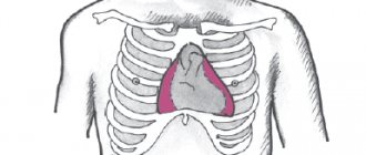
Transesophageal pacing
– a non-invasive procedure aimed at obtaining a recording of biological potentials from the outer surface of the heart, using special esophageal electrodes and recording equipment.
Carrying out special types of stimulation to study the electrophysiological properties of the conduction system, atrial and ventricular myocardium. Identification of arrhythmia substrates, their localization and electrophysiological characteristics. Monitoring drug and/or non-pharmacological therapy.
Non-invasive electrophysiological examination of the heart (TEPS)
The experience of using TEES in cardiology spans more than 30 years.
In our country, the first report on the use of TEES in patients with coronary heart disease appeared in the scientific medical literature more than 10 years ago.
Over this period of time, the attitude towards any research method has already become stable, and the capabilities of the method itself have been well studied.
It should be said that the attitude of cardiologists to the TEES method during this time changed depending on the development of cardiology itself and the technical capabilities of the stimulators used.
The increased interest in this method is currently due, on the one hand, to the rapid development of cardiology itself as a science, in particular its arrhythmology, as well as the emergence of modern stimulators with good technical characteristics that allow the study to be carried out with minimal discomfort for the patient.
The use of TEE helps solve three main problems: diagnosis, treatment
(therapeutic, selection of antiarrhythmic drugs) and
prognosis
in many clinical situations.
Preparation for transesophageal echocardiography
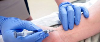
Before a transesophageal echocardiogram is ordered, the patient must undergo a series of laboratory tests.
Among them: biochemical blood test, OAC and OAM, coagulogram. In this case, changes such as an increase in leukocytes, platelets, and acceleration of ESR may be observed. In addition, before this examination, transthoracic ultrasound diagnostics is often performed. In addition, if a heart pathology is suspected, an ECG must be taken. Transesophageal echocardiography is a safe procedure and does not require special preparation. If the patient is taking any medications, there is no need to stop them before the study. A prerequisite for the procedure is refusal to eat for 6 hours prior to TEE. If the patient has dentures, they must be removed. Sometimes premedication is given before the test. To reduce salivation, the drug Atropine is administered intravenously. If the patient is in an excited state, tranquilizers (the drug “Diazepam”) are prescribed.
Assessment of coronary circulation using the TEES method
This stimulation program is the basis from which the TEES method began as one of the stress tests (rhythm load) in cardiology.
The use of a stimulation program allows you to gradually and dosedly increase the frequency of ventricular contractions with constant assessment of changes in the final part of the ventricular complex on the monitor and when recording an ECG.
The use of TEES to assess coronary circulation allows the cardiologist to solve a number of important problems:
1. establish the very fact that the patient has coronary heart disease (CHD) and its forms, in particular, determine painless myocardial ischemia; 2. determine the degree of coronary insufficiency; 3. determine the optimal effective dose of the antianginal drug and the frequency of its administration; 4. to identify a group of patients with coronary heart disease with severe coronary insufficiency, for whom coronary angiography and possible surgical treatment of coronary artery disease are strongly recommended; 5. determine the prognosis and management tactics for patients with coronary artery disease after myocardial infarction; 6. identify a group of patients with coronary artery disease who have a high risk of sudden cardiac death for the purpose of preventive treatment; 7. carry out differential diagnosis of post-infarction cardiosclerosis in patients with Wolff-Parkinson-White syndrome; 8. identification of hidden forms of rhythm and conduction disorders; 9. when dynamically performing TES in the same patient with coronary artery disease, indirectly judge the rate of progression of coronary atherosclerosis and the effectiveness of the treatment.
In addition to general contraindications, performing TES according to the coronary circulation assessment program is not advisable in the following cases:
1. in the presence of persistent complete blockade of the left bundle branch; 2. when recording an obvious (manifesting) syndrome of premature excitation of the ventricles caused by the functioning of the Kent bundle on a resting ECG; 3. a patient with coronary artery disease with 3-4 functional class; 4. in a patient with new-onset angina during the first 4 weeks and in a patient with unstable angina; 5. during the first 3 weeks of uncomplicated myocardial infarction; 6. with pronounced hypertrophy of the left ventricular myocardium with secondary changes in the final part of the ventricular complex on the ECG. TEES is carried out as prescribed by the attending physician 2 hours after a meal against the background of withdrawal of coronary drugs in the case of a diagnostic study. In this case, the patient’s consent to perform TES, recording a resting ECG and echocardiogram (EchoCG) is required.
The stimulation program for the purpose of assessing coronary circulation is quite simple. After obtaining a stable pacemaker rhythm that exceeds the natural heart rate by 20 pulses/min, continuous stepwise stimulation is performed. The duration of each stage is 1 minute.
If there are no changes at the end of the step, the stimulation frequency is increased by 10-20 pulses/min. until a maximum frequency of 160 pulses/min is reached. After each stage of stimulation, the coronary circulation is assessed by changes in the ST segment on the ECG.
If, after reaching the maximum frequency (160 pulses/min.), no ischemic changes are observed on the ECG, then the stimulation time is extended to 2 minutes, after which a final assessment of the study is carried out. It is considered inappropriate to assess coronary circulation at a stimulation frequency of more than 160 pulses/min. at the same time, the number of false-positive results increases significantly, which is partly due to the so-called post-depolarization syndrome.
Post-depolarization syndrome is expressed in the occurrence of ST segment depression and T wave inversion on the ECG after the cessation of high ventricular frequency. The development of this syndrome after relief of paroxysm of ventricular tachycardia is well known.
In the absence of verified coronary artery disease in a patient, this syndrome indicates disturbances in the processes of ventricular repolarization not associated with deterioration of coronary circulation.
to the top of the page
Preparing for the study
Preparation for performing transesophageal ultrasound of the heart includes a number of features:
- You should not eat 6 hours before the procedure, do not drink 3 hours before;
- if necessary, take the medicine, wash it down with a few sips of water;
- remove existing removable dentures from the mouth;
- promptly notify the doctor about previously identified allergic reactions and pathologies of the esophagus.
1.General information
Electrocardiostimulation (ECS) – conduction of additional, external electrical impulses to the heart muscle. As is known, the rhythmic contractile activity of the myocardium is controlled and controlled in the body by a bioelectrical method, which has made it possible to develop a number of well-known diagnostic, therapeutic and resuscitation methods built precisely on electrophysiological principles.
Modern high-tech pacemakers, implanted inside and designed for long-term automatic operation, are equipped with a microprocessor controller that records the main indicators of the electrical activity of the heart and connects additional stimulating discharges only if necessary.
Transesophageal cardiac pacing is one of the non-invasive modifications of the pacemaker method, which is used both for diagnostic purposes and for the relief of certain dangerous conditions.
A must read! Help with treatment and hospitalization!
What can be revealed by transesophageal echocardiography?
Thanks to echocardiography performed through the cavity of the esophagus, it is possible to assess the condition of the heart muscle, endocardium and valve apparatus. This study is decisive in making a diagnosis. Given the high information content of the method, even minor damage to the heart cavity can be detected. TEE can detect the presence of blood clots, inflammatory changes, and aortic dissection. Three-dimensional research is particularly informative. Thanks to 3D ultrasound cardiography, it is possible not only to assess the condition of the heart muscle, but also to prepare the patient for valve replacement surgery. This method is a high-tech research and is carried out in specialized clinics.
Application of TEES for cardiac arrhythmias
The use of TEES for cardiac arrhythmias was justified as a result of the rapid development of arrhythmology and based on its problems.
The use of TEES in this category of patients solved many problems of supraventricular arrhythmias and fully replaced such a research method as intracardiac electrophysiological study (EPS).
Therefore, indications for EPI are currently narrowed and can be determined by the following conditions:
1 Clarification of the diagnosis and selection of therapy for patients with ventricular tachycardia. 2. Syncope, unknown etiology. 3. Before surgical treatment of arrhythmias. 4. Before implantation of a pacemaker or cardioverter-defibrillator in patients with tachyarrhythmias. 5. To carry out cryodestruction of abnormal conduction pathways in case of their right-sided passage. 6. Patients with WPW syndrome and paroxysmal atrial fibrillation, which occurs with loss of consciousness and the threat of transformation into ventricular fibrillation.
Rice. Transesophageal electrogram, bipolar recording
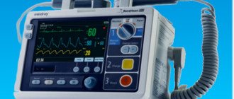
When starting a study on the rhythm disturbance program using the TEES method, the doctor conducting it must be sure that everything is prepared in case of resuscitation measures: the defibrillator is prepared and turned on, there is the necessary set of resuscitation drugs and supplies.
In addition, the doctor conducting the study must be well trained in such disciplines as clinical cardiology with arrhythmology, clinical electrocardiography, know the electrophysiology of the myocardium, issues of emergency cardiology and resuscitation.
It should be especially emphasized that the study of PPES according to the program for cardiac arrhythmias must be carried out in the presence of at least two medical workers - a doctor and a nurse who have undergone special training.
TEES can be performed for diagnostic and therapeutic purposes. In the event of a diagnostic study, all antiarrhythmic drugs should be discontinued.
Rice. A. Paired stimulation of the atria causes paroxysm of AV reciprocal orthodromic tachycardia. The RP'-interval on TEE is less than 1/2 the RR-interval and = 120 ms. B. Relief of paroxysm by paired stimulation.
to the top of the page
Features of transesophageal cardiac examination
Transesophageal echocardiography shows even minor damage to the heart cavity, myocardium, endocardium and valves, the presence of blood clots, inflammation, and aortic dissection.
Three-dimensional diagnostics are highly informative, which can show not only the condition of the heart, but also prepare the patient for valve replacement. The examination is carried out in dispensaries where the cardiology department is located, in private clinics equipped with modern medical equipment.
The price of the procedure varies from 3.5 to 33 thousand rubles.
The method involves the use of a special miniature sensor, which is practically the same in size as a fiber gastroscope. The procedure includes the following steps:
- Premedication. At this stage, tranquilizers are administered parenterally to the patient. Diazepam is widely used in a dose of 2-5 mg. In some cases, the administration of narcotic analgesics is indicated, which significantly improves the tolerability of subsequent manipulations. However, drugs should be prescribed with caution to patients with lung diseases. Atropine is used to reduce saliva production.
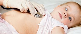
- Esophageal intubation. The patient is advised to lie on his left side. The doctor irrigates the pharyngeal mucosa with a 10% lidocaine solution to reduce the severity of the gag reflex. If there is a history of atrial fibrillation, it is recommended to avoid using local anesthetics. The patient is put on a special mouthpiece that prevents damage to the endoscope. The sensor is lubricated with a hypoallergenic gel and slowly inserted into the oral cavity, holding the patient’s tongue. If an obstacle appears, the doctor asks the patient to swallow so that the endoscope can enter the esophagus. If the doctor is unable to insert the sensor, the procedure is repeated under general anesthesia.
- The doctor places the sensor behind the heart, after which an image of the heart muscle appears on the monitor. The specialist constantly changes the position of the endoscope to assess the structure and functionality of the organ being examined.
This stage takes no more than 15 minutes.
In rare cases, transesophageal echocardiography leads to the development of dangerous complications: atrial fibrillation, anaphylactic shock due to the use of drugs, esophageal rupture.
- Echocardiography of the heart: principle of operation, capabilities and indications for the study
Therefore, the room in which diagnostic procedures are carried out must be equipped with equipment for oxygen inhalation, a defibrillator, and a standard resuscitation kit. During the procedure, the resuscitator must constantly monitor the patient's condition.
After completing the examination, it is recommended to refrain from drinking and eating for 1.5-2 hours so that the pharyngeal reflexes are restored. During an Echo KG emergency, small wounds may appear in the esophagus, so you should avoid eating hot foods and drinks for several days.
Scope of application of TEES in cardiology
In patients with coronary heart disease:
1) diagnosis of coronary insufficiency; 2) determination of the degree (functional class) of coronary insufficiency, 3) diagnosis of silent myocardial ischemia; 4) identification of a group of patients with coronary heart disease (CHD) who have a high risk of sudden cardiac death; 5) selection of the optimal effective dose of the antianginal drug and the most rational use of it during the day; 6) identifying a group of patients for whom it is most appropriate to undergo coronary angiography and subsequent surgical treatment of coronary artery disease; 7) verification of large focal scar changes in the myocardium in patients with WPW syndrome; simultaneous registration of an echocardiogram according to the stress echocardiogram program during dosed frequency loading during TEES makes it possible to diagnose latent forms of coronary and myocardial insufficiency.
In patients with cardiac arrhythmias:
1) assessment of sinus node function: - diagnosis of sick sinus syndrome (SSNS); — diagnosis of functional dysfunction of the sinus node (most often associated with increased activity of the Vagus node) — assessment of the functional state of the myocardium before installing a permanent endocardial pacemaker;
2) assessment of atrioventricular (AB) node function;
3) differential diagnosis of paroxysmal supraventricular tachyarrhythmias using the method of tachyarrhythmia provocation and subsequent registration of the esophageal electrogram (PE);
4) diagnosis and study of the electrophysiological properties of additional, abnormal conduction pathways (Kent's bundle and James' bundle); — diagnosis of the syndrome of premature excitation of the ventricles in the case of the functioning of the Kent or James points; — diagnosis of paroxysmal tachyarrhythmias in Wolff-Parkinson-White (WPU) or Clerk-Levy-Cristescu (CLC), Lown-Ganong-Levin (LGL) syndrome; — identification of a group of patients with VPU syndrome and atrial fibrillation at risk of developing ventricular fibrillation;
5) selection of the optimal effective dose of an antiarrhythmic drug: - to relieve paroxysm of tachyarrhythmia; - to prevent the occurrence of paroxysmal tachyarrhythmia; — identification of the arrhythmogenic effect of the drug;
6) relief of paroxysmal supraventricular tachyarrhythmias (except for atrial fibrillation);
7) maintaining the required heart rate (HR) during surgery in case of initial bradycardia;
8] study of the electrophysiological properties of the supraventricular zone: atria, AV node, additional conduction pathways (refractory periods of structures);
9) registration of tachycardia-dependent extrasystole and intraventricular blockades;
CPES has a wide range of applications from outpatient clinics to inpatient units. The method for clinicians in their clinical activities is the most accessible and less burdensome for the patient.
The diagnostic capabilities of TEE are limited to stimulation of the left atrium. In some cases, stimulation of the left ventricle can be achieved, but for this it is necessary to apply a voltage with an amplitude of 30-60 V (mA), which is practically impossible without the use of anesthesia.
to the top of the page
How to prepare for the examination
To obtain accurate research results, you must adhere to simple rules:
- as with gastric endoscopy, cardiac ultrasound with a transesophageal sensor is performed strictly on an empty stomach (6–8 hours of fasting);
- It is recommended to take herbal sedatives to relieve anxiety and agitation of the patient;
- remove dentures;
- wear comfortable, comfortable clothes;
- refuse neck jewelry;
- tell your doctor about allergic reactions;
- report any discomfort.
We advise you to follow all the specialist’s recommendations to avoid pain and negative consequences.
Complications during TEES
It should be noted right away that paroxysmal rhythm disturbances, which were previously considered as a complication of the TEES method, are not currently such.
This is due to the rapid development of arrhythmology and changes in ideas. However, it must be borne in mind that the doctor conducting the study may encounter two problems:
1. when conducting TEES, the doctor consciously tries to provoke a paroxysm of supraventricular tachyarrhythmia using various stimulation modes, i.e. induction of tachycardia is the goal of the study itself. It is unlikely that this situation should be considered as a complication of the method;
2. when conducting TEES, the induction of tachycardia is a surprise for the doctor conducting the study and occurs for the first time in the patient’s life. Paroxysmal tachycardia can occur when using any stimulation mode. In this situation, the issue is not resolved unambiguously, but depending on the specific form of paroxysm.
The very fact of the possibility of provoking tachycardia indicates that all the necessary prerequisites for the implementation of the re-entry mechanism have been formed in the patient’s heart and only a trigger is needed for the occurrence of tachycardia.
This triggering mechanism was PPES. However, the same paroxysm can occur independently when certain conditions arise - most often the appearance of a supraventricular extrasystole in a certain phase of the cardiac cycle. Thus, provoking tachycardia for the first time in a patient’s life confirms only what may happen to him in the future and, to a certain extent, helps the doctor adjust treatment taking this fact into account.
The specific form of provoked paroxysm should also be taken into account, based on the complexity of treatment and possible complications of this paroxysm. Thus, a paroxysm of nodal AV tachycardia or a paroxysm of reciprocal AV tachycardia in a patient with WPW syndrome (orthodromic or antidromic variant) can be quite easily stopped using the TEES method and, as a rule, does not require intravenous administration of drugs (we must not forget about the possibility of stopping these paroxysms with using vagal tests).
Inducing a paroxysm of atrial fibrillation for the first time in a patient’s life requires further use of medications to stop it, since the TEES method does not stop atrial fibrillation.
Provocation of atrial fibrillation in a patient for the first time in his life often indicates either critical dilatation of the atria or pronounced dystrophic changes in the atrial myocardium [after myocarditis or chronic alcohol intoxication), after which maintaining sinus rhythm becomes a difficult task.
Particular attention should be paid to the development of two forms of paroxysmal tachyarrhythmia - ventricular tachycardia and atrial fibrillation in a patient with WPW syndrome, which, if they develop, should undoubtedly be considered as a complication of the method, associated in turn with a possible error in the study.
These situations should be reviewed and studied in detail to avoid their recurrence in the future. Provoking ventricular tachycardia using the TEES method is a difficult task due to many circumstances. It must be remembered that when affecting the atria, TES can provoke the development of ventricular tachycardia only indirectly through a combination of two unfavorable factors - deterioration of coronary circulation and electrical instability of the ventricles.
Knowing this, the doctor should be especially careful when, with the appearance of significant ischemic depression of the ST segment, ventricular extrasystole of high gradations (according to Lown) appears - frequent, paired, early. In this situation, the study should be stopped immediately, and the identified violations are reflected and emphasized in the study protocol as dangerous in terms of the development of paroxysm of ventricular tachycardia, and the patient himself should be considered as at risk of sudden cardiac death. Thus, the situation with the development of paroxysm of ventricular tachycardia during TEES can and should be controlled, the main thing is that the doctor conducting the study knows about it.
Complications of the TEES method
The problems that a doctor conducting a study may encounter are:
1. Insertion of the esophageal electrode into the trachea instead of the esophagus.
This complication usually occurs among doctors who are just beginning to independently conduct TEES research and is usually associated with their haste when inserting the probe into the esophagus. This complication extremely rarely occurs when the probe is slowly inserted at the moment of swallowing saliva, when the epiglottis rises and the trachea closes.
If the probe gets into the trachea, the patient experiences sudden suffocation, coughing, and redness of the face - the probe should be immediately removed, and the probe should be reintroduced only a few minutes after the described symptoms cease. In extremely rare cases, the erroneous insertion of a probe into the trachea is not accompanied by pronounced reactions on the part of the patient and stimulation is carried out from the trachea. In our practice, we observed one such patient (a former mountaineer, Honored Master of Sports).
To avoid such cases, it is necessary to pay attention to the extremely low amplitude of the “P” wave on the esophageal electrogram at the beginning of the study, as well as the appearance of cough when stimulation is turned on.
2. A burning sensation in the esophagus, behind the sternum.
The appearance of these sensations is mandatory when stimulation is turned on, and their absence in the patient should be associated with a malfunction of the equipment. The patient's level of sensation is usually low or easily tolerated. If the sensations are pronounced and the patient refuses the study, the study is stopped.
3. During stimulation, the patient feels pain in the back associated with contraction of the spinal muscles. In case of severe pain, the study is stopped.
4. When the stimulator is turned on, effective stimulation of the diaphragm occurs, which is accompanied by its rhythmic contractions with the frequency that the stimulator gives at a given time. The patient feels frequent hiccups or shortness of breath, and the doctor conducting the study observes frequent contractions of the diaphragm, which disappears immediately after the stimulator is turned off.
This complication most often occurs in patients with a hypersthenic physique or obesity, because the location of the heart in the chest in such patients is such that the heart practically lies on a highly located diaphragm and the poles of the esophageal electrode during TEES are located in close proximity not only to the atria, but also to the diaphragm. As a rule, the study is stopped when this complication occurs.
A technique in which the esophageal electrode is advised to be pulled up, thereby removing the electrode poles from the diaphragm, rarely gives a positive result, because by removing the probe from the diaphragm, we thereby remove the electrode poles from the zone of their optimal location in relation to the left atrium, and this in turn will require an increase in current strength and, as a result, the re-emergence of effective stimulation of the diaphragm. We observed a patient in whom contraction of the diaphragm during TEES occurred at the very minimum voltage and from the upper part of the esophagus.
5. Electrode getting stuck in the nose is the most unpleasant complication during TEES, as it injures the patient and leads to loss of the electrode.
The esophageal electrode gets stuck in the nose, usually when it is removed after stopping the study. The electrode gets stuck in the area of their poles, because their diameter is larger than the diameter of the probe itself. Electrode getting stuck is associated with injury to the nasal mucosa and its swelling when the probe is inserted. If it is impossible to remove the electrode after applying vasoconstrictor drops into the nose, it is removed as follows: holding the distal part of the probe through the mouth with a special clamp (used in the ENT office), bite the probe near the wings of the nose and remove the probe through the patient’s mouth. It should be emphasized that this complication occurs only among doctors who begin to independently perform TEES, who often forget the “golden rule” - insertion of the probe through the nose should be free and painless for the patient, and therefore atraumatic for the nasal mucosa. This is why anesthesia of the nasal mucosa not only does not help, but also harms when inserting a probe, since it is very important to know about pain when inserting a probe.
In conclusion of the presentation of this topic, it should be said that if the doctor conducting the study has good knowledge of TEES, arrhythmology, issues of cardiology, myocardial electrophysiology, the TEES method is safer compared to other methods of functional diagnostics that use physical activity or the administration of drugs as a load .
to the top of the page
Detailed examination - TEE of the heart
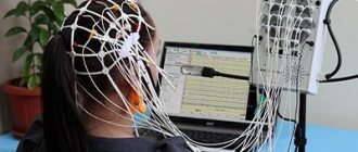
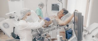
The proximity of the esophagus and the heart is used to study the contractile function and rhythm of the myocardium. To do this, small doses of electric current are applied to the nearest area of the muscle layer.
In patients with latent forms of arrhythmia, pathological contractions occur under the influence of impulses supplied through the electrode. They can be detected by sensors attached to the chest. A transesophageal electrophysiological study (TEPE) is used if a conventional ECG or Holter monitoring does not reveal any abnormalities, and the patient has symptoms of irregular heartbeat.
Advantages and disadvantages of the method
The advantages of this diagnostic method are the following:
- examination of rare and short forms of tachycardia of supraventricular origin;
- studying the exact place where it is possible to generate frequent signals or block the passage of impulses;
- can be used as an alternative to stress tests if the patient is unable to complete tasks for such studies;
- minor trauma;
- availability;
- no penetration into the vascular bed is required;
- There are no contraindications for repeated use.
What symptoms may indicate the need for TEE?
Rhythm disturbances requiring transesophageal pacing can be suspected based on the presence of the following signs in the patient:
- Attacks of sudden rapid heartbeat with rapid pulse (more than 100-120 per minute), also suddenly stopping spontaneously or stopping only after the administration of antiarrhythmic drugs,
- Feelings of irregular heartbeat with a rare pulse (less than 50 per minute),
- Attacks of loss of consciousness that are not associated with neurological problems or with situationally occurring conditions (stuffiness in the room, stress, etc.), but are caused by rhythm disturbances and are called Morgagni-Edams-Stokes attacks (MES episodes).
If a doctor, while examining a patient, hears about the presence of the symptoms described above, then he should think about a more accurate diagnosis of heart rhythm disturbances. And if the ECG and 24-hour monitor do not reveal any types of arrhythmias, and the patient’s complaints persist, it is necessary to perform TEE. If it was possible to register an arrhythmia on the ECG, for example, a delta wave, characteristic of latent SVC syndrome, the patient should be further examined.
In any case, the need for this technique is determined only by a doctor during an in-person examination of the patient.
4.Contraindications
Transesophageal pacing is not performed in the presence of confirmed diseases of the esophagus, including neoplasms, strictures (narrowings with limited patency), inflammatory processes (esophagitis), etc. Contraindications also include a number of cardiac diseases, including severe atrioventricular block, heart attack, thrombosis, aneurysms , as well as the presence of implanted artificial valves. Finally, TEE is contraindicated for respiratory or other acute infections.
3.Indications
TEE as a diagnostic procedure is used to identify some pathological phenomena that cannot be recorded with a conventional electrocardiograph with percutaneous electrodes: arrhythmias, tachycardias, disorders in the conduction system of the heart, latent pathology of the coronary arteries, etc. In certain diagnostically difficult situations, electrical stimulation provocation is also indicated.
The therapeutic value of transesophageal pacing is to relieve attacks of supraventricular tachycardia and atrial flutter.
About our clinic Chistye Prudy metro station Medintercom page!