What is the aorta
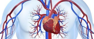
The aorta is the largest vessel in the human body, which carries blood from the heart to the organs and limbs. The upper section of the aorta runs inside the chest, this section is called the thoracic aorta . The lower part is in the abdominal cavity and is called the abdominal aorta . It delivers blood to the lower part of the body. In the lower abdomen, the abdominal aorta divides into two large vessels - the iliac arteries , which carry blood to the lower extremities.
The aortic wall consists of three layers: internal (intima), middle (media), external (adventitia).
Preparation for aortography
In preparation for angiography, the patient must donate blood for a general analysis, a biochemical analysis to exclude kidney and liver pathologies, and a coagulogram (blood test for clotting).
It is necessary to notify your doctor about allergic reactions, especially to iodine-containing drugs.
Aortography is best performed on an empty stomach to avoid nausea and vomiting. Fluid intake is not limited. If endovascular intervention is planned, you may be given antithrombotic drugs before the angiography.
Immediately before the procedure, you must sign an informed consent for the procedure.
Before the examination, the patient must go to the toilet, remove all metal objects (since they are reflected on x-rays and can distort the picture) and change into special clothes
During angiography, the patient is in a supine position and fixed to the table, since movements during the examination can distort the results.
Abdominal aortic aneurysm
Abdominal aortic aneurysm is a chronic degenerative disease with life-threatening complications. An abdominal aortic aneurysm is defined as an increase in its diameter by more than 50% compared to the norm or a local bulging of its wall. Under the pressure of blood flowing through this vessel, the dilatation or bulging of the aorta may progress. The diameter of a normal aorta in the abdominal region is approximately 2 cm. However, at the site of an aneurysm, the aorta can be dilated to 7 cm or more.
Why is an aortic aneurysm dangerous?
An aortic aneurysm poses a major health risk as it can rupture. A ruptured aneurysm can cause massive internal bleeding, which in turn leads to shock or death.
An abdominal aortic aneurysm can cause other serious health problems. Blood clots (thrombi) often form in the aneurysm sac or parts of the aneurysm tear off, which move with the blood flow along the branches of the aorta to the internal organs and limbs. If one of the blood vessels becomes blocked, it can cause severe pain and lead to organ death or loss of a lower limb. Fortunately, if an aortic aneurysm is diagnosed early, treatment can be timely, safe and effective.
Complications of aortography
In order to avoid the development of complications, it is necessary to carefully prepare for the study and choose the least dangerous method of conducting it.
- Bleeding, hematoma, pain or swelling at the site of catheter insertion is very rare when performing arterial puncture under ultrasound guidance and using suturing devices.
- Allergic reaction to iodine, which is part of the X-ray contrast agent - it is necessary to carefully determine the history of allergies and exclude such patients.
- Damage to the vessel wall can occur during complex or rough puncture. In our clinic, if there are difficulties with arterial puncture, we use an ultrasound scanner to guide the needle into the vessel.
- The development of acute liver or kidney failure may occur due to the toxic effects of the contrast agent. With adequate fluid loading and minimal use of contrast, this complication is uncommon.
- Heart rhythm disturbances are rare and are associated with irritation of the reflexogenic zones in the aorta by the conductor or catheter.
Our specialists do everything possible to prevent these complications, and also know the necessary treatment methods in case of development.
Types of aortic aneurysms
There are “true” and “false” aortic aneurysms. A true aneurysm develops due to the gradual weakening of all layers of the aortic wall. A false aneurysm is usually the result of trauma. It is formed from the connective tissue surrounding the aorta. The cavity of the false aneurysm is filled with blood through a crack in the aortic wall. The aortic walls themselves do not participate in the formation of an aneurysm.
Depending on the form there are:
- saccular aneurysm - expansion of the aortic cavity on only one side;
- spindle-shaped (fusiform) aneurysm - expansion of the aneurysm cavity on all sides;
- mixed aneurysm - a combination of saccular and fusiform forms.
Causes and risk factors for the development of abdominal aortic aneurysm
The causes of the development of abdominal aortic aneurysms are very diverse. The most common cause of aneurysm development is atherosclerosis. Atherosclerotic aneurysms account for 96% of the total number of all aneurysms. In addition, the disease can be either congenital (fibromuscular dysplasia, Erdheim's cystic medianecrosis, Marfan syndrome, etc.) or acquired (inflammatory and non-inflammatory). Inflammation of the aorta occurs when various microorganisms invade (syphilis, tuberculosis, salmonellosis, etc.) or as a result of an allergic-inflammatory process (nonspecific aortoarteritis). Non-inflammatory aneurysms most often develop with atherosclerotic lesions of the aorta. Less commonly, they are the result of injury to its wall.
Risk factors for developing an aneurysm
- Arterial hypertension;
- Smoking;
- The presence of aneurysms in other family members. Which indicates the role of hereditary factors in the development of this disease;
- Gender: men over 60 years of age (abdominal aortic aneurysms occur less frequently in women).
Statistics and facts
An abdominal aortic aneurysm
(AAA) is a local or diffuse expansion of the aortic diameter by more than 2 times.
Abdominal aortic aneurysms account for 95% of all aneurysms
, while approximately 9% of people over 65 years of age are asymptomatic. The incidence of the disease is 2-5% among men over 60 years of age. The peak incidence in men is observed after 80 years of age, and in women after 90 years of age, the ratio of men to women with AAA is 4:1.
Unfortunately, the course of the disease is progressive. Aneurysm size
increases by an average of 10% per year, often without symptoms and the only manifestation of the disease can be its rupture with a fatal outcome.
Every year, up to 200,000 cases of abdominal aortic aneurysms
.
In the United States, up to 15,000 people a year die from ruptures and postoperative complications of AAA, with 30 to 50% of patients dying from rupture of abdominal aortic aneurysms before medical care is provided. Abdominal aortic aneurysm
ranks 15th among all causes of death and 10th among causes of death in men over 60 years of age.
There are several classifications of abdominal aortic aneurysms
.
The most widely used anatomical classification is that aneurysms
located below the renal arteries are infrarenal (95% of AAA) and aneurysms located above the renal arteries are suprarenal.
In about a quarter of AAA cases, the iliac arteries (pelvic arteries that arise from the abdominal aorta) are also involved. In 12% of cases, AAA is combined with an aneurysm above the thoracic aorta. The main risk factors for the occurrence of an aneurysm are: male gender (5-6 times risk), age (1.7 times risk). Having an aneurysm in family members doubles the risk of developing an AAA. For example, 15 to 25% of patients with AAA have or have had a first-degree relative with an abdominal aortic aneurysm.
An important factor in the occurrence and development of abdominal aortic aneurysm is smoking. Thus, there are five times more smokers with AAA than non-smokers; 75% of patients with AAA 4 cm or more are smokers. Risk of abdominal aortic aneurysm
increases depending on the length of smoking and the daily number of cigarettes smoked.
Risk of aneurysm
increases with arterial hypertension, the presence of chronic lung diseases, and certain forms of aneurysmal sac (symmetrical aneurysms are less susceptible to rupture than asymmetrical ones). However, the main factor in rupture is the size of the aneurysm sac.
The mortality rate for an abdominal aortic aneurysm with a diameter of 4 cm is less than 5% per year, and the mortality rate for an AAA with an internal diameter of more than 9 cm is more than 75% per year.
Symptoms and signs of abdominal aortic aneurysm
In most patients, abdominal aortic aneurysms occur without any symptoms and are an incidental finding during examinations and operations for other reasons.
When signs of an aneurysm develop, the patient experiences one or more of the following symptoms:
- A feeling of pulsation in the abdomen, similar to a heartbeat, an unpleasant feeling of heaviness or fullness.
- Dull, aching pain in the abdomen, in the navel area, usually on the left.
Indirect signs of abdominal aortic aneurysm are important :
- Abdominal syndrome. Manifested by the appearance of belching, vomiting, unstable stool or constipation, lack of appetite and weight loss;
- Ischioradic syndrome. Manifested by lower back pain, sensory disturbances and movement disorders in the lower extremities;
- Syndrome of chronic ischemia of the lower extremities. Manifests itself in the appearance of pain in the muscles of the lower extremities when walking, sometimes at rest, coldness of the skin of the lower extremities;
- Urological syndrome. It manifests itself as pain and heaviness in the lower back, difficulty urinating, and the appearance of blood in the urine.
Harbingers of rupture may be increased abdominal pain.
When an aneurysm ruptures, the patient suddenly feels an increase or appearance of abdominal pain, sometimes “radiating” to the lower back, groin area and perineum, as well as severe weakness and dizziness. These are symptoms of massive internal bleeding. The development of such a situation is life-threatening! The patient needs emergency medical care!
Structure [edit]
The abdominal aorta begins at the level of the diaphragm, crossing through the aortic hiatus, technically behind the diaphragm, at the level of the T12 vertebra. [1] It runs along the back wall of the abdomen, anterior to the spine. Thus, it follows the curvature of the lumbar vertebrae, that is, convex anteriorly. The peak of this convexity is at the level of the third lumbar vertebra (L3). It runs parallel to the inferior vena cava, which is located to the right of the abdominal aorta and becomes smaller in diameter as branches arise from it. This is believed to be due to the large size of its main branches. The 11th rib has a diameter of 122 mm in length and 55 mm in width, and this is due to constant pressure. [2] The abdominal aorta is clinically divided into 2 segments:
- The adrenal abdominal cavity
, or
paravisceral
segment, is inferior to the diaphragm but superior to the renal arteries. - INFRARENAL
segment, inferior to the renal arteries and superior to the iliac bifurcation.
Branches [edit]
The abdominal aorta supplies blood to most of the abdominal cavity. It begins at T12 and ends at L4, branching into the common iliac arteries [1] and usually has the following branches:
| Artery branch | Vertebra | Type | Paired with? | A/P | Description |
| inferior diaphragmatic | T12 | Parietal | Yes | mail. | Arises above the celiac trunk, below the diaphragm. It passes upward and inward to the adrenal glands and crosses the crus of the diaphragm of the corresponding side. Supplies the diaphragm and gives rise to the superior adrenal arteries. |
| celiac disease | T12 | Visceral | No | ant. | Greater anterior branch |
| superior mesenteric | L1 | Visceral | No | ant. | The large anterior branch arises just below the celiac trunk. |
| middle adrenal gland | L1 | Visceral | Yes | mail. | Crosses the crus of the diaphragm laterally on each side; supplies the adrenal gland. |
| renal | Between L1 and L2 | Visceral | Yes | mail. | Arises just below the superior mesenteric artery. The right renal artery passes deep into the inferior vena cava, into the right kidney; here it is divided into branches. The left renal artery passes deep into the left renal vein. Divides at the hilum of the kidney. Both arteries give rise to the inferior adrenal arteries and branches of the ureter. |
| gonadal | L2 | Visceral | Yes | ant. | Ovarian artery in women; testicular artery in men |
| lumbar | L1-L4 | Parietal | Yes | mail. | Four on each side, supplying the abdominal wall and spinal cord. The fifth pair is the lumbar branches of the iliopsoas arteries. They pass deep into the calf on the side of the vertebral bodies and pass deep into the psoas and quadratus lumborum muscles to enter the space between the internal oblique and transverse abdominis muscles. Each artery gives off a small dorsal branch, which gives a spinal branch to the spinal canal and then continues to supply the muscles of the back. |
| inferior mesenteric | L3 | Visceral | No | ant. | Greater anterior branch |
| midsacral | L4 | Parietal | No | mail. | An artery that arises from the middle of the aorta in its lower section. It is a continuation of the primitive dorsal aorta; quite large in animals with a tail, but smaller in humans. |
| common ilium | L4 | Terminal | Yes | mail. | Branches (bifurcations) to supply blood to the lower extremities and pelvis, ending in the abdominal aorta |
Note that the bifurcation (junction) of the inferior vena cava is at the level of L5 and therefore inferior to the aortic bifurcation.
Volumetric computed tomography of the blood vessels of the abdominal cavity and pelvis.
- lower diaphragmatic a.
- celiac disease a. left stomach a.
- splenic a. short gastric arteries (6)
- splenic arteries (6)
- left gastroepiploic a.
- pancreatic arteries
- right stomach a.
- cystic a.
- lower pancreaticoduodenal a.
- left colic a.
- external ilium a.
Relationships [edit]
The abdominal aorta is located slightly to the left of the midline of the body. In front it is covered by the lesser omentum and stomach, behind which are the branches of the celiac artery and the celiac plexus; Below them, the splenic vein(s), are the pancreas, left renal vein, lower part of the duodenum, the mesentery and the aortic plexus.
Posteriorly, it is separated from the lumbar vertebrae and intervertebral fibrocartilages by the anterior longitudinal ligament and the left lumbar veins.
On the right side it is in connection above with the azygos vein, cisterna chylae, thoracic duct and the right leg of the diaphragm - the latter separates it from the upper part of the inferior vena cava and from the right celiac ganglion; the inferior vena cava is in contact with the underlying aorta.
On the left are the left crus of the diaphragm, the left celiac ganglion, the ascending part of the duodenum and some spirals of the small intestine.
3D illustration of the abdominal aorta at the iliac junction.
Connection with the inferior vena cava [edit]
The venous counterpart of the abdominal aorta, the inferior vena cava (IVC), runs parallel to it on the right side.
- Above the level of the umbilicus, the aorta lies slightly behind the IVC, sending behind it the right renal artery. The IVC also sends its opposite side, the left renal vein, crossing in front of the aorta.
- Below the level of the umbilicus, the situation is usually reversed: the aorta directs its right common iliac artery to cross its opposite side (the left common iliac vein) forward.
Collateral circulation[edit]
Collateral circulation will be maintained by anastomoses between the internal mammary artery and the inferior epigastric artery; free communication between the superior and inferior mesenteries, if a ligature was placed between these vessels; or by an anastomosis between the inferior mesenteric artery and the internal pudendal artery, when (as is most common) the point of the ligature is below the origin of the inferior mesenteric artery; and possibly by anastomoses of the lumbar arteries with branches of the internal iliac artery.
Diagnosis of abdominal aortic aneurysms
Most often, abdominal aortic aneurysms are detected by ultrasound examination of the abdominal organs. Typically, the discovery of an aneurysm is an incidental finding. If a doctor suspects a patient has an aortic aneurysm, modern diagnostic methods are used to clarify the diagnosis.
Methods for diagnosing abdominal aortic aneurysm
- Computed tomography in angio mode;
- Magnetic resonance imaging in angio mode;
- X-ray contrast aorto- and angiography;
- Ultrasound duplex or triplex angioscanning of the abdominal aorta.
If necessary, the abdominal and thoracic aorta is examined.
Triplex scanning of the abdominal aorta and its visceral branches
What is the abdominal aorta?
The abdominal (abdominal) aorta is the descending part of the aorta, located in the retroperitoneal space between the abdominal organs and the spinal muscles. The visceral branches of the abdominal aorta are arteries extending from it to the internal organs: to the kidneys, to the spleen, to the intestines, etc.
What is triplex scanning?
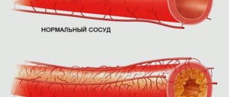
This is a comprehensive non-invasive ultrasound examination that allows you to obtain images of blood vessels and assess the condition of the vascular wall and indicators of intravascular blood flow. The study allows you to evaluate the shape, course, diameter of blood vessels, their patency, identify narrowed or dilated areas in them, congenital anomalies, determine the presence of atherosclerotic plaques, blood clots, and changes in blood flow.
What is the difference between triplex scanning and duplex scanning?
Duplex scanning uses two ultrasound modes:
- Ultrasound examination (US) in grey-scale scanning mode (B-mode);
- Ultrasound spectral Dopplerography (USDG).
Triplex scanning is supplemented with a color Doppler mapping (CDC) mode of blood flow, which makes it possible to more clearly judge the direction of blood flow and its speed, and more accurately assess the patency of blood vessels and the degree of stenosis.
In what cases can triplex scanning of the abdominal aorta and its branches be indicated?
- Atherosclerosis of the abdominal aorta and vessels supplying the gastrointestinal tract, liver, spleen and kidneys.
- Suspicion of the presence of congenital anomalies and developmental features of the aorta and its visceral branches.
- Suspicion of impaired blood flow in the aorta and its visceral branches as a result of narrowing, blockage of the lumen, kinking, and pathological tortuosity of the artery.
- Suspicion of an aneurysm (local expansion) of the abdominal aorta and visceral vessels, its dissection, vascular malformation (congenital disorder of blood flow), or rupture of a vessel.
- Vasculitis (inflammatory vascular disease) of various types, incl. infectious origin.
- Suspicion of thrombosis of intestinal vessels, incl. after injury, against the background of inflammatory vascular diseases.
- Arterial hypertension associated with pathology of the renal vessels - vasorenal hypertension.
- The presence of risk factors for the development of arterial pathology: smoking, diabetes, hypercholesterolemia, excess weight, etc.
- Preparation for surgical intervention on the aorta and its visceral branches, as well as monitoring the performed surgical intervention on blood vessels.
- Aortic injury.
- The patient's desire to undergo examination for preventive purposes.
Are there any contraindications to triplex scanning?
The study has no contraindications. The negative impact of diagnostic ultrasound on humans has not yet been identified.
Is it possible to conduct other methods of studying the abdominal aorta and its visceral branches?
To study the abdominal aorta and its visceral branches, it is possible to perform computed tomography (CT) and angiography, magnetic resonance imaging (MRI). These methods have a number of restrictions on use and certain contraindications. Triplex scanning has no contraindications and is the optimal examination in terms of price-quality ratio in comparison with CT and MRI. In some cases, in addition to triplex scanning, CT or MRI is necessary. The choice of an additional method, as well as the indications for its implementation, are determined by the doctor.
Preparation for triplex examination of the abdominal aorta and its branches
- The study is carried out in the morning, strictly on an empty stomach.
- On the day of the study, it is necessary to avoid heavy physical activity.
- You should not smoke or chew chewing gum before the test.
- 2-3 days before the study, it is necessary to exclude the consumption of black bread, dairy products, raw vegetables, legumes, carbonated and alcoholic drinks, because this can lead to increased gas formation and reduce the information content of the study. Last meal the day before 7:00 p.m.
- If you are overweight, in addition to dietary restrictions, your doctor may recommend that you take medications that suppress gas formation for two to three days before the examination.
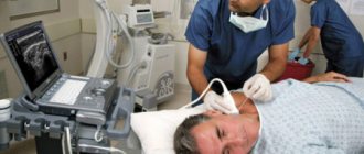
How is the study conducted and how long does it last?
To conduct the study, you need to undress in the office from the waist up (down to your underwear), lower your trousers or skirt and lie down on the couch on your back. During the examination, the doctor may ask you to turn on your left or right side, on your stomach, and hold your breath. The device's sensor is lubricated with a specialized gel and applied to the area under study. The examination is painless. Its duration on average is 30 – 40 minutes. A conclusion based on the results of the study is formed within a few minutes after its completion.
The quality of the study performed is determined by the quality of the ultrasound equipment and the experience of the diagnostician , with the exception of certain cases when there are additional factors that affect the ability to visualize blood vessels (excess body weight, patient body size, increased gas formation in the intestines, inability to hold one’s breath).
Our equipment
Our clinic uses modern diagnostic equipment, including Accuvix A30 ultrasound scanners manufactured by Samsung Medisson, South Korea. The study is carried out by highly qualified specialists with extensive experience in diagnosing vascular pathologies.
Treatment methods for aortic aneurysm
There are several methods for treating aortic aneurysm. It is important to know the advantages and disadvantages of each of these techniques. Approaches to the treatment of abdominal aortic aneurysms:
Monitoring the patient over time
If the aneurysm is less than 4.5 cm in diameter, the patient is recommended to be monitored by a vascular surgeon, since the risk of surgery exceeds the risk of rupture of the aortic aneurysm. Such patients should undergo repeated ultrasound examinations and/or computed tomography at least once every 6 months.
When the aneurysm diameter is more than 5 cm, surgical intervention becomes preferable, since as the size of the aneurysm increases, the risk of aneurysm rupture increases.
If the size of the aneurysm increases by more than 1 cm per year, the risk of rupture increases and surgical treatment also becomes preferable.
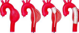
Open surgery: aneurysm resection and aortic replacement
Surgical treatment is aimed at preventing life-threatening complications. The risk of surgery is associated with possible complications that include heart attack, stroke, limb loss, acute intestinal ischemia, male sexual dysfunction, embolization, prosthetic infection, and renal failure.
The operation is performed under general anesthesia. The essence of the operation is to remove the aneurysmal expansion and replace it with a synthetic prosthesis. The average mortality rate for open procedures is 3-5%. However, it may be higher if the renal and/or iliac arteries are involved in the aneurysm, as well as due to the patient’s concomitant pathology. Observation in the postoperative period is carried out once a year. Long-term treatment results are good.
Endovascular repair of aortic aneurysm: installation of a stent graft
Endoprosthetics of aortic aneurysm is a modern alternative to open surgery.
The operation is performed under spinal or local anesthesia through small incisions/punctures in the groin areas. Through the above approaches, catheters are inserted into the femoral artery under X-ray control. According to which, in the future, the endoprosthesis will be brought to the aneurysmal extension. An endoprosthesis or stent-graft of the abdominal aorta is a mesh frame made of a special alloy and wrapped in synthetic material. The last stage of the operation is the installation of a stent graft at the site of the aneurysmal expansion of the aorta. Eventually, the aneurysm is “switched off” from the bloodstream and the risk of rupture becomes unlikely. After aortic replacement, the patient is observed in the hospital for 2-4 days and discharged.
This technique allows to reduce the incidence of early complications, shorten the length of patient stay in the hospital and reduce the mortality rate to 1-2%. Observation in the postoperative period is carried out every 4-6 months using ultrasound techniques, CT angiography, X-ray contrast angiography. The endovascular treatment method is certainly less traumatic. About 40,000 such operations are performed annually in the United States alone.
Thus, the choice of treatment method for abdominal aortic aneurysm is based on the individual characteristics of the patient.
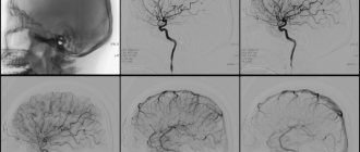
Aortography at the Innovative Vascular Center
The approach to performing aortography in our clinic is based on our belief that angiography is the final stage of diagnosis and should be performed only after receiving sufficient information from other, minimally invasive treatment methods. Most often we combine angiography and endovascular surgery.
The study is performed in a specialized X-ray operating room, where a Philips Allura Xper FD20 angiographic complex is installed, equipped with a software package for high-speed recording of the movement of contrast in vessels and a number of programs that allow the calculation of many parameters. The modern method of computational (subtraction) angiography allows us to obtain additional information about hemodynamics and the condition of blood vessels.