Abdominal aortic aneurysm
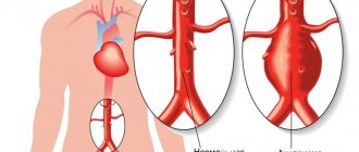
An abdominal aortic aneurysm is an enlarged section of the lower aorta, the main blood vessel that carries blood throughout the body. The diameter of the aorta is about 2-3 cm, it passes through the center of the chest and abdomen. Because the aorta is the body's main blood supply, a ruptured aortic aneurysm can cause life-threatening bleeding.
Depending on the size and rate at which the aneurysm is growing, treatment can range from observation to emergency surgery. Once an abdominal aortic aneurysm is detected, your doctor will monitor its condition so that surgery can be planned if it is needed. Emergency surgery for a ruptured aortic aneurysm is very dangerous.
Symptoms
Abdominal aortic aneurysms often grow slowly and are usually asymptomatic, making them difficult to identify. Some aneurysms may never rupture. Many start out small in size and remain that way, although many expand over time. Others are expanding quickly. It is difficult to predict how quickly an abdominal aortic aneurysm will grow.
As the aneurysm gets larger, some people may notice:
- feeling of pulsation near the navel;
- deep, constant pain in the abdomen or side of the abdomen;
- lower back pain.
Diagnosis of abdominal aortic aneurysms
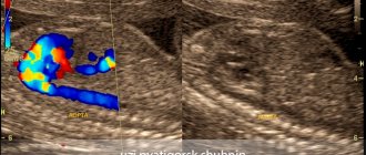
Most often, abdominal aortic aneurysms are detected by ultrasound examination of the abdominal organs. Typically, the discovery of an aneurysm is an incidental finding. If a doctor suspects a patient has an aortic aneurysm, modern diagnostic methods are used to clarify the diagnosis.
Methods for diagnosing abdominal aortic aneurysm:
- Computed tomography in angio mode;
- Magnetic resonance imaging in angio mode;
- X-ray contrast aorto- and angiography;
- Ultrasound duplex or triplex angioscanning of the abdominal aorta.
Causes
Most aortic aneurysms appear in the part of the aorta that belongs to the abdominal cavity. Although the exact cause of abdominal aortic aneurysms is unknown, many factors may play a role, including:
- Smoking tobacco.
Cigarette smoking and other forms of tobacco use increase the risk of aortic aneurysms. In addition to the harmful effects that smoking has directly on the arteries, smoking contributes to the accumulation of lipid plaques in the arteries (atherosclerosis) and high blood pressure. Smoking can also cause rapid growth of the aneurysm and subsequent damage to the aorta. - Hardening of the arteries (atherosclerosis).
Atherosclerosis occurs when lipids and other substances deposit on the inner wall of a blood vessel, increasing the risk of an aneurysm. - Infection of the aorta (vasculitis).
In rare cases, an abdominal aortic aneurysm may be caused by an infection or inflammation that weakens part of the aortic wall. - Aneurysms can develop anywhere along the aorta, but when they form in the upper part of the aorta, they are called thoracic aortic aneurysms. Most often, aneurysms form in the lower part of the aorta and are called abdominal aortic aneurysms.
Types of open surgical operations for aortic aneurysms:
- Bentalla-De Bono operation (replacement of the ascending aorta with a valve-containing conduit with a mechanical prosthetic aortic valve);
- David's operation (replacement of the ascending aorta while preserving the native aortic valve);
- Supracoronary aortic replacement;
- Prosthetics of the ascending aorta and its arch (Borst technique, the use of oblique aggressive anastomosis and other techniques);
- Thoracic aortic replacement;
- Abdominal aortic replacement.
Risk factors
Risk factors for abdominal aneurysm include:
- Age.
Abdominal aortic aneurysms occur most often in people over 65 years of age. - Smoking tobacco.
Tobacco smoking is a significant risk factor for the development of abdominal aortic aneurysm. The longer you smoked or chewed tobacco, the more likely you were to develop an aneurysm. - Atherosclerosis.
Atherosclerosis, the deposition of lipids and other substances that can damage the inner wall of a blood vessel, increases the likelihood of an aneurysm. - Male gender.
Men develop abdominal aortic aneurysms much more often than women. - Family history.
People who have a family history of abdominal aortic aneurysm are at increased risk. People who have a family history of aneurysms tend to develop aneurysms at a younger age and are at increased risk of aneurysm rupture.
Complications
Dissecting aneurysm - rupture of the aortic wall (dissection) is the main complication of an abdominal aortic aneurysm.
A ruptured aortic aneurysm can lead to life-threatening internal bleeding. In general, the larger the aneurysm, the greater the risk of rupture.
Signs and symptoms indicating a ruptured aneurysm:
- sudden intense and constant pain in the abdominal cavity or lower back;
- pain that radiates to the back or legs;
- sweating;
- sticky sweat;
- dizziness;
- nausea;
- vomit;
- low pressure;
- rapid pulse;
- loss of consciousness;
- dyspnea.
Another complication of aortic aneurysms is the risk of blood clots. Small blood clots may develop in the area of the aortic aneurysm. If a clot breaks away from the inside wall of the aneurysm and closes a blood vessel elsewhere in your body, it can cause pain or block blood flow to the legs, toes, kidneys, or abdominal organs.
Modern approaches to the treatment of abdominal aortic aneurysm
- Abdominal aortic aneurysm is detected annually in approximately 12 to 15 people out of 100,000, and due to the potential for rupture, increases mortality;
- Ultrasound or CT screening is recommended for men over 65 years of age who smoke, and for men over 50 years of age and women over 60 years of age whose parents or siblings have had an aortic aneurysm;
- It is recommended to operate on aneurysms with a diameter greater than 5 cm in women and 5.5 cm in men, or if the aneurysm has increased by 5 mm (or more) in less than 6 months;
- Endovascular surgery for abdominal aortic aneurysm is the treatment of choice in patients over 65 years of age, at high risk due to other diseases, and with previous aortic surgery.
In the hands of experienced surgeons, open aortic aneurysm surgery is safe and long-lasting in most cases, and is well suited for younger patients. During this operation, the aorta is clamped above and below the aneurysm, and the damaged area is replaced with a polyester patch.
Perioperative complications (including cardiac and pulmonary complications, incisional hernias, sexual dysfunction, paraplegia, and death) and recovery time when choosing traditional open surgery may have a poorer outcome in patients who are elderly or at high surgical risk.
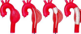
Endovascular treatment of aortic aneurysm using an implantable stent is a safer alternative to open surgery. This procedure produces excellent results in patients with suitable anatomy.
Who is in more danger?
- Age - 50-79 years
- Gender – male (3 times more often)
- Smoking (increases risk 4-5 times)
- Patients with uncontrolled arterial hypertension (high blood pressure);
- Overweight patients;
- Patients with atherosclerotic lesions of blood vessels supplying the brain;
- Patients with impaired blood cholesterol metabolism (dyslipidemia);
- Patients whose relatives also had an aneurysm (the risk is doubled).
Men who smoke in adulthood are at greatest risk (statistically, every 10 have an aneurysm).
Control and treatment
- aneurysms are often asymptomatic;
- Abdominal aortic aneurysms are often discovered by chance during examination of the abdomen during radiography, computed tomography, ultrasound, performed for another purpose;
- if the aneurysm is small (4.5 cm in diameter) and there are no symptoms, annual duplex ultrasound examination is recommended;
- quitting smoking and controlling hypertension is the optimal prevention of the appearance and growth of aneurysms;
- It is recommended to operate on aneurysms with a diameter greater than 5 cm in women and 5.5 cm in men, or if the aneurysm has increased by 5 mm (or more) in less than 6 months.
The procedure is performed through small incisions in the femoral arteries. After puncture of the femoral artery, the guidewire is passed through the dilated part of the aorta, then the stent is advanced along the guidewire. Once the stent is positioned correctly, the balloon expands and the stent pushes the vessel wall apart to prevent the aneurysm from spreading below the renal arteries.
To ensure proper sealing between the implanted stent and the aorta, most currently available stents require that the aneurysm have a proximal isthmus at least 1.0 to 1.5 cm below the renal arteries. However, surgery can be done in patients with aneurysms whose aneurysm isthmuses are shorter or absent altogether, by implanting a stent with multiple holes and branches into the arteries of the kidneys or intestines.
Suitable iliac artery conditions are required for guidewire placement, but the potential placement of a stent through a small retroperitoneal incision has increased the number of candidates for endovascular surgery
Compared to traditional open surgery, endovascular surgery has several advantages:
- reduction of operation time;
- reducing blood loss and the need for infusions;
- reducing the time of treatment in the intensive care unit and hospital stay;
- reduced risk of complications;
- Less contrast is used and often less than 60 ml of contrast is required during the procedure;
- intraoperative and early (up to 30 days) mortality are also lower for endovascular operations than for open ones.
The average stay after open surgery is approximately 3 days in the intensive care unit, followed by 7-10 days in the ward, with recovery taking 8-12 weeks.
Most patients undergoing endovascular surgery do not require hospitalization in the IT department and can be sent home the next day after surgery. A larger percentage of patients who have undergone endovascular surgery are sent straight home rather than to rehabilitation sanatoriums. These patients return to normal levels of physical activity more quickly; the rehabilitation period is 1-2 weeks.
In patients with minor comorbidities who are candidates for endovascular intervention, some complications specific to this method may also occur. It is extremely rare to switch to open surgery. Problems associated with stent placement occur in 5-10% of patients and require CT or ultrasound monitoring
Displacement of the stent is rarely possible, because Newer generation stents have hooks and deploy above the level of the renal arteries for better fixation. “Leaks”—when blood leaks between the stent and the aortic wall—occur in 5-10% of cases. Most of them are type II “leaks” - blood continues to accumulate in the aneurysm from the lumbar artery. If there is growth of the aneurysm, patients are not considered cured; in this case, under local anesthesia, the lumbar arteries are closed by embolization on an outpatient basis.
With proper monitoring, the risk of subsequent rupture is extremely low. Therefore, patients should be prepared to undergo further evaluation, which includes computed tomography of the endovascular stent 4 to 6 months after surgery and annually thereafter. Other less common complications are stent failure or infection.
The current prognosis for healthy patients who undergo aneurysm surgery is excellent. Endovascular surgery is a remarkable advance in the treatment of patients with suitable anatomy and is the preferred treatment option for high-risk and elderly patients.
Treatment and drugs
Here are the general guidelines for treating abdominal aortic aneurysms.
Small aneurysm
If you have a small abdominal aortic aneurysm—about 4 cm in diameter or smaller—and you have no symptoms, your doctor may suggest observation rather than surgery. In general, small aneurysms do not require surgery because the risk of surgery may outweigh the risk of rupture.
If you choose this approach, your doctor will monitor your aneurysm with periodic ultrasounds, usually every 6-12 months; You should report immediately if you experience abdominal tenderness or lower back pain—potential signs of rupture.
Average aneurysm
The average aneurysm measures between 4 and 5.3 cm. It is impossible to say for sure in the case of a medium-sized abdominal aortic aneurysm the risk ratio between rupture and surgery. You will need to discuss the pros and cons and make a decision with your doctor. If you choose surveillance, you will need to have ultrasound scans every 6 to 12 months to monitor your aneurysm.
A large, fast-growing, or “leaky” aneurysm. If you have an aneurysm that is large (greater than 5.6 cm) or rapidly growing (growing more than 0.5 cm in six months), then you will likely need surgery. In addition, a “leaking”, thinned or painful aneurysm requires treatment. There are two types of surgery for abdominal aortic aneurysms.
Open abdominal surgery for an abdominal aortic aneurysm involves removing the damaged section of the aorta and replacing it with a synthetic tube (prosthesis) that replaces the damaged section through an open approach to the abdominal cavity. With this type of intervention, you will likely need a month or more to recover.
Endovascular surgery is a less invasive procedure and can be used to repair an aneurysm in some cases. The doctor uses a synthetic implant, which, with the help of a guide (catheter), is passed through an artery in the thigh into the aorta. The implant—a fabric tube covered with a metal support mesh—is placed at the site of the aneurysm and secured with small hooks. The tissue implant strengthens the weakened part of the aorta and prevents rupture of the aneurysm.
Recovery time for people who undergo endovascular surgery is shorter than for people who have open abdominal surgery. However, more frequent examinations will be required in the future because an endovascularly placed implant may leak. Follow-up ultrasounds should be performed every six months for the first year, and then once a year thereafter. Long-term survival rates are similar for both endovascular surgery and open surgery.
Treatment options for your aneurysm will depend on many factors, including the location of the aneurysm, your age, kidney function, and other conditions that may increase your risk of endovascular or open surgery.
Diagnosis of the disease
The initial stages of diagnosis are an examination by a doctor. The specialist, performing palpation, will detect pulsation in the peritoneum and will probably suspect an aneurysm. The next stage is research to confirm or refute your assumptions. This can only be done by visualizing the processes in the patient’s abdominal cavity. Methods used:
- Ultrasound.
- Computed tomography (MRI).
- Multislice computed tomography of the aorta (MSCT).
Ultrasound allows one to diagnose or refute an aneurysm with almost one hundred percent certainty. If the disease is confirmed, the ultrasound will show the exact location of the disease, the condition of the vessel walls, and the site of rupture (if any).
If the above studies are not enough, aortography is not prescribed. This method makes it possible to check the aorta and all its branches by introducing a special fluid into the system. Such a study is prescribed if there is a suspicion of damage to the visceral and renal arteries, as well as to evaluate the distal bloodstream.
Pulsation of the abdominal aorta, heaviness and bloating, pain, and other discomfort are symptoms for which Doppler ultrasound (USDG) is usually prescribed. The Doppler ultrasound technique makes it possible to determine the nature of the disease and the degree of damage to the aorta and its branches. The technique is based on different frequency sound waves that are reflected by blood cells. The radiation range is individually selected for each patient, which is what determines the high efficiency and effectiveness of the study. The data received on the device’s dashboard looks like high-definition images, which are entered into the protocol.
Combined method for studying pathologies of the abdominal aorta. If making a diagnosis using standard methods is difficult for some reason, then duplex scanning of blood vessels is prescribed. This method includes two methods - ultrasound and Dopplerography. Duplex diagnostics is an ultrasound examination. It allows you to evaluate the echostructure of the walls, the condition of the vessels, the length of the affected area, establish the stage of the disease, as well as information about the speed and intensity of blood flow.
The clinical picture of the aneurysm will become extremely clear. Using duplex diagnostics, aortic insufficiency is determined, which cannot be identified with any other study. This disease is characterized by incomplete closure of the valve flaps, as a result of which reverse blood flow is disrupted.
Lifestyle and Home Remedies
The best way to prevent an aortic aneurysm is to keep your blood vessels as healthy as possible. To do this, you can take the following steps:
- quit smoking;
- keep your blood pressure under control;
- get checked regularly;
- reduce the amount of cholesterol and fat in your diet.
If you think you may have an abdominal aortic aneurysm, or are concerned about your risk of an aneurysm due to a family history, see your doctor. If an aneurysm is detected early, treatment may be easier and more effective.