Reticular (network) varicose veins are an enlargement of small intradermal and subcutaneous veins visible to the eye. The problem of reticular varicose veins is given considerable attention in modern phlebology. This is a “cosmetic” type of venous pathology, which is more often observed in women and is manifested by varicose dilation of small intradermal vessels and the appearance of telangiectasis on the lateral surface of the leg and less often along its inner part. Reticular varicose veins cause nothing but a cosmetic defect. Not dangerous, but not pleasing to the eye. The risk group consists of young women. After the first pregnancy and childbirth, a pathological process often develops in their intradermal vessels due to hormonal changes.
What is telangiectasia or spider veins?
Telangiectasia is a vascular formation, which is a persistent dilatation of skin vessels of small diameter (arterioles, venules, capillaries) of a non-inflammatory nature, morphologically manifested by spider veins or meshwork. The diameter of dilated vessels is 0.5-1.5 mm.
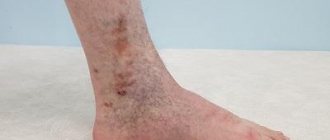
This is what spider veins on the legs look like
What is a reticular (varicose) vein?
Reticular (intermediate) veins are venous vessels of medium caliber (diameter from 1.5 to 4 mm), they are larger than venules and capillaries, but smaller than the main saphenous veins. Quite often these veins become varicose and develop valvular insufficiency. This condition is called reticular varicose veins.
Reticular varicose veins
It can be either an independent disease or combined with varicose veins of the main saphenous veins. In almost 100% of cases, reticular varicose veins will be accompanied by multiple telangiectasias. Moreover, the latter often occur without the presence of reticular varicose veins. Both of these pathological conditions are combined into class C1 of the international classification CEAP and, as a rule, have close pathogenetic connections.
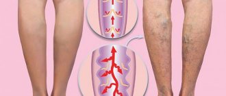
Variants of intradermal varicose veins
Most often, reticular veins are visible as small blue vessels under the skin. They have small one-way valves, and when these valves stop working, the veins begin to dilate. Problematic vessels have poor blood flow and become twisted and distorted due to increased venous pressure, leading to the formation of red or purple spider veins. When dilated veins have a diameter of more than 3 mm and begin to bulge above the surface of the skin, they are called varicose veins. There are three types of varicose veins: linear (lines are formed on the surface of the skin), stellate (rays extend from a darker center, similar to a web or mesh) and tree-like (vessels have a branching appearance).
Telangiectasis and reticular veins - cosmetics or a serious problem?
Many women paid attention to spider veins and stars on their legs, which clearly did not add to their beauty. Often these formations significantly worsen the appearance of the lower extremities. Quite often, despite the pronounced disfiguring veins in the legs, patients have no signs of main varicose veins. In this case, the problem becomes primarily cosmetic. But despite the fact that the likelihood of thrombophlebitis and thrombosis is quite low (due to the small diameter of the affected veins), blood stagnation occurs in telangiectasis and reticular veins. Of course, the symptoms of CVI will be much more modestly expressed than with the main form of varicose veins, but they will manifest themselves to one degree or another. Therefore, reticular varicose veins and telangioctases cannot be attributed to a purely cosmetic problem. In most public hospitals, the treatment of this pathology is not regulated and patients must go to private clinics.
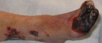
Signs of the disease
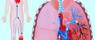
Such pathologies themselves may be symptoms of rapidly progressing chronic venous insufficiency. In turn, these diseases are accompanied by symptoms that, in essence, are characteristic of other pathologies of the veins of the lower extremities - for example, post-thrombotic disease:
- leg cramps during night sleep;
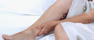
- feeling of heaviness in the legs;
- swelling of the ankles and ankles.
However, in some cases, these diseases are also characterized by other symptoms, which, by strange chance, the vast majority of patients do not recognize as something serious enough to see a doctor:
- hypersensitivity of the skin of the legs;
- periodic burning sensation;
- “creepy crawling” on the lower limbs, etc.
If you notice at least one of the above symptoms, then under no circumstances let the situation take its course and do not put off seeing a doctor. The phlebology department of ON CLINIC in Ryazan sees experienced specialists who can quickly diagnose and prescribe effective treatment for any venous disease.
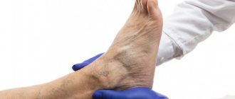
In what places on the legs do spider veins and reticular veins usually appear?
Telangiectasia and reticular veins can appear in any area, but they mainly affect the skin:
- upper thighs;
- above the knee joint;
- supramalleolar region.
Common location of spider veins on the legs
Telangiectasia and reticular varicose veins may be associated with acquired causes:
- Acne-rosacea.
- Systemic scleroderma.
- Exposure to cold, sun, radiation, chemotherapy.
- Mechanical damage to the skin.
- Corticosteroid therapy.
- Pregnancy, childbirth.
- Cirrhosis of the liver.
- Smoking.
- Prolonged standing or sitting position.
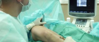
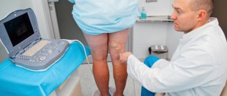
The main reason for the appearance of telangiectasias, according to the modern opinion of leading European scientists, is the pathological discharge of blood (reflux) in the venous system. It can form not only in these vessels, but also in larger ones: reticular (intermediate) and varicose veins. Therefore, even if the patient only has spider veins and spider veins (in the absence of other complaints), a good duplex ultrasound scan of the venous system of the lower extremities is necessary. Only a complete information picture allows you to select an effective modern treatment regimen. Unfortunately, patients in most public medical institutions and some private clinics are often deprived of the possibility of a full diagnosis. And even good treatment of telangiectasia and reticular varicose veins without an accurate understanding of the disturbance of venous blood flow often becomes ineffective.
Why does reticular varicose veins develop?
Hereditary factors play a major role in the development of varicose veins, including reticular varicose veins. At the same time, it is not the disease itself that is inherited from parents to child, but certain structural features of the vascular wall, which result in the veins losing their tone. In addition, there are a number of other provoking factors for reticular varicose veins:
- various metabolic disorders;
- endocrine diseases and hormonal therapy;
- some cardiovascular diseases;
- liver diseases (especially cirrhosis);
- poor nutrition, abuse of “wrong” foods;
- presence of bad habits;
- excess body weight;
- physiological hormonal changes associated with pregnancy or menopause;
- childbirth;
- hormonal contraception;
- features of work activity (sedentary, standing);
- abuse of wearing high-heeled shoes, tight clothing (stockings, leggings);
- excessive sports or work physical activity.
What methods exist for removing spider veins and reticular veins?
Leading innovative European clinics use the following techniques to remove telangiectasias:
- Sclerotherapy is based on the introduction of a special substance (sclerosant) into the lumen of the vein, which causes gluing of the walls and subsequent elimination of the vessel. Depending on the diameter of the vein, a liquid or foam form of the drug can be used. This method is the “gold standard of treatment” not only for telangiectasias, but also for reticular veins and has the highest efficacy and safety profile.
- Percutaneous laser coagulation. The method is based on the impact of a directed beam of high-intensity light, which causes thermal destruction of the sprockets. The method is highly effective, but side effects include burns and skin depigmentation.
- Electrocoagulation is based on contact thermal effects on a vessel using a special electrode.
- Combined techniques: ClaCS, ELASTIK combine sclerotherapy and percutaneous laser exposure. The methods are gaining popularity, but their effectiveness and safety are still comparable to isolated sclerotherapy.
The “gold standard”, rightfully the best, in the treatment of telangiectasia and reticular veins is sclerotherapy (scleroobliteration). Decades of practical application by leading progressive specialists around the world have determined methodological approaches and made the technology the most effective tool in the fight against telangiectasia and reticular varicose veins.
Sclerotherapy (removal) of spider veins in our Moscow center
Phlebologists at our innovation center in Moscow have significant experience in performing this procedure (one of the largest not only in our country, but also in European practice) and cope excellently with even the most complex cases.
Treatment of cosmetic varicose veins
Once diagnosed, the patient may be offered several treatment regimens. The choice of the optimal method depends on the stage of reticular varicose veins, the location of the problem and the characteristics of the human body. The doctor, based on practical experience, chooses the most effective treatment option.
- Neodymium laser (the most modern method) involves transdermal action of high-energy pulses on the vascular wall, no compression stockings are worn, and there is no drug load during the procedure.
- Microsclerotherapy under the control of a venovisor . This method consists of introducing a sclerosant into the lumen accompanied by a vein visor, because the vessels have a small diameter and without good visualization, a high percentage of the solution may not get inside.
- CLaCS therapy (a combination of microsclerotherapy and neodymium laser under the influence of cold).
- Aesthetic miniphlebectomy . This is a minimally invasive surgical procedure aimed at achieving a cosmetic effect. There are no scars left after this procedure.
Sclerotherapy (removal of asterisks) - price of the procedure
One of the advantages of our phlebology center in Moscow is the prices for sclerotherapy; they are quite affordable and accessible to any category of patients.
| Service provided | Cost (price) of the service |
|
|
|
|
|
|
Prevention
One of the features of reticular varicose veins is that the development of the disease (primary or after a course of treatment) can be prevented. To do this you need:
- do light physical exercises daily, putting moderate stress on the lower limbs;
- when working for long periods of time sitting or standing, take short breaks and take a walk every hour;
- give up high heels and give preference to shoes with a wide last;
- control body weight, establish proper nutrition, give up bad habits.
To summarize, we note that reticular varicose veins, although not a critically dangerous disease, requires timely and professional treatment. An integrated approach and care for your feet will save you from possible complications and the development of more serious diseases.