Phlebothrombosis of deep veins is today considered one of the most complex pathologies of modern angiology.
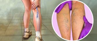
This disease is characterized by the formation of a thrombus (blood clot) in the lumen of the deep veins, which partially or completely blocks the blood flow in the vessel. The most common location of blood clots is the large veins of the leg.
The main distinguishing feature of phlebothrombosis is the weak fixation of the thrombus in the vessel, associated with the addition of inflammation of the vein, so its separation and further travel through the vascular system is difficult to predict.
The disease has a long latent course and is often accompanied by a dangerous and complex complication - thromboembolism of various vessels (pulmonary artery, coronary vessels of the heart, renal vessels, arteries and veins of the brain), which occurs after the separation of a blood clot or part of it.
Phlebothrombosis of the veins of the lower extremities occurs quite often, so it is necessary to know the causes, symptoms, diagnosis and treatment methods of the disease.
Pulmonary embolism is a dangerous complication of phlebothrombosis of the veins of the lower extremities
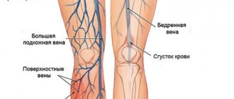
Deep vein thrombosis of the lower extremities (clinical and anatomical forms)
- Deep vein thrombosis of the leg
Complaints of swelling of the foot, pain and tension in the calves, pain when pressing on the calf muscles. If thrombosis does not spread, it is almost asymptomatic. Sometimes there is thromboembolism of small branches of the pulmonary artery with cough and the development of pneumonia (pneumonia). Treatment of thrombosis of the veins of the leg can be carried out on an outpatient basis, under the supervision of a phlebologist with control ultrasound examinations.
- Thrombosis of the popliteal vein
Has a clear clinical picture. Severe swelling and tension of the lower leg, swollen saphenous veins, severe pain when walking. Thrombosis of the popliteal vein is very dangerous due to frequent pulmonary embolism, so treatment is best carried out in a vascular hospital. Most often, conservative therapy is carried out with antithrombotic drugs (heparin). If the patient had a thromboembolism, then urgent surgical treatment is necessary - ligation of the femoral vein above the thrombus.
- Clinic for deep vein thrombosis of the thigh and iliofemoral segment (ileofemoral phlebothrombosis)
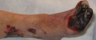
It is characterized by a severe general condition, pronounced swelling of the entire lower limb, and severe pain. The saphenous veins are sharply dilated, the leg takes on a bluish color. With ascending deep venous thrombosis, thrombosis of the entire venous bed is possible with a block of venous outflow and the development of venous gangrene (blue phlegmasia), which is accompanied by high mortality. Pulmonary embolism often occurs with a fatal outcome. Treatment of ileofemoral phlebothrombosis is only in a hospital. For occlusive thrombosis, conservative treatment is possible, but it is better to remove the thrombus so that post-thrombotic disease does not develop. In case of floating thrombosis, urgent removal of the thrombus (thrombectomy) using innovative methods is necessary. A vena cava filter can be installed in cancer patients.
- Thrombosis of the inferior vena cava
The most dangerous disease. Clinically, it is manifested by a severe general condition, swelling of both legs. Kidney failure and blood in the urine often develop. With thrombosis of the liver segment, liver failure develops resulting in Budd-Chiari syndrome. Treatment of acute thrombosis of the inferior vena cava should be active. It is necessary to remove thrombotic masses, as surviving patients may develop severe inferior vena cava syndrome. For this, it is good to use our innovative methods and systemic thrombolysis. The effectiveness of this treatment
- Asymptomatic thrombosis
It should be said right away that there are silent thromboses, that is, completely asymptomatic. Therein lies a great danger. This problem is becoming more and more acute, because with the expansion of ultrasound examination of veins, signs of previous thrombosis are being found more and more often. According to some phlebologists, by old age, most people experience such asymptomatic deep vein thrombosis. In quantity they even exceed those that can be diagnosed without the use of ultrasound methods. The patient does not even feel any health problems, and serious complications occur in the midst of complete well-being, in the event of an increase in blood clot and closure of the main veins. It is not uncommon for the disease to be discovered only after the patient dies from these complications. From this position, if there are no signs of the disease and you are at risk, there is only one way out - you need to direct all your efforts to prevention.
Diagnosis of acute deep vein thrombosis of the lower extremities is very difficult. Signs of deep vein thrombosis appear only in certain localizations of the process. This is primarily due to the absence of clinical symptoms. According to some data, out of 1000 venous thromboses, only 100 have any clinical manifestations. Of these, 60 patients will develop PE, but only 10 will have clinical signs.
It should be recognized that today there is not a single clinical symptom, laboratory or instrumental sign that would indicate with absolute certainty the presence of PE and DVT. Clinical manifestations of thrombosis and ultrasound results can be the basis for the correct diagnosis of venous thrombosis. The clinical picture of deep vein thrombosis consists of a complex of symptoms that characterize a sudden disturbance of venous outflow with preserved arterial blood flow to the limb. Swelling, cyanosis of the limb, bursting pain, local increase in skin temperature, overflow of the saphenous veins, pain along the vascular bundle are characteristic to one degree or another for thrombosis of any localization. Movements in the joints of the limb and sensitivity remain virtually unchanged. General signs such as low-grade fever, weakness, adynamia, and slight leukocytosis occur in most patients. The diagnosis of thrombosis largely depends on the location of the lesion and the level of distribution of thrombotic masses.
History of the study of phlebothrombosis
The study of deep vein phlebothrombosis dates back more than 400 years.
Occlusion of the great veins as a cause of gangrene was first described by F. Hildanus in 1593. The first mention of ileofemoral phlebothrombosis appeared in the medical literature 300 years ago, it was made by Mauriceau.
The concept of “thrombophlebitis” was first introduced into medicine by the English surgeon John Hunter (1728-1793), who performed many operations on gunshot and other wounds and noted the frequency of inflammatory processes combined with the formation of blood clots in the veins.
Interest in deep vein phlebothrombosis increased significantly after the creation of the theory of venous thromboembolism by the outstanding German pathologist R. Virchow. While autopsying the corpse of a young man in 1844 who had suddenly died after developing pain in his thigh, Virchow discovered a blood clot in the right femoral vein and a twisted blood clot in the pulmonary artery. After that, he introduced the concepts of “thrombus” and “embolus” into medical terminology. In 1845, having discovered venous thrombi in 18 cases from 76 autopsies, and in 11 cases revealing the presence of thromboembolism in the pulmonary artery, he came to the conclusion that blood clots form in the veins and are transported by the bloodstream to the pulmonary artery. He also formulated the classical triad, which is still the most complete reflection of the links in the pathogenesis of local vascular thrombus formation.
The first Russian-language monograph devoted to this problem was the work of I.F. Klein "On thrombosis, embolism and ichorremia", published in 1863.
Despite the fact that acute deep phlebothrombosis in various variants of localization and clinical course differ significantly from each other, they are united by the commonality of basic etiopathogenetic processes. The concept of phlebothrombosis as a nosological group is based on the classical Virchow triad.
More than 150 years ago, Rudolf Virchow described the basic mechanisms of intravascular thrombus formation. Its classic triad includes hypercoagulability, vessel wall damage, and slowing of blood flow. Sometimes, for this pathology to occur, a pathological change in only one of these factors is sufficient.
Despite the fact that the thrombotic process can develop at any level of the main veins, in more than half of the cases the starting point of its development in the centripetal direction is the veins of the leg. In the vast majority of cases, thrombosis is primarily localized in the veins of the leg, and subsequently grows proximally into the popliteal, femoral and iliac veins.
It is this type of development that is very often embologenic, since the growth of a thrombus occurs in the direction of veins with an increasing internal diameter, where thrombus masses are not always fixed along the entire perimeter of the vein. Such blood clots are called floating.
One of the main reasons for slow blood flow is immobilization. Under normal conditions, the outflow of blood from the lower extremities is carried out by contraction of the calf muscles, which act as a peripheral pump, pushing blood in a proximal direction, facilitated by the function of the valves. Limiting physical activity significantly disrupts this mechanism. In this case, blood is retained in the venous sinuses of the leg.
The question of the importance of risk factors and trigger factors for thrombosis has been studied in sufficient detail:
- Congenital thrombophilias (deficiencies of various factors of the hemostatic system or their pathological changes)
- Activation of coagulation factors and fibrinolysis disorders (trauma, surgery, neoplasms, pregnancy, childbirth, etc.).
- Pathology of platelets.
- Slowing and/or disruption of blood flow (age over 40 - 45 years, immobilization, pathology of central circulatory mechanisms, obesity, etc.).
- Changes in the rheological properties of blood.
- Damage to the endothelium and vascular wall (contrast agents, intravascular devices, venous catheters, dilatation of veins, etc.).
- Drug therapy (anaesthetics, muscle relaxants, chemotherapy, contraceptives, contrast agents). For example, the incidence of postoperative thrombosis after various surgical interventions can reach 20-59%.
Contrast venography
A method of direct staining of deep veins using the injection of a contrast agent under x-ray control. Phlebography is performed immediately before endovascular intervention for venous thrombosis. In our clinic, the study is carried out with a safe contrast agent - carbon dioxide, which does not have a harmful effect on the kidneys. Phlebography allows you to answer questions about the localization of blood clots, the mechanical reasons for their formation, and the condition of bypass tracts. During phlebography, the surgeon can perform interventions such as installing a vena cava filter to prevent pulmonary embolism, dissolving blood clots, or installing a stent in the area of narrowing of the deep vein.
Causes of phlebothrombosis
There are three main provoking factors for the development of phlebothrombosis of large veins of the leg (Virchow’s triad):
- Damage to the vascular wall (without rupture of the vessel) is long-term mechanical compression of a vein or its varicose veins.
- Changes in the activity of the body's coagulation system (hypercoagulation).
- Reduced blood flow in the vessel.
Predisposing factors include:
- surgical interventions, injuries (severe bruises and fractures) of the lower extremities;
- sedentary lifestyle, long flights;
- prolonged immobilization of the limb or strict bed rest;
- dyshormonal disorders, long-term use of oral contraceptives;
- all conditions and diseases that disrupt the activity of the coagulation system (genetic diseases, autoimmune processes, blood diseases);
- smoking, drug use, frequent alcohol consumption;
- oncological diseases;
- overweight;
- pregnancy (the increasing size of the uterus compresses the inferior vena cava, causing blood stagnation).
If long-term bed rest is necessary, it is imperative to take measures to prevent thrombosis of the veins of the legs
Treatment of phlebothrombosis: traditional and folk medicine
All of us know the simple truth that treatment of any disease should begin as early as possible. For patients diagnosed with phlebothrombosis of the lower extremities, doctors will provide assistance at any stage from the formation stage to the recanalization stage.
Timely treatment makes the prognosis more favorable - the risk of thromboembolism is significantly reduced, further spread of the blood clot is stopped, the blood clot is resolved with the restoration of the vein lumen to one degree or another, which means that the manifestations of post-thrombophlebitis syndrome are minimized.
How to treat phlebothrombosis? Therapy for this disease is always complex. As mentioned earlier, it depends on the etiology and degree of development of the pathology. The treatment regimen involves conservative therapy and, if necessary, surgical intervention.
Conservative methods include:
- Taking medications. To improve the rheological properties of blood and increase the elasticity of vessel walls, various groups of medications are used: phlebotonics, antiplatelet agents, anti-inflammatory drugs and anticoagulants.
- Carrying out local therapy. The use of ointments and gels that have anti-inflammatory, venoprotective, decongestant and other effects.
- Wearing medical compression stockings or bandaging limbs.
- Nutrition. The diet for phlebothrombosis of the lower extremities excludes alcohol and spicy foods. You should drink at least 2 liters of fluid and eat foods rich in antioxidants.
If conservative therapy methods fail to achieve a positive result, then surgical methods will be used.
In case of acute development of the disease at home, an ambulance call is required as early as possible.
Expert opinion
Until the doctors arrive, it is important to try to fix the limb in a motionless state. This will reduce the risk of a blood clot breaking off.
Vascular surgeon, phlebologist
Osipova Ekaterina Yakovlevna
Phlebothrombosis - herbal baths
Treatment of ileofemoral thrombosis
All patients without exception with an established diagnosis of “ileofemoral thrombosis” are required to be hospitalized in an angiosurgical hospital. The patient must be transported in a supine position. Until medical assistance is provided, he must adhere to the strictest bed rest. If it is not possible to perform a qualitative examination of the victim, then he is prescribed anticoagulants, fibrinolytics and thrombolytics for up to 10 days.
General recommendations for the management of patients with acute ileofemoral thrombosis:
- Anticoagulant drugs: low molecular weight Heparin, Logiparin, Fraxiparin.
- Eliminating pain, removing the patient from a state of shock.
- Relieving spasm from blood vessels, normalizing hemodynamics.
- Drugs for thrombolysis: Streptokinase or Urokinase. However, it should be remembered that the use of thrombolytic drugs is always associated with the risk of bleeding and death of the patient. Therefore, thrombolysis drugs are prescribed only to patients under the age of 50 years in whom acute thrombosis occurred no later than 7 days before visiting the doctor. In this case, patients must have vena cava filters installed, otherwise there is a high probability of small particles of a blood clot spreading through the bloodstream and developing a pulmonary embolism.
- Fibrinolysis activator drugs: Complamin, Teonicol, Nicotinic acid (intravenous administration), Pyrogenal (intramuscular administration).
- Normalization of rheological blood parameters is carried out with the help of drugs Trental, Eufillin, Actovegin, etc.
- If inflammation develops, antibiotics are indicated.
Surgery for ileofemoral thrombosis is prescribed only for vital indications: if the patient is diagnosed with floating blood clots that pose a threat to pulmonary embolism, or if complications of thrombosis develop. These include: embologenic thrombosis, a high risk of developing gangrene against the background of blue phlegmasia, ascending thrombosis.
There are also relative indications for surgery, including:
- Lack of effect from drug treatment for 2-3 days.
- The duration of thrombosis is more than 8 days.
- Senile age.
Thrombectomy is the main method of surgical intervention for ileofemoral thrombosis. It should be remembered that with blue phlegmasia, conservative therapy is useless in 100% of cases. The prognosis for blue phlegmasia is largely determined by how timely the surgical intervention was performed (before the development of gangrene). In this case, patients are indicated for radical throbectomy. The risk of pulmonary embolism increases when thrombectomy is performed on the right iliac vein.
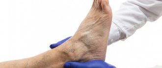
Diagnostics
Diagnosis of the disease begins with examination of the patient. Based on the characteristic set of symptoms, the doctor will be able to suspect ileofemoral thrombosis.
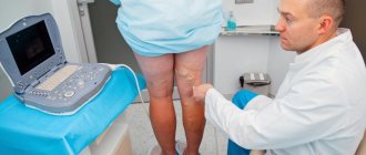
To confirm the diagnosis, the following instrumental techniques are used:
- Duplex or triplex scanning.
- X-ray contrast venography.
- Radionuclide phlebography.
- Labeled fibrinogen scan.
It is imperative to distinguish between ileofemoral thrombosis and erysipelas, renal and heart failure, radiculitis, arthritis, and bursitis.
Prevention
- Maintaining a balanced diet and maintaining normal body weight.
- Eliminate or minimize alcohol, fatty, fried, salty and smoked foods, vinegar, coffee.
- It is necessary to lead a moderately active lifestyle. It is recommended to perform gymnastic exercises without lifting heavy weights or stretching. Walking, swimming, dancing, cycling, and cross-country skiing are beneficial.
- Wear comfortable clothes that do not restrict movement, use comfortable shoes with heels no higher than four centimeters.
- People with signs of varicose veins and a family history should undergo regular examinations by a phlebologist.
- With forced long-term static loads (air travel, long trips by car, etc.) as well as during surgical interventions.
You can make an appointment with a phlebologist to make a diagnosis and prescribe therapy by calling a single multi-channel phone number 676-25-25 or leaving a request on the website.
Diet for thrombophlebitis
A large amount of fresh vegetables, fruits, fiber, nuts, cereals, whole grain breads. Among products of plant origin, the following also have a beneficial effect on venous diseases:
- ginkgo biloba,
- ginger,
- red capsicum,
- valerian root,
- garlic,
- hawthorn berries.
Nutritional supplements and vitamins:
- vitamins C, A, E and B6,
- linseed oil,
- magnesium and calcium (in combination),
- Pycnogenol
The risk group for the development of phlebothrombosis includes:
- Pregnant women, especially in the second - early third trimesters, and women in the postpartum period.
- People who are overweight and lead a sedentary lifestyle and sedentary work.
- Elderly people especially with diseases of the musculoskeletal system.
- Patients with cancer.
- Patients with decompensated forms of cardiovascular diseases.
- Persons whose activities involve heavy lifting or changes in atmospheric pressure.
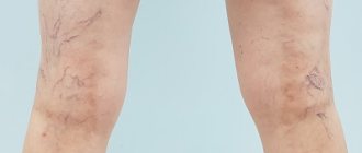
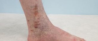
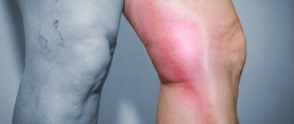
Symptoms of thrombophlebitis
- soreness,
- the appearance of local swelling of the limb,
- temperature rise,
- thickening and redness along the affected vein.
In the case of thrombophlebitis of the superficial veins, the patient may notice a gradual increase in the intensity of the changes described above over several days.
If you have these symptoms, you should definitely consult a specialist.
However, with deep vein thrombosis (phlebothrombosis), the main symptom is most often swelling of the affected limb, although it should be remembered that many patients with this form of the disease may not have any symptoms for a long time.
If swelling and pain are severe, and are also accompanied by high fever or shortness of breath with coughing attacks or chest pain, it is necessary to call an ambulance team.
These symptoms may indicate the development of deep vein thrombosis, which significantly increases the likelihood of a blood clot breaking off and migrating into the vessels of the lungs.