Relevance
Pulmonary arterial hypertension (PAH) is a heterogeneous, often multifactorial condition that can be either an independent, isolated pathology or a complication of a wide range of diseases, including congenital heart defects, bronchopulmonary dysplasia (BPD), bronchial asthma, genetic and chromosomal diseases, diseases connective tissue and other pathologies.
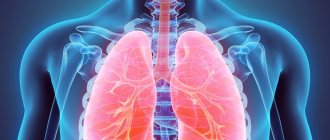
According to various estimates, the prevalence of PAH in childhood is rare, approximately 2–16 cases per 1 million children. However, in some risk groups this pathology occurs many times more often [1, 2]. A significant portion, more than 30% of cases, of PAH is associated with pathology of the respiratory system, including BPD [3, 4]. It should be recognized that the prevalence of pulmonary hypertension is often underestimated. It is usually regarded as a long-term dynamic process that can manifest itself only in the late stages of chronic bronchopulmonary disease. The initial stage of PAH occurs with a small, often intermittent increase in pressure in the pulmonary artery, which does not require a significant increase in the work of the right ventricle and, as a rule, does not have early clinical manifestations [5, 6]. Therefore, the diagnosis of PAH in the initial stage depends on many factors, including the patient’s age, the severity of concomitant pathology, the type of heart damage and, first of all, on the awareness of specialists about this pathology, which occurs in very early childhood. In children in the first months of life, the leading place is given to secondary pulmonary hypertension [7]. The most pronounced changes in pulmonary hemodynamics in infants, as a rule, occur with BPD, chronic bronchiolitis and other chronic obstructive pulmonary pathologies [8–10]. In patients with BPD, pulmonary hypertension may be associated with intermittent chronic hypoxia, hypercapnia, lung and airway tissue damage, diastolic dysfunction, vascular growth retardation, and pulmonary vein stenosis [11]. One of the main causes is arterial hypoxemia due to hypoventilation. Pulmonary artery pressure is influenced by changes in pulmonary blood flow due to damage to the pulmonary vascular endothelium. Dysfunction of the endothelium of these vessels leads to disruption of the synthesis of prostaglandins and prostacyclin, as well as to a marked increase in the production of thromboxane and endothelin-1 against the background of a significant decrease in the formation of nitric oxide. These disorders contribute to additional vasoconstriction, increased pulmonary vascular resistance, and PAH becomes chronic [12].
As described back in the last century by WJ Northway et al. (1967), BPD can occur after prolonged artificial pulmonary ventilation (ALV) and oxygen therapy, mainly in premature newborns. However, in recent years, against the backdrop of successes and achievements in perinatology, the problem of caring for premature babies, especially those born with extremely low body weight (ELBW), has become even more relevant due to high mortality in the neonatal period. During this period, in children with low birth weight, the risk of mortality is aggravated by the development of diseases such as sepsis, congenital infections, pneumonia and BPD, the prevalence of which varies significantly, according to various sources, and is up to 30% among children receiving mechanical ventilation, especially long-term [13]. The incidence of BPD in newborns has an inverse correlation with birth weight and depends on the timing and modes of mechanical ventilation and oxygen therapy. I would like to emphasize that in children with ELBW, the severity of changes in the cardiovascular system and BPD, as well as the progression of retinopathy, as a rule, are links in one chain, which is based on deep morphofunctional immaturity of organs and systems [14, 15].
In modern conditions, despite the optimization of the timing of mechanical ventilation and the rational use of surfactant, a high risk of mortality remains, due, in particular, to progressive pulmonary hypertension. Along with this, there are cases of PAH in which it is possible to achieve a less severe course of the disease and, with timely corrective therapy, obtain the outcome in the form of significant improvement or even recovery. There is still a lack of data on the individualized approach and optimal management of children with pulmonary hypertension [1, 16]. All this makes the problem of early diagnosis and adequate treatment of children with PAH due to BPD and congenital heart defects urgent.
Symptoms
When an attack of stenosis occurs in a child, it is very important to recognize it in time and, if possible, provide first aid until professional medical help arrives. Since there are many variants of the clinical course of this pathology, the signs of stenosis in children will differ. They depend primarily on the cause of the disease. Also influencing factors are the child’s age and the presence of other concomitant chronic diseases.
As the glottis narrows and its lumen decreases, the intensity of symptoms of laryngeal stenosis in children increases. The development of this pathological condition has several stages.
- 1st degree – accompanied by breathing problems in the child. Another name for this stage is compensated, since this stage has a good prognosis for treatment and complete elimination of symptoms. A characteristic sign for this stage is a change in the baby’s voice, it becomes more hoarse.
- Grade 2 – adverse symptoms are more pronounced. The second name of the stage is subcompensated. The child's breathing quickens, he becomes overly excited, and the skin is characterized by acquiring a bright red color. It is possible that some areas of the chest located between the ribs may “sink.”
- 3rd degree - in this condition there is a sharp change in the color of the skin. The skin becomes very pale, and the lips and the area of the nasolabial triangle acquire a bluish tint. The child himself can be either overly excited or, on the contrary, overly inhibited. Possible loss of consciousness. The stage is also called decompensated.
- Asphyxia is the most extreme degree of development of pathology. The most dangerous condition and primarily for infants and children under 3 years of age. The child stops breathing completely. Resuscitation is required, otherwise, without oxygen supply, brain cell death will begin to occur. Without emergency medical care, death from acute cardiac and respiratory failure is possible.
Clinical observation
In order to focus the attention of pediatricians, pediatric cardiologists and pulmonologists on this problem, we present an interesting clinical case of PAH against the background of a combination of BPD and congenital heart disease in a child with ELBW at birth.
Child A., female, from the 1st pregnancy, which proceeded with the threat of termination in the first and second half, was born from 1 premature birth at the 26th week. gestation, by Caesarean section, with a weight of 870 g and a length of 34 cm, with an Apgar score of 4/6 points.
From the first hours of her life, the girl had a serious condition with the manifestation of respiratory distress syndrome. Resuscitation measures were carried out using mechanical ventilation, oxygen therapy, surfactant replacement therapy, and incubation regimen. On the 2nd day of life, a hemodynamically significant patent ductus arteriosus (PDA) was detected, 3 doses of ibuprofen were administered intravenously according to the usual regimen, without a positive effect. Over time, the manifestations of heart failure increased. In this regard, the child received cardiac glycosides, dopaminomimetics, β-adrenergic agonists (dopamine, digoxin, dobutamine). With this therapy, the condition stabilized somewhat, although it remained serious. Persistent respiratory failure of the 3rd degree was noted, from the 3rd day anemia (Hb 102 g/l), hematuria were noted, fine rales and crepitus were heard on auscultation in the lungs on both sides. On the 8th day of life, radiological changes were revealed in the form of a bilateral decrease in pneumatization due to gentle infiltrative shadowing in both lungs with an increase in the bronchovascular pattern due to the interstitial component (Fig. 1), leukocytosis (21.0 × 10⁹/l), there was a negative dynamics in blood tests on the 1st week. life (increase in leukocytosis, ESR, decrease in hemoglobin).
A diagnosis was made: Neonatal pneumonia, bilateral, focal, severe, infiltration stage. DN III degree. Microbiological culture of throat and endotracheal aspirate revealed Klebsiella pneumoniae
. In addition, in a child with a burdened obstetric history, arteritis and phlebitis of the umbilical vessels, anemia, and pneumonia, signs of SIRS (thrombocytopenia, impaired thermoregulation, inflammatory changes in the blood) were recorded, which suggested management as being at risk of neonatal sepsis. Due to many diseases, the girl received complex therapy (including antibacterial) and continued to be on mechanical ventilation for a long time (FiO2 0.45; f 40 in 1 min; pi. 19 mm Hg; SaO2 90–92%) . Periodically, attempts were made to reduce the parameters of mechanical ventilation, however, due to persistent hypercapnia (pCO2 55 mm Hg) and hypoxia (pO2 24 mm Hg), acidosis, according to the acid-base composition of the blood (lactate 1.1 mmol/ l; cHCO3 38.2 mmol/l; BE 12.3 mmol/l), low levels of oxygen saturation, it was not possible to extubate the child.
From the 28th day of life, in accordance with a set of criteria [14], a diagnosis was made: Bronchopulmonary dysplasia of prematurity, a new form, severe. The main point for making the diagnosis was the need for oxygen concentration of more than 21% on the 28th day of life, oxygen dependence at 36 weeks. postconceptional age indicated severe BPD. A computed tomogram revealed the main signs of BPD—fibrous cords, ground glass, and bullous swellings (Fig. 2). For the disease, the child received therapy with inhaled glucocorticosteroid (ICS) budesonide at a dose of 1.0 mg/day, muco- and bronchodilator therapy through a nebulizer (ambroxol, fenoterol, ipratropium bromide).
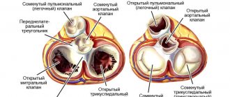
At the age of 2.5 months. Life diagnosis confirmed: Congenital heart defect (CHD): PDA. On ECHO-CG: PDA 3.5 mm with blood discharge into the pulmonary artery with a gradient of 4 mm Hg. Art., aneurysm of the interatrial septum (ASA) and a patent foramen ovale 3.5 mm with right-to-left shunting, high PAH 89 mm Hg. Art. with expansion of the right atrium (RA) (Fig. 3).
The ECG showed signs of RA overload. X-ray: signs of enlargement of the right chambers of the heart. In the hemogram: anemia Hb 90 g/l, Er 3.88 × 10¹²/l, CP 0.9, leukocyte count without signs of inflammation. Blood glucose was 5.0 mmol/L, creatinine 51.5 mmol/L, ALT 0.5 µmol/L, AST 0.94 µmol/L. At the age of 3.5 months. An operation for PDA (ligation) was performed at the Federal Center for Cardiovascular Surgery (Astrakhan). The early postoperative period passed without complications.
For the identified pathology of congenital heart disease + PAH, the child received complex therapy, including oxygen therapy, diuretics (spironolactone under the control of creatinine and potassium), sildenafil (at a dose of 3.5 mg × 4 times / day), digoxin at a maintenance dose (10 mg/kg /day), dobutamine (1.5 mcg/kg/min). The child's condition stabilized, shortness of breath decreased, appetite improved, and the girl began to gain body weight of 350 g or more per week.
The diagnosis was made: Deep prematurity (26 weeks of gestation). BPD of prematurity, a new form, severe severity, respiratory failure of II degree. Congenital heart disease: PDA, stage IIA heart failure, FC II, minor cardiac anomaly: patent foramen ovale. Consequences of hypoxic damage to the central nervous system, depression syndrome. Early anemia of prematurity of moderate severity. Retinopathy of the retina of both eyes, stage III, active phase.
Further, against the background of treatment and positive clinical and laboratory dynamics (increase in weight and height indicators, absence of inflammatory changes in the hemogram, negative CRP and PCT indicators, absence of pathological deviations of other biochemical indicators, improvement in SaO2 indicator: 94–95%) on the 65th day of life, the child was extubated and transferred to spontaneous breathing. Inhaled therapy for severe BPD with ICS (budesonide) was continued at the same volume.
Against the background of stabilization of the condition at the age of 5 months. Surgical treatment of retinopathy of prematurity was carried out (at the St. Petersburg State Pediatric Medical University of the Ministry of Health of Russia, Department of Pathology of Newborns and Infants). After the operation, the dynamics of the child remained persistent respiratory failure (RR 42–44, with anxiety up to 85 per minute; SaO2 90–94%), which was associated with prolonged oxygen support, the total duration of receiving the air-oxygen mixture was about 6 months.
On an outpatient basis, the child was observed by a pediatrician, pulmonologist, and cardiologist with a diagnosis of Bronchopulmonary dysplasia of prematurity, a new form, severe, complicated by secondary high pulmonary hypertension (89 mm Hg). Condition after correction of congenital heart disease (ligation of the PDA, AMP, patent foramen ovale, ectopic chord in the cavity of the left ventricle). Paresis of the left vocal cord. Consequences of hypoxic damage to the central nervous system. Delayed physical and motor development. Stage III retinopathy of the retinas of both eyes, active phase (surgical treatment: transpupillary laser coagulation of the retina). Moderate anemia of prematurity. Protein-energy deficiency of the third degree.
During the first year of life, the child had extremely low physical development, disharmonious due to weight deficiency (at 1 year, weight 6.0 kg and body length 58 cm) -6.2SD and -3.2SD in height and weight, respectively [17]. Noteworthy were increased weakness, fatigue, dry cough during the day, periodic stridor breathing, shortness of breath at rest of a mixed nature from 65 respiratory movements per minute at rest, remission and up to 82 per minute during periods of exacerbation of BPD. Oxygen saturation in peripheral blood by pulse oximetry was 90–95%. During the observation period, the most severe deterioration of the condition was observed twice at the age of 6 months. and 8 months, which was associated with respiratory infections.
The girl received basic therapy for BPD with the drug budesonide at a dose of 1.0 mg/day from the moment of diagnosis continuously for 6 months, then 0.5 mg 1 time/day for 6 months. During the period of exacerbation of the disease, the dose of ICS was increased to 2.0 mg/day, and ipratropium bromide was used at a dose of 0.075 mg (up to 6 drops) to relieve shortness of breath.
During outpatient treatment for pulmonary hypertension, a course of sildenafil was continued at a dose of 4 mg/kg/day × 4 times/day for 2 weeks, then 1 mg/kg/day for 6 months, spironolactone 2 mg/kg × 2 rubles/day
During observation for 1 year, systolic pressure in the pulmonary artery (SPAP) had a positive trend. The ECG recorded sinus rhythm, pacemaker migration, the electrical axis of the heart was sharply deviated to the right, â=+150°, heart rate 120–150 per minute, PQ 0.1. ECHO-CG shows: the condition after ligation of the PDA, the duct is hermetically closed, the borderline dimensions of the right chambers of the heart (right ventricle 1.77 cm; RA 1.76 cm). Global contractility of the left ventricle is not impaired. Emission fraction SF 60%. Ectopic chord in the cavity of the left ventricle. Mitral regurgitation grade 0–1. Tricuspid regurgitation of the first degree. Pulmonary artery 1.46 cm, MPAP 46 mm Hg. Art. The pericardium is without features. The maximum pressure gradient at Ao is 8.1 mm Hg. Art. O2 saturation 94%.
During treatment, a positive dynamics of MPAP was noted from 89 to 46 mm Hg. Art., clinical signs of PAH have decreased significantly, signs of BPD and the consequences of hypoxic damage to the central nervous system, delayed physical and psychomotor development, and correction of congenital heart disease remain.
When to see a doctor
If symptoms of laryngeal stenosis are observed in a child, then before taking first aid measures yourself, you must call emergency medical help. Such services are offered by JSC “Medicine” (clinic of academician Roitblerg), located in the center of Moscow. Competent doctors work here. All studies are carried out in the shortest possible time using modern equipment.
First aid from parents in this case may include only non-specific actions. If possible, it is necessary to calm the child (if he is conscious), pick him up or help him take a position in which he is most comfortable breathing. It is important not to panic, so that the child feels supported and confident from adults that everything will end soon and everything will be fine. Under no circumstances should you delay contacting a doctor, otherwise the consequences may be irreversible.
Also, an undoubted advantage of contacting “Medicine” is the presence of highly qualified doctors and experienced medical staff who have many years of experience and always apply an individual approach.
Diagnosis of pathology
During an attack in an acute form of pathology, there is simply no time for research and analysis. Doctors who arrive on call diagnose the condition based on the description of the situation by the parents (or other persons present), an external examination of the child and palpation in the throat area.
After providing the patient with medical care and eliminating the threat to life, the child will have to undergo a detailed examination, possibly in a hospital. The purpose of the examination is to identify the cause of the attack and eliminate it by prescribing the most effective treatment.
The main diagnostic measures are as follows.
- Examination of the larynx using visual methods - laryngoscopy. Allows you to determine the degree of narrowing of the larynx, the absence or presence of a neoplasm.
- Fibrolaryngoscopy - the study is carried out using a flexible endoscope, at the end of which a video camera is attached. The image is displayed on the computer screen, and the doctor is able to accurately determine the condition and size of the lumen of the larynx.
- A chest x-ray is performed to rule out other concomitant diseases with similar symptoms. For example, with heart disease, shortness of breath is a characteristic symptom.
- Various radiological research methods - computed tomography, MRI. They can be prescribed if difficulties arise with an accurate diagnosis.
- Ultrasound of the thyroid gland.
- Laboratory studies of throat smears are necessary to accurately determine the nature of the infectious disease that provoked laryngeal stenosis.
How to prevent laryngeal stenosis in children and adults - prevention
Prevention of laryngeal stenosis consists of:
- competent and timely treatment of diseases that can cause narrowing of the airways;
- exclude neck injuries;
- timely diagnosis and treatment of upper respiratory tract infections;
- refusal of long intubation (no more than 3 days);
- strict adherence to the timing of tracheostomy;
- observation by an otolaryngologist after laryngeal surgery;
- avoiding inhalation of caustic smoke, ingestion of acids and alkalis into the respiratory tract;
- carrying out allergen-specific and immune therapy;
- avoiding contact with allergens.
This article is posted for educational purposes only and does not constitute scientific material or professional medical advice.