Deep vein thrombosis of the lower extremities is a disease in which blood clots form in the lumen of the vein. The thrombus can completely cover the lumen of the vessel or only partially, settling on one of its walls. Vein thrombosis can occur in every patient with varicose veins of the lower extremities; in this case, the superficial and communicating (connecting) veins are more likely to thrombose. Deep vein thrombosis affects not only patients with chronic venous disease of the lower extremities, but also patients with blood diseases, diabetes mellitus, kidney disease, neurological pathology, pregnant women, cancer patients, people with disorders in the blood coagulation system or the so-called hypercoagulation syndrome.
The causes of deep vein thrombosis are as follows:
- expansion of the vein lumen and decrease in blood flow speed,
- violation of the integrity of the inner lining of the vessel, which creates an obstacle to blood flow and causes a delay of blood cells in this area, subsequently a blood clot forms there;
- Blood viscosity decreases, that is, the blood becomes thick, this occurs due to dehydration, fever, and endocrine disorders.
But the main cause of deep vein thrombosis is stagnation of venous blood in the legs or impaired venous outflow from the lower extremities. Against the background of the described reasons, after infectious diseases, surgical treatment, injuries of the lower extremities or significant physical activity, a blood clot forms. And the cause of stagnation can be a prolonged static position of the legs; this happens if a person is in a stationary position for a long time, standing on his feet or with his legs down. This applies mainly to representatives of the following professions: drivers, salesmen, teachers, machine operators, surgeons, hairdressers. Blood clots can often form during long flights, in those who work at a computer or sit in front of the TV for a long time.
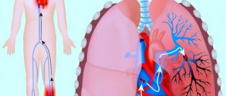
The main danger of deep vein thrombosis of the lower extremities is that part of the thrombus, especially when its uppermost part or “thrombus head” is not attached to the wall, but is in a free position (floating thrombus), can come off and then, with the blood flow enter the pulmonary artery and from there into the lungs. This condition is called pulmonary embolism (PE). Against the background of this complication, blood flow in the lungs is disrupted, which is manifested by difficulty breathing, shortness of breath, a feeling of interruptions in the heart, dizziness, and a cough with streaks of blood in the sputum. If the necessary treatment measures are not started in time, the patient may die.
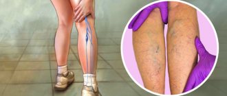
Deep vein thrombosis has very specific symptoms. The disease usually develops acutely, especially if there are provoking factors. First of all, swelling of the limb affected by thrombosis appears. If this is acute thrombosis of the deep vein of the thigh, then pronounced swelling of the thigh occurs, but more often thrombosis of the deep veins of the leg occurs and then the lower leg swells from the ankle to the knee. Then the skin turns blue, and when an infection occurs, the skin of the legs becomes crimson red. Bursting pain and itching of the skin can accompany this pathology. Superficial veins swell and protrude from the skin, leading to an increase in skin temperature. If treatment is not started on time or is started incorrectly, the blood clot can grow and reach the uppermost parts of the thigh, this is called total thrombosis of the limb.
Get an online consultation
right now.
Get
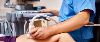
Diagnosis of deep vein thrombosis is based on Doppler ultrasound during angioscanning. Multislice computed tomography with contrast helps establish the topical picture. A biochemical blood test, called a coagulogram, helps to assess the state of the blood coagulation and anticoagulation systems. All additional research methods, as well as the diagnosis itself, are prescribed by a phlebologist.
Treatment of deep vein thrombosis is a long and labor-intensive process. First of all, it is necessary to stop the spread of thrombosis and try to dissolve already formed thrombotic masses, in order to prevent pulmonary embolism and fatal complications.
In the acute period, treatment is best done in a hospital. An elastic bandage is applied to the limb or compression stockings with maximum compression are put on. During a period of unstable swelling on the leg, when it is difficult to choose knitwear in size, it is wiser to use elastic bandages, but at the same time apply them in two layers at the site of blood clot formation. Compression therapy is not used in patients with atherosclerosis of the vessels of the lower extremities, due to the risk of ischemia, and who have eczema or dermatitis of the skin of the legs.
Anticoagulants are prescribed - drugs that thin the blood and dissolve the blood clot. They are usually given as a subcutaneous injection once a day. The drugs in this group are low molecular weight heparins: Clexane, Fraxiparine, Fragmin. These drugs inhibit the activity of the thrombin enzyme or coagulation factor Xa, resulting in increased anticoagulant and thrombotic effects. The blood becomes more fluid, and the formation of a blood clot stops. The state of the coagulation system is monitored by assessing coagulogram parameters. After 2-5 days, the patient is transferred to oral coagulants, which are prescribed for at least one month. The most common drug from this group of rivaroxaban is Xarelto, its course dose is 15 mg 2 times a day for 3 weeks, followed by a reduction in dosage to 20 mg per day in a single dose. Previously used blood clot traps - vena cava filters - are no longer used to prevent pulmonary embolism due to many dangerous complications. Their use can only be justified in cases of high risk and bleeding when anticoagulant therapy is prescribed.
For deep vein thrombosis, treatment is carried out with phlebotonics, drugs that normalize the tone of the venous vessel and improve venous outflow. “Antistax”, “Phlebodia”, “Detralex”, “Venarus” - this is a far from complete list for the treatment of this complication, which occurs in more than half of the cases in patients with varicose veins.
Deep vein thrombosis of the lower extremities (clinical and anatomical forms)
- Deep vein thrombosis of the leg
Complaints of swelling of the foot, pain and tension in the calves, pain when pressing on the calf muscles. If thrombosis does not spread, it is almost asymptomatic. Sometimes there is thromboembolism of small branches of the pulmonary artery with cough and the development of pneumonia (pneumonia). Treatment of thrombosis of the veins of the leg can be carried out on an outpatient basis, under the supervision of a phlebologist with control ultrasound examinations.
- Thrombosis of the popliteal vein
Has a clear clinical picture. Severe swelling and tension of the lower leg, swollen saphenous veins, severe pain when walking. Thrombosis of the popliteal vein is very dangerous due to frequent pulmonary embolism, so treatment is best carried out in a vascular hospital. Most often, conservative therapy is carried out with antithrombotic drugs (heparin). If the patient had a thromboembolism, then urgent surgical treatment is necessary - ligation of the femoral vein above the thrombus.
- Clinic for deep vein thrombosis of the thigh and iliofemoral segment (ileofemoral phlebothrombosis)
It is characterized by a severe general condition, pronounced swelling of the entire lower limb, and severe pain. The saphenous veins are sharply dilated, the leg takes on a bluish color. With ascending deep venous thrombosis, thrombosis of the entire venous bed is possible with a block of venous outflow and the development of venous gangrene (blue phlegmasia), which is accompanied by high mortality. Pulmonary embolism often occurs with a fatal outcome. Treatment of ileofemoral phlebothrombosis is only in a hospital. For occlusive thrombosis, conservative treatment is possible, but it is better to remove the thrombus so that post-thrombotic disease does not develop. In case of floating thrombosis, urgent removal of the thrombus (thrombectomy) using innovative methods is necessary. A vena cava filter can be installed in cancer patients.
- Thrombosis of the inferior vena cava
The most dangerous disease. Clinically, it is manifested by a severe general condition, swelling of both legs. Kidney failure and blood in the urine often develop. With thrombosis of the liver segment, liver failure develops resulting in Budd-Chiari syndrome. Treatment of acute thrombosis of the inferior vena cava should be active. It is necessary to remove thrombotic masses, as surviving patients may develop severe inferior vena cava syndrome. For this, it is good to use our innovative methods and systemic thrombolysis. The effectiveness of this treatment
- Asymptomatic thrombosis
It should be said right away that there are silent thromboses, that is, completely asymptomatic. Therein lies a great danger. This problem is becoming more and more acute, because with the expansion of ultrasound examination of veins, signs of previous thrombosis are being found more and more often. According to some phlebologists, by old age, most people experience such asymptomatic deep vein thrombosis. In quantity they even exceed those that can be diagnosed without the use of ultrasound methods. The patient does not even feel any health problems, and serious complications occur in the midst of complete well-being, in the event of an increase in blood clot and closure of the main veins. It is not uncommon for the disease to be discovered only after the patient dies from these complications. From this position, if there are no signs of the disease and you are at risk, there is only one way out - you need to direct all your efforts to prevention.
Diagnosis of acute deep vein thrombosis of the lower extremities is very difficult. Signs of deep vein thrombosis appear only in certain localizations of the process. This is primarily due to the absence of clinical symptoms. According to some data, out of 1000 venous thromboses, only 100 have any clinical manifestations. Of these, 60 patients will develop PE, but only 10 will have clinical signs.
It should be recognized that today there is not a single clinical symptom, laboratory or instrumental sign that would indicate with absolute certainty the presence of PE and DVT. Clinical manifestations of thrombosis and ultrasound results can be the basis for the correct diagnosis of venous thrombosis. The clinical picture of deep vein thrombosis consists of a complex of symptoms that characterize a sudden disturbance of venous outflow with preserved arterial blood flow to the limb. Swelling, cyanosis of the limb, bursting pain, local increase in skin temperature, overflow of the saphenous veins, pain along the vascular bundle are characteristic to one degree or another for thrombosis of any localization. Movements in the joints of the limb and sensitivity remain virtually unchanged. General signs such as low-grade fever, weakness, adynamia, and slight leukocytosis occur in most patients. The diagnosis of thrombosis largely depends on the location of the lesion and the level of distribution of thrombotic masses.
Treatment of the disease
There are many reasons for the occurrence of pathology: a sedentary lifestyle, little physical activity, infections, vascular injuries, changes in blood composition, as well as high blood clotting, a hereditary tendency to form blood clots. All these factors require special monitoring on the part of the patient and the doctor prescribing therapy.
The principles of therapeutic measures aimed at eliminating pathology are:
- taking anti-inflammatory drugs;
- elevated position of injured limbs;
- prescribing ointments that improve blood flow;
- warm compresses on affected areas;
- elastic leg bandaging.
It is necessary to do therapeutic exercises for thrombophlebitis of the legs; therapeutic exercise is one of the main stages of disease therapy, leading to an improvement in the patient’s condition, allowing to avoid the formation of new lesions.
Physical therapy for this disease is an important component that plays a big role in the full recovery of the human body:
- prevents the occurrence of recurrent foci of the disease:
- helps prevent the occurrence of pulmonary embolism;
- helps improve blood flow in the affected part;
- helps prevent the occurrence of post-thrombophlebic syndrome;
- reducing the risk of serious complications.
Contrast venography
A method of direct staining of deep veins using the injection of a contrast agent under x-ray control. Phlebography is performed immediately before endovascular intervention for venous thrombosis. In our clinic, the study is carried out with a safe contrast agent - carbon dioxide, which does not have a harmful effect on the kidneys. Phlebography allows you to answer questions about the localization of blood clots, the mechanical reasons for their formation, and the condition of bypass tracts. During phlebography, the surgeon can perform interventions such as installing a vena cava filter to prevent pulmonary embolism, dissolving blood clots, or installing a stent in the area of narrowing of the deep vein.
Contraindications
Therapeutic gymnastic exercises for diseases of the veins in the legs should be performed in a fairly gentle manner. In addition, there are a number of contraindications that limit the circle of people allowed to practice exercise therapy for this pathology. Contraindications to gymnastics for thrombophlebitis:
- patients who are in serious condition or have postthrombophlebitic syndrome;
- elevated body temperature in the patient;
- complications after the main treatment process;
- bleeding from dilated vessels;
- early postoperative period (1-2 days after surgery).
These contraindications require additional measures to improve the general condition of the patient. After taking additional measures and restoring the patient’s acceptable condition, gymnastics can be started and performed as prescribed by a specialist.
Preventive actions
The formation of blood clots in the vascular bed can be prevented. To do this you need:
- Normalize physical activity . It is necessary to perform daily exercises aimed at restoring blood circulation in the lower extremities. If you have a sedentary job, you should take a break and stretch once an hour. It is also recommended to walk more and visit the pool.
- Completely give up bad habits . Alcohol and tobacco have a destructive effect on the walls of blood vessels and negatively affect all systems of the body.
- Avoid wearing clothes that are too tight or too tight . Especially if you have to walk in it all day. Things can injure capillaries, which impairs blood circulation in the vascular bed.
Thrombosis is not an independent disease, but a sign of a more serious pathology. At the first sign of it, you should seek qualified medical help. It is very easy to worsen the situation by using folk remedies or completely ignoring the symptoms of the disease. But getting rid of the consequences of thrombosis is extremely difficult. Timely diagnosis is the key to successful treatment.
Exercises
A set of exercises allowed for thrombophlebitis of the lower extremities
With this pathology, patients are advised to do activities such as swimming in the pool and light exercise. These tasks have a minimal, gradual impact on the body, do not require sudden and intense movements, which are prohibited in this pathology, and help the body quickly get used to the load received.
The main methods allowed for the disease include:
- Measured walking with high knees and arms swing. On average, about 100 steps are performed. The main criterion for completing the task is correct breathing - slow, deep, it is recommended to inhale through the nose and exhale through the mouth.
- Running at an average or slow pace for short distances.
In a supine position
Exercises in a lying position:
- Legs and arms straight, make bending movements of the legs towards the chin. Perform about 10-15 times.
- Raise your legs straight up and stay in this position for a few seconds. Quantity – 5-10 times.
- “Bicycle” - performed lying on your back, arms parallel to the body. Alternately bending and straightening your knees, make circular movements, as when riding a bicycle. Execution time is within half a minute.
- Flexion and circular movements of the feet. Perform for half a minute.
- Alternately bend and straighten your knees, pressing them to your stomach. Performed 5-15 times on each leg.
- Lying on your side, raise your straight leg up about 10 times, then roll over to the other side and repeat the exercise on the other leg.
Exercises while sitting
Exercises in a sitting position:
- Pressing your legs together, slide your feet along the floor at a distance equal to the length of your foot. Repeat 10-15 times.
- Rest your heels and then relax your muscles – 10-15 times.
- While sitting, raise your legs on your toes, while tilting your body forward and putting pressure on your toes, hold in the position for 10 seconds, then relax.
- Make circular movements with your feet, first alternately, then with both feet together - 10-15 times in one approach. Can be repeated several times, resting between sets.
During training, the complexity of the exercises should be gradually increased - from the simplest to the more complex. The exercises should not be too difficult and should not cause discomfort.
Primary risk factors for thrombosis:
- frequent or extensive surgical operations;
- wearing a central venous catheter;
- severe injuries to the legs or pelvis;
- prolonged immobility (bed rest, wearing a cast);
- individual blood characteristics (high blood clotting, high levels of homocysteine or fibrinogen);
- oncology;
- heredity (antithrombin deficiency, pathologies of the system, blood circulation or homeostasis, problems with the secretion and absorption of proteins C and S).
Important:
congenital hereditary and individual characteristics are the most dangerous risk factors for thrombosis.
Literature
1. Side-effects and complications of foam sclerotherapy of the great and small saphenous veins: a controlled multicentre prospective study including 1025 patients / JL Gillet // Phlebology. – 2009. – Vol. 24, N4. – P. 131–138.
2. Russian clinical recommendations for the diagnosis and treatment of chronic venous diseases // Phlebology. – 2013. – T. 7, No. 2. – P. 1–47
3. Kirienko, A.I. Compression sclerotherapy (a practical guide for doctors) / A.I. Kirienko, V.Yu. Bogachev, I.A. Zolotukhin. Ed. Academician of the Russian Academy of Sciences and Russian Academy of Medical Sciences V.S. Savelyeva. – M.: Publishing house NTsSSKh im. A.N. Bakuleva RAMS, 2004. – 40 p.
How it appears and develops
About 50 percent of patients initially do not feel any signs of thrombosis. The disease manifests itself when there is more pronounced inflammation and problematic blood flow. The calves, lower leg, foot or thigh begin to swell, depending on the location of the affected vessel. All of the above occurs due to the formation of blood clots, which at any moment can break away from the vascular walls and penetrate into the lungs.
Some of them turn into scar tissue, damaging the valves of the venous vessels. This causes even more swelling. Therefore, by the evening the patient can barely move. This is due to gravity, which further affects the swelling. It subsides during the night, as the blood vessels function better with the legs in a horizontal position.
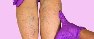
One of the late signs of the disease is a brownish tint to the skin (above the ankle). The change in color is a consequence of the penetration of red blood cells into the skin from distended veins.
Symptoms
The disease has a number of characteristic symptoms:
- pain in the leg, aggravated by walking and prolonged standing;
- feeling of heaviness, fullness;
- swelling of soft tissues;
- local increase in temperature;
- change in skin color in the affected area - it becomes bluish and shiny;
- the network of superficial veins becomes visible.
Often, blood clots in deep veins do not manifest themselves for a long time - problems with blood flow are partially compensated by other elements of the vascular bed. But even slight swelling in the ankle area may indicate the presence of thrombosis.
If you have any of the above symptoms, you should immediately consult your doctor. He will conduct an examination, make a preliminary diagnosis and issue a referral to a phlebologist.