Diseases of the vascular system are considered the most dangerous pathologies, because they are the leading causes of sudden death. To study the condition of blood vessels, a highly informative and safe diagnostic method is used - ultrasound. Unlike conventional ultrasound, when examining blood vessels, special devices with the Doppler effect are used, which make it possible to assess not only the condition of the vessel, but also the nature of the blood flow.
During the study, a diagnosis of the size of the vessel, its shape, tortuosity, condition of the walls, the presence of various neoplasms and other features is carried out. This gives the doctor a detailed clinical picture of the existing pathologies and helps to choose the most optimal and effective method of treatment. In addition to ultrasound-guided diagnostics, a wide range of therapeutic procedures on arterial and venous vessels is performed.
How is ultrasound of blood vessels performed?
The operating principle of the device is based on the effect of reflecting sound waves, thanks to which the doctor can obtain a visual image of the internal cavities of the body. To study blood vessels, ultrasound machines with the Doppler effect are used, thanks to which you can see the movement of blood in the vessel. The doctor can determine the speed and direction of blood flow, vessel fullness, level of blood supply to tissues and other indicators of the circulatory system.
Special preparation is needed only before the abdominal vascular ultrasound procedure. For 3 days you must follow a special diet that reduces gas formation in the intestines and take sorbents. Before the examination, you need to take a shower; you cannot use medicated creams and ointments on the examination sites. The day before you should not drink alcohol, energy drinks, strong tea and coffee, or smoke. In consultation with the attending physician, it is advisable to discontinue vascular medications.
Types of vascular studies:
- Doppler ultrasound (USDG). The study allows you to obtain a two-dimensional image of the vessels being examined.
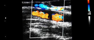
- Ultrasound duplex scanning (USDS). During the study, the condition of the vessels and the nature of blood flow are assessed.
How is an ultrasound of the veins of the lower extremities performed?
An ultrasound examination is important if you suspect the presence of serious diseases, such as thrombosis or varicose veins. A specialist will determine with 100% accuracy the area where pathological processes occur (formation of blood clots, slowing of blood flow, expansion and inflammation, damage to the walls).
To perform an ultrasound, you do not need to prepare and shave hair from the area, limit physical activity, follow a diet, etc. The procedure does not take much time and does not require complex and uncomfortable preparation.
Since ultrasound of the veins of the lower extremities is performed to determine the disease, excellent quality equipment is required. The beam must be accurately reflected from the red blood cells. Thanks to this, the doctor sees what is happening in the veins, vessels and arteries. Therefore, in a few minutes you can assess the condition of valves, deep and superficial veins, blood flow speed, etc.
What is duplex scanning?
The best modern diagnostic method is ultrasound scanning, since the quality of the results obtained is maximum. In the West, preference is given to color mapping. It increases the accuracy of the information if you need to track the speed of blood flow towards and away from the sensor (marking is done in red and blue). The method allows you to perform the following tasks:
- Monitor damage and deformation of the venous walls.
- Find out the speed of blood flow in the deep and superficial veins.
- Check the functionality of the valves.
- Assess the dimensions and density of the thrombus (determine the degree of thrombosis).
Classical and minimally invasive operations are always performed with 100% accuracy, as ultrasound of the veins of the lower extremities is performed in combination with duplex scanning.
Before visiting a specialist, be sure to take a shower. There is no need to shave your leg hair or deep clean your skin. It is advisable not to drink alcohol on the eve of an ultrasound examination. The procedure is performed as follows:
- In the office you undress from the waist down (down to your underwear).
- Ultrasound gel is then applied to the legs.
- The specialist checks the condition of the veins, valves, vessels and arteries using a device with a sensor.
- If necessary, the intensity of pressure and the power of the beam are increased to more clearly see the condition of the deep veins.
- Your body position changes—your doctor may ask you to stand up or lie down to see how the blood flows. For more information, you will need to hold your breath (take a deep breath).
Now you know exactly how an ultrasound scan of the veins of the lower extremities is performed. The session lasts a few minutes and you will not experience any discomfort. There is no pain. During the change in ultrasound intensity, you will not feel anything (many people fear pain or discomfort).
Do not forget that only a competent phlebologist with extensive experience can decipher the result. Therefore, immediately after a duplex scan or ultrasound, go to an appointment with a specialist so that he can make a final conclusion.
The First Phlebology Center will determine the condition of the veins of the lower extremities and prescribe an effective course of treatment for you. You can sign up for an examination right now by phone.
What veins and arteries are examined using ultrasound?
- Doppler ultrasound of the vessels of the neck, brain, eyes.
- Ultrasound diagnostics of veins and arteries of the lower and upper extremities.
- Ultrasound scanning of the vessels of the pelvis, abdominal cavity and kidneys.
- Ultrasound examination of heart vessels.
- Duplex scanning of the embryo.
- Ultrasound scanning/ultrasound scanning of the penis, scrotum.
During an ultrasound examination, the doctor can identify a wide range of vascular pathologies - aneurysm of the aorta or other vessels, varicose veins, phlebitis and thrombophlebitis, dissection and occlusion of vessels, atherosclerosis, etc.
In a medical clinic you can do ultrasound of blood vessels at the best price in St. Petersburg. The study is carried out using modern expert-level ultrasound equipment, and the clinic’s specialists have extensive experience in performing ultrasound diagnostics. Based on the results of the examination, you can get advice from a specialized specialist at the clinic. You can make an appointment by phone: +7.
Who is indicated for vein ultrasound?
An ultrasound examination is performed if a person has venous disease or symptoms associated with it:
- Large protruding veins are visible under the skin on the lower extremities;
- A characteristic star-shaped vascular pattern appeared;
- Severe swelling is observed in the mornings or evenings;
- After physical activity, your legs hurt, there is heaviness and discomfort;
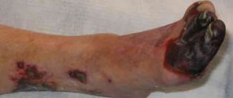
- Wounds appear on the skin that do not heal for a long time;
- Feet are constantly cold;
- The skin on the legs changes color.
Experts recommend that you undergo an ultrasound examination of the veins in your legs once a year. It is recommended to undergo diagnostics twice a year if you have diabetes or obesity, if you have bad habits, as well as if you are taking combined oral contraceptives or if there have been cases of vascular diseases in your family.
Typically, a referral for an ultrasound is given for existing diseases:
Vein thrombosis
This is a pathological process that is characterized by an increased risk of blood clots.
This disease is dangerous - it can even be fatal. To avoid this, you need to start treatment immediately and be constantly monitored by a doctor. Postthrombophlebitic disease.
It is a complication of thrombosis.
Varicose veins and venous insufficiency.
These diseases are characterized by impaired circulation in the veins.
Varicothrombophlebitis.
It manifests itself in severe pain and, if left untreated, leads to serious complications.
Angiodysplasia.
Loss of vascular functionality. It can only be treated surgically. Ultrasound in this case is necessary to monitor the effectiveness of treatment.
Congenital anomalies of vascular development on ultrasound
Hypoplasia, that is, a small diameter of the vertebral arteries, is a fairly common finding in the practice of ultrasound diagnosticians. The most typical form of its manifestation is hypoplasia of one of the vertebral arteries with a decrease in blood flow in its basin. Often there is a high entrance of one or two arteries into the spinal canal, while part of the path of the vertebral artery is not protected by the bone canal and can be compressed when turning the neck. Less common are the absence of any of the vertebral arteries and hypoplasia of the carotid arteries. An ultrasound of the neck vessels will clearly show the presence of hypoplasia, but we will not always be able to say whether this is a congenital anomaly or an acquired narrowing of the vessel due to osteochondrosis. This pathology requires conservative vascular treatment in order to increase the general blood flow of the body so that compensation occurs at the expense of other arteries. The effectiveness of the treatment is usually monitored using a series of control ultrasounds of the neck vessels.
Deep vein thrombosis
Thrombosis in the early stages may be asymptomatic.
The main classic symptoms of deep vein thrombosis are: increased temperature in the affected area. The general body temperature may rise to 39 degrees. Bursting pain and heaviness in the legs appear, the color of the skin is bluish. During the first two days after the formation of thrombosis, the symptoms are mild. As a rule, this is not pronounced pain in the calf muscle, intensifying during movement and when touched, and swelling. Subsequently, the condition worsens and swelling increases. The risk of blood clot rupture increases. Within a few days after the formation of a blood clot, superficial veins appear. Indications for ultrasound scanning for the following symptoms: - Presence of swelling of the legs; -Cold feet; -Pain in the calf and feeling of “heavy legs”; - Convulsions; - Suspicion of changes in the blood vessels of the legs (especially during pregnancy); - Periodic numbness of the legs; -Itching in the legs, without external skin changes; -Diabetes; -Decreased vascular tone; - Suspicion of atherosclerosis; - Suspicion of varicose veins or other changes in venous blood flow; - Trophic changes in the skin; -Heart problems (angina or heart attack); - Migraines and fainting; -Pain when walking or exercising; -Increased cholesterol levels.
How to study the vessels of the neck
When visiting an ultrasound diagnostician’s office, the doctor usually asks you to remove jewelry from your neck and ears and put your long hair in a ponytail so that nothing interferes with the examination process. Next, the patient lies on a flat couch, with his head towards the doctor. The gel is applied to the skin of the neck and the sensor is moved over it. The head is thrown back or tilted. First, the carotid artery is examined, then all other vessels located in the neck.
The procedure lasts no more than 45 minutes. The doctor may also conduct the examination in a sitting or standing position and ask you to tilt your head. At the end of the procedure, the doctor gives the results of the study to the patient.
ADVANTAGES AND DISADVANTAGES OF THE METHOD
Patients who have been prescribed an ultrasound examination of leg veins may be interested in learning information about the disadvantages and advantages of this method:
- The procedure is informative and allows to identify a wide range of pathological conditions of the vessels of the lower extremities;
- It is completely safe for the body, ultrasound can be performed an unlimited number of times;
- It is not painful and does not even cause discomfort;
- Suitable for people of any age.
- The only disadvantage we can note is the relatively high cost, on average from 1000 rubles.
How is ultrasound of the vessels of the brain and neck performed?
Diagnostics together with decoding takes about 40-50 minutes. The patient is asked to lie on the couch and tilt his head back slightly so that the doctor can comfortably reach the vessels being examined. A conductor gel must be applied to the skin. Next, a sensor transmitting ultrasonic waves is applied to certain areas on the head and neck. First, the condition of the vessels of the brain is assessed, and then the cervical spine. The received data is projected onto the monitor screen.
During an ultrasound of the cervical vessels, the patient should generally lie still and not move, although the doctor may ask you to turn your head or hold your breath. After the procedure, the uzist will issue a sheet with the examination results, based on which the attending physician will be able to make the correct diagnosis.
What is examined during ultrasound of cerebral vessels
Ultrasound scanning of the vessels of the brain of the head is carried out in segments. For the doctor, the reference points are the cervical cartilage, vertebrae, and skull bones.
What can be seen during diagnosis:
- Condition of the vertebral arteries.
- Causes of development of vascular diseases.
- Structure of the carotid arteries.
- The nature of the venous outflow: through the sinuses of the brain and veins.
- Blood flow through the main vessel of the brain (basilar artery).
- The condition and structure of the entire arterial chain of the head and neck.
- Pathologies of arterial trunks, etc.
Each section of the arterial system has its own designation, for example, the vertebral artery is marked with the letter V and has 5 segments to be studied.
When interpreting ultrasound and making a diagnosis, the linear velocity of blood flow must be taken into account. If the condition of the veins is assessed, then indicators of the phasing of blood flow are taken into account (depending on the diameters of the vessels, lumen and other indicators). The gaps are also checked for the presence of neoplasms: plaques in the arteries, blood clots in the veins.
The diagnostic results obtained are always compared with generally accepted standards. Only after a thorough examination of the entire venous-arterial network of the brain can the doctor find the cause of the development of the ailment and select an effective treatment.