Types and purposes of ultrasound of the veins of the lower extremities
In recent decades, ultrasound diagnostics has become a simple, inexpensive, and most importantly, accessible research method.
The quality and capabilities of ultrasonic devices are constantly being improved, their classification is constantly expanding. The great advantage of the technique is its non-invasiveness. Vein ultrasound is no exception. Modern ultrasound scanners allow a thorough diagnosis of the venous system.
The main goals of ultrasound of the veins of the lower extremities are to assess the condition of the vessels (their diameter, course, relationship with other structures, wall condition), examine blood flow parameters, and identify blood clots. The article will talk about the types and purposes of ultrasound examination of blood vessels, as well as how ultrasound of the veins of the lower extremities is performed.
Due to the constant improvement of devices, many terms have appeared to denote various components of ultrasound examinations and their combinations.
Let's try to figure out what ultrasound, DS, ultrasound, ultrasound of veins are.
- Doppler ultrasound (Doppler ultrasound) is a method based on the Doppler effect. This effect consists of changing the frequency of an ultrasonic wave when reflected from a moving object. In the lumen of the vessel, such objects are formed elements of blood, for example, red blood cells. Computer processing of reflected signals allows one to obtain a curve of the speed characteristics of blood flow. Currently, ultrasound scanning in its pure form is not used, but is included in the structure of other studies.
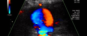
- DS (duplex scanning (USD)) - allows you to visually evaluate the vessel - its wall, course, lumen, as well as conduct an ultrasound scan of any part of the vessel - i.e. in addition to examining the vessel, show the characteristics of blood flow at any point. Modern devices also use coloring of blood flows in blue and red colors, based on the Doppler effect (the so-called color Doppler or color Doppler mapping (CDC)). Combining duplex scanning with color flow is also called triplex scanning.
- USAS (ultrasound angioscanning) - a synonym for DS, is the same.
- Ultrasound of veins (ultrasound examination of veins) - this term refers to the entire combination of the above methods. Modern devices provide all the components of the study - ultrasound, DS, and color Doppler mapping (CDC).
Currently, when a doctor prescribes an ultrasound scan of the veins of the lower extremities, an ultrasound scan of the veins of the lower extremities, he means that the patient will undergo an ultrasound scan of the veins according to all possible parameters.
Duplex scanning
This is one of the types of ultrasound examination. The result is a color image of the blood flow in the vessel. The method is completely safe, non-invasive and does not expose humans to x-ray radiation.
Since the combination of Dopplerography and B-mode has more capabilities than Doppler ultrasound. This diagnostic method can be performed an unlimited number of times.
The examination takes place in an outpatient clinic, as with other ultrasound examinations, a gel is applied to the patient’s skin, the procedure lasts about half an hour. However, the resulting picture is highly informative. The vessels are visible in both longitudinal and transverse sections, but only in an area that is not very significant in area. Both veins and arteries are equally clearly visible.
Experts believe that the only competitor to such scanning can be X-ray contrast angiography, which is invasive and is not suitable for everyone due to health reasons.
If the goal is to track the dynamics of recovery and it is necessary to determine the condition of the vessels several times, then it is better to resort to the duplex method.
Contraindications to the use of in-depth studies
Hardware methods, despite their information content and the most gentle approaches, still have a number of contraindications. These include:
•Allergies to contrast solution;
•Kidney, heart, liver failure;
•Infectious diseases and inflammations;
•Chronic diseases in the acute stage;
•Psychical deviations.
Diagnosing the condition of blood vessels in pregnant and lactating women, elderly patients, and people with bleeding disorders requires extreme caution.
Warning: Undefined array key “module” in /customers/0/e/9/germanklinik.de/httpd.www/control/config/inc/templates_c/front^7198fc69815638cccb44869b7c86cae91ff823b0_0.file.DefaultGK.tpl.php on line 488 Warning: Attempt to read property “value” on NULL in /customers/0/e/9/germanklinik.de/httpd.www/control/config/inc/templates_c/front^7198fc69815638cccb44869b7c86cae91ff823b0_0.file.DefaultGK.tpl.php on line 488 Warning : Undefined array key “extension” in /customers/0/e/9/germanklinik.de/httpd.www/control/config/inc/templates_c/front^7198fc69815638cccb44869b7c86cae91ff823b0_0.file.DefaultGK.tpl.php on line 490 Warning: Attempt to read property “value” on NULL in /customers/0/e/9/germanklinik.de/httpd.www/control/config/inc/templates_c/front^7198fc69815638cccb44869b7c86cae91ff823b0_0.file.DefaultGK.tpl.php on line 490
Thank you! We have received your request for consultation.
If you want to fill out a Request for Treatment, continue filling out. Having all the necessary data, we will be able to find the best solution to your problem in the shortest possible time
Who needs to undergo Doppler ultrasound of the extremities?
An ultrasound examination of the veins is usually prescribed by a doctor (surgeon, phlebologist, therapist). Indications for it can be a wide variety of conditions. Most often, the doctor prescribes a study if there are complaints and suspicions of diseases of the venous system. In other cases, ultrasound of the veins is prescribed to exclude hidden pathology of the venous system (for example, before operations, during pregnancy).
The main indications for prescribing ultrasound examination of veins are:
- Swelling of one or both legs
- Leg pain
- Feeling of heaviness and fatigue in the legs
- Night cramps in the legs
- Varicose veins, spider veins
- Before any operations
- Late pregnancy
Indications for use
There are three groups of indications for diagnosis.
Suspicious symptoms, the origin of which needs to be clarified. Among them:
- Pain of unknown origin in the legs.
- Heaviness in the lower extremities.
- Dilated veins. Tortuosity of blood vessels. The appearance of spider veins. The described signs are early visual symptoms of varicose veins.
- Exercise intolerance.
- Intermittent claudication. A typical symptom of atherosclerosis. When an attack of acute pain begins in response to physical activity.
- Edema. In the morning or evening, after a hard day at work.
- Inadequate temperature perception. Chilliness or excessive sweating of the feet, a feeling of heat regardless of the ambient temperature.
- Cramps.
- Paresthesia. Feeling of goosebumps, tingling, numbness of the skin.
- Pallor of the skin of the legs.
The second group of indications are controversial diagnostic findings obtained as a result of other examinations. Including laboratory tests: blood biochemistry, blood lipid studies.
Phlebologists and vascular surgeons prescribe a systematic examination for patients from high-risk groups:
- Persons over 35 years of age.
- People who smoke and drink.
- Patients with diabetes mellitus, hypertension.
- People with a family history.
- Persons with physical inactivity and lack of physical activity.
- Patients who are overweight.
The frequency of preventive examinations depends on the severity of the clinical case. On average, we are talking about a procedure every 6-12 months.
How is the examination done?
Many patients are concerned about the question of how ultrasound examination is performed, whether there are any contraindications or painful sensations during the examination.
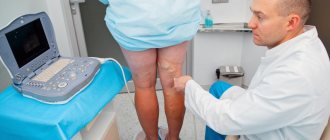
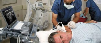
Ultrasound diagnostics is non-invasive, painless, does not cause any discomfort, and has no contraindications. An ultrasound can be performed either by an ultrasound diagnostics doctor or by a surgeon or phlebologist who knows this method and has undergone appropriate training.
The examination of the veins of the lower extremities is carried out from the inguinal fold to the ankles. The patient completely frees his legs (panties can be left on) and lies down on the couch. The doctor applies a special gel to the lower limbs, along which the sensor will be moved during the examination. The gel is needed to ensure good contact between the skin and the sensor so that there is no obstacle to the passage of ultrasonic waves. The examination can be carried out both lying down and standing. At certain moments, the doctor may ask you to hold your breath, strain, bend your leg, etc.
The study lasts from 10 to 40 minutes (depending on the structural features of the veins and the identified pathology). For example, detected varicose veins increase the examination time, because it is necessary to carry out a number of additional tests. At the end of the study, the doctor gives the patient paper napkins to remove any remaining gel. The gel is absolutely harmless and does not leave stains on clothes.
Then the doctor writes a conclusion and hands it to the patient. It should be remembered that the conclusion itself is not a diagnosis, but only describes the identified ultrasound signs. The diagnosis can only be made by a clinician (surgeon, phlebologist), based on a comparison of the ultrasound picture, examination data and other examinations. In many cases, an ultrasound of the veins is performed not by an ultrasound diagnostics doctor, but by a phlebologist who is proficient in vein ultrasound. Then a clinical diagnosis can be made immediately.
Advantages of the method
There are several advantages:
- Information content. The method is suitable for diagnosing most vascular pathologies.
- Complete security. There is no harmful burden on the body.
- Possibility of repeated implementation.
- No discomfort or pain.
- Availability. The devices are available in most private and public clinics.
- Relatively low cost.
The examination is carried out according to indications, as many times as the situation requires. It is used for monitoring the condition, dynamic observation, and assessing the quality of treatment.
How to decrypt data
Only a doctor can determine what an ultrasound examination of the veins of the lower extremities shows.
The doctor evaluates:
- Vein diameter
- Condition of the vascular wall (irregularities, thickening)
- The condition of the veins of the right and left legs is compared
- Valve condition
- Presence of pathological blood flows (refluxes)
- Vessel lumen (presence of blood clots, their structure, level of extent, apex)
- Presence of varicose veins
- Presence of incompetent perforators (veins connecting superficial and deep veins)
If during the examination the doctor notices changes in other structures that are not related to the veins (for example, pathology of the arteries, Baker's cyst, altered lymph node), he may note this in the protocol or recommend additional research.
Based on the data received, the doctor writes a conclusion; if necessary, photographs are attached to the conclusion.
Example of a conclusion: “Echo signs of varicose veins of the lower extremities. Insufficiency of the great saphenous vein on the left. Incompetent perforator of the left leg." It should be noted that the classification of varicose veins according to this conclusion is impossible - the stage is determined by the attending phlebologist based on a comparison of the ultrasound picture with clinical signs.
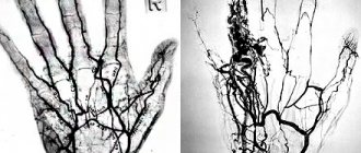
Angiography
Angiography is used to examine arterial dysfunction. This type of diagnosis will be called arteriography. If the goal is to determine the condition of the veins, then they talk about phlebography.
In any case, the procedure takes place in a hospital where there is an X-ray angiography room.
In order to determine the condition of the veins and arteries, it is necessary to inject a contrast agent and take x-rays of its progress through the body. Atherosclerosis, thrombosis, dilation of the vessel, aneurysm, thrombophlebitis will be clearly visible.
Where to take the study
Currently in Moscow there are many medical centers that offer ultrasound diagnostics. What should you look for in order to get to a good specialist?
The doctor must have extensive experience, work experience, and have positive reviews from other patients. It is very good to have an ultrasound of the veins done by a doctor who not only knows ultrasound diagnostics, but is also a surgeon or phlebologist. Such a doctor has the most complete understanding of the anatomy and functioning of the venous system, knows well how ultrasound of the veins of the lower extremities is performed, and can also make a clinical diagnosis and prescribe treatment immediately after the ultrasound.
Dr. Elshansky Igor Vitalievich fully meets these requirements, is a phlebologist surgeon, and also has advanced training in ultrasound diagnostics in angiology, has been involved in the diagnosis and treatment of vascular diseases of the lower extremities for many years, Ph.D.
Diagnostic results
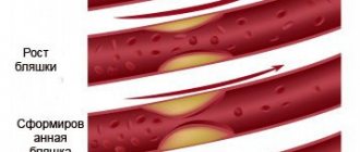
In pathologies, the technique demonstrates a narrowing of the vascular lumen. It is due to the following problems:
presence of atherosclerotic plaques
assessment of the speed of blood movement, study of blood filling of vessels, narrowing of the vascular lumen due to thickening of the walls, presence, localization of parietal thrombi, assessment of the functional ability of venous valves, assessment of the phasing of blood flow during exhalation, inhalation, pathological tortuosity, vasospasms, aneurysms, various damage to the arterial wall.
The technique is completely reliable, harmless and painless. This allows you to repeat the procedure as necessary.